[box type=”bio”] Learning Point of the Article: [/box]
It is important to remember the importance of sending neoplastic samples for histopathology as various tumors can often mimic each other on clinical exam.
Case Report | Volume 9 | Issue 1 | JOCR January – February 2019 | Page 3-5 | Jackson R. Staggers, Jeffrey M. Pearson, Nileshkumar M. Chaudhari. DOI: 10.13107/jocr.2250-0685.1282
Authors: Jackson R. Staggers[1], Jeffrey M. Pearson[1], Nileshkumar M. Chaudhari[1]
[1]Department of Orthopaedics, University of Alabama at Birmingham, 1313 13th St South, Birmingham, AL 35205.
Address of Correspondence:
Dr. Jackson R. Staggers,
Department of Orthopaedics, University of Alabama at Birmingham, 1313 13th St South, Birmingham, AL 35205
E-mail: rucker@uab.edu
Abstract
Introduction: Capillary hemangiomas and pyogenic granulomas are benign vascular neoplasms that are usually identified clinically by their characteristic features. Capillary hemangiomasmost commonly develop in infancy on the head and neck and nearly all spontaneously ingress by the teenage years. Pyogenic granulomas, however, typically present in adults and can be induced by trauma. It is exceedingly rare for capillary hemangiomas to present in adulthood or after trauma. We present an extremely unusual case of capillary hemangioma on the tip of the finger of an adult male presenting immediately after a burn. The mass was clinically diagnosed as pyogenic granuloma but histopathologically diagnosed as a capillary hemangioma. To our knowledge, this is the only presentation of its kind.
Case Report: A 29-year-old African American, right-hand-dominant male laborer presented to the outpatient orthopedic hand clinic with a 2–3-week-old growing mass on the tip of the right small finger. A clinical diagnosis of pyogenic granuloma was made. Silver nitrate therapy was ineffective, though surgical excision resulted in complete resolution of the mass. Surprisingly, the histopathological diagnosis was instead consistent with capillary hemangioma.
Conclusion: Clinicians should maintain a high clinical suspicion for both pyogenic granulomas and capillary hemangiomas in children and adults with a vascular soft tissue mass, even after trauma. With this in mind, health-care providers should maintain a low clinical threshold to send soft tissue masses for histopathology to obtain an accurate diagnosis and to provide the best care possible.
Keywords: Adult, benign vascular neoplasm, capillary hemangioma, hand, pyogenic granuloma, vascular malformation.
Introduction
Capillary hemangiomas and pyogenic granulomas are well-known benign vascular neoplasms. Although they have macroscopic features that can typically allow them to be identified clinically, a diagnosisis most accurately madehistologically [1]. Pyogenic granulomas, also known as lobular capillary hemangiomas, can appear at any age, though they are most common in children and young adults. They are most commonly found on fingers and mucousmembranes [1]. Several predisposing factors have been identified for pyogenic granulomas, though as many as 76.7% may occur spontaneously. These predisposing factures includetrauma, foreign body reactions such as bug bites, and some dermatologic conditions [2, 3]. They are identified clinically as solitary red papules or polyps that grow rapidly over weeks to months. These lesions rarely resolve spontaneously, and therefore, surgical removal is often required as they can bleed [4, 5]. By comparison, capillary hemangiomas are the most common vascular tumors of infancy and can be found on skin, mucous membranes, and internal viscera [1]. Many occur sporadically, though there is likely a genetic association. They almost always present before the age of one, and it is extremely rare for them to develop past the teenage years [6]. Clinically, they are identified as bright red papules, nodules, or plaques. These lesions can grow rapidly, but most do not require treatment, as 90% will regress by age 9 [1, 2, 7]. Capillary hemangiomas can also be treated with laser therapy and beta-blockers; surgery is rarely indicated [8, 1]. We present an extremely unusual case of capillary hemangioma on the tip of the finger of an adult male presenting immediately after a burn. The mass was clinically diagnosed as pyogenic granuloma but histopathologically diagnosed as a capillary hemangioma. To our knowledge, this is the only presentation of its kind.
Case Report
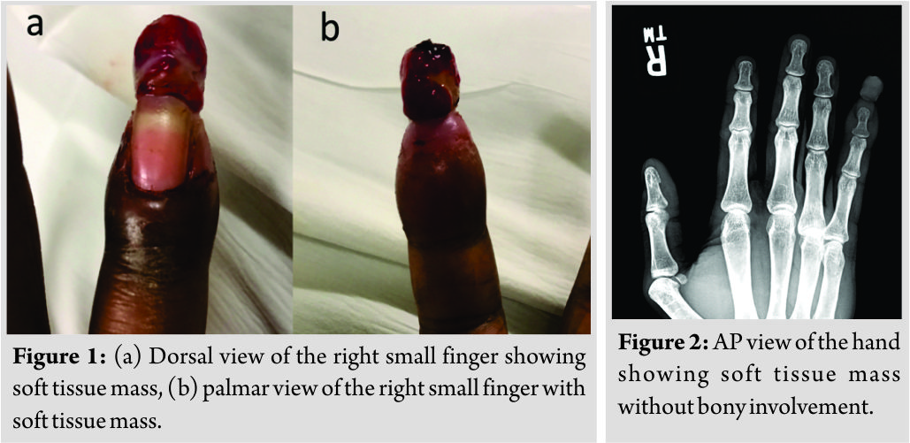
A 29-year-old African American, right-hand-dominant male laborer presented to the outpatient orthopedic hand clinic with a 2–3-week-old growing mass on the tip of the right small finger. He first noticed the mass after burning the tip of the finger. Over the next 2 days, he developed a blister that ruptured and bled profusely. Soon after, a soft tissue mass began growing at this location, prompting him to come to the clinic. His only other complaint was swelling of the finger over the past 2 days. Otherwise, the patient denied pain, fevers, weakness, or numbness in the hand, or other complaints. Physical examination showed a 1.6 cm × 1.1 cm × 1.1 cm reddish-brown, oval-shaped soft tissue mass extending from the right small finger on a broad-based stalk (Fig. 1). There was mild swelling of the finger. The mass was painful at its base if moved, but sensation and motor function were intact throughout. Examination of the hand was otherwise normal. Multiview radiographs of the hand showed a soft tissue mass extending from the distal aspect of the right small finger without bony involvement (Fig. 2). 
Discussion
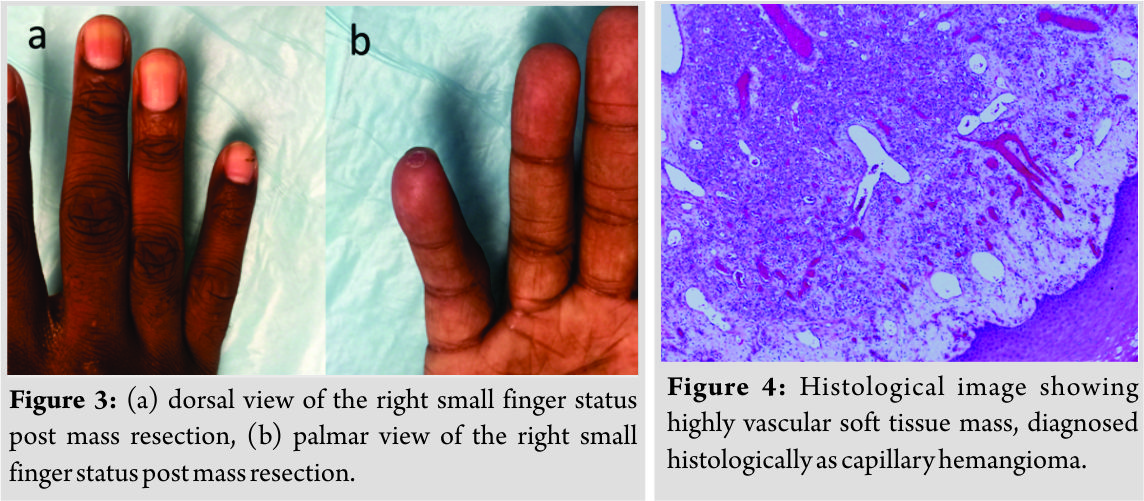
Previous reports have shown that capillary hemangioma is often confused with pyogenic granulomas [2]. A report by Jananni et al. described two cases of adult capillary hemangioma where both clinically resembled pyogenic granuloma[4].The first case was a 24-year-old male with a longstanding sessile mass of the mouth, and the second case was a 50-year-old female who developed a sessile mass in the mouth after trauma. Both cases were treated successfully with surgical excision. Similarly, our case was difficult to identify clinically and required histological examination for proper diagnosis, though these cases occurred within the mouth and not the hand. Post-traumatic capillary hemangiomas of the hand are exceedingly rare and have only been described in a few previous reports. Habibi et al. described the case of a 12-year-old who developed multiple hand nodules after trauma [9]. The only other case of capillary hemangioma of the hand after trauma was published in 1974 by Ben-Menachem and Epstein [10]. In this case, an adult formed nodular masses in the hand soon after a crush injury. Our case is unique in that it occurred in an adult, was exophyticin nature, and occurred after burn. The ambiguity that is associated with these lesions can be rather confusing and can lead to erroneous diagnosis of other more serious conditions, such as squamous cell carcinoma, Kaposi’s sarcoma, and bacillary angiomatosis. Capillary hemangioma and pyogenic granuloma have characteristic features that typically allow for them to be diagnosed clinically, though histopathology is the most accurate means of diagnosis. Microscopic examination of pyogenic granuloma shows the lobular arrangement of capillaries surrounded by stoma that may contain inflammation and granulation tissue. Plump endothelial cells line the capillaries, and vessel lumen may vary in size [5]. Capillary hemangiomas also contain lobules of thin-walled capillaries, though they express glucose transporter 1 which differentiates them from other lesions, including pyogenic granuloma [7].
Conclusion
Clinically, a vascular soft tissue mass in an adult that presents after trauma represents several key characteristics of a pyogenic granuloma. Interestingly, histological examination, in this case, showed the mass to be a capillary hemangioma. Multiple rounds of silver nitrate therapy were ineffective, though surgical resection provided excellent results without recurrence. This report highlights the importance of having a high clinical suspicion for both pyogenic granulomas and capillary hemangiomasin children and adults with a vascular soft tissue mass somewhere on the body. With this in mind, health-care providers should maintain a low clinical threshold to send soft tissue masses for histopathology to obtain an accurate diagnosis and to provide the best care possible.
Clinical Message
Physicians should have a high index of suspicion for both pyogenic granulomas and capillary hemangiomas in children and adults with a vascular soft tissue mass somewhere on the body, as they can closely resemble each other clinically.
References
1. Kumar V, Abbas AA. Robbins and Cotran Pathologic Basis of Disease. 9th ed. Philadelphia, PA: Elsevier Saunders; 2005.
2. Rachappa MM, Triveni MN. Capillary hemangioma or pyogenic granuloma: A diagnostic dilemma. Contemp Clin Dent 2010;1:119-22.
3. Pagliai KA, Cohen BA. Pyogenic granuloma in children. Pediatr Dermatol 2004;21:10-3.
4. Akbulut F, Akbulut T, Kucukdurmaz F, Sonmezay E, Simsek A, Gurbuz G, et al. Huge pyogenic granuloma of the penis. Case Rep Urol2015;2015:263168.
5. Lawley L. Pyogenic Granuloma (Lobular Capillary Hemangioma). 2016. Available from: https://www.uptodate.com/contents/pyogenic-granuloma-lobular-capillary-hemangioma?source= search_result&search=pyogenic granuloma& selectedTitle=1~45. [Last accessed on 2017 Jan 01].
6. Mass DM, Littner DM. On the Lips in Children. 1998. p. 188-91. Available from: http://www.aapd.org/assets/ 1/25/Kaplan-20-03.pdf. [Last accessed on 2017 May 19].
7. Metry D. Infantile Hemangiomas: Epidemiology, Pathogenesis, Clinical Features, and Complications. 2017. Available from: https://www.uptodate.com/ contents/infantile-hemangiomas- epidemiology-pathogenesis-clinical-features-and-complications? source=search_result& search=capillary hemangioma&selected Title=1~39. [Last accessed on 2017 May 14].
8. Jananni M, Gubernath U, Mahendra J, Sivaramakrishnan M. Capillary hemangioma of gingiva mimicking as pyogenic granuloma: Report of two cases. J Interdiscip Dent 2012;2:218.
9. Habibi S, Agrawal S, Kadel JK, Narsimulu G. Post-traumatic capillary hemangiomas: A rare cause of hand nodules. Clin Rheumatol 2009;28 Suppl 1:S51-2.
10. Ben-Menachem Y, Epstein MJ. Post-traumatic capillary hemangioma of the hand. A case report. J Bone Joint Surg Am 1974;56:1741-3.
 |
 |
 |
| Dr. Jackson R. Staggers | Dr. Jeffrey M. Pearson | Dr. Nileshkumar M. Chaudhari |
| How to Cite This Article: Staggers J R, Pearson J M, Chaudhari N M. Capillary Hemangioma of the Finger in an Adult after a Burn: A Unique Case Mimicking Pyogenic Granuloma. Journal of Orthopaedic Case Reports 2019 Jan-Feb; 9(1): 3-5. |
[Full Text HTML] [Full Text PDF] [XML]
[rate_this_page]
Dear Reader, We are very excited about New Features in JOCR. Please do let us know what you think by Clicking on the Sliding “Feedback Form” button on the <<< left of the page or sending a mail to us at editor.jocr@gmail.com


