[box type=”bio”] Learning Point of the Article: [/box]
Although, parosteal lipoma accounts for less than 0.3% of all lipomas it is imperative to diagnose and treat early so as to alleviate future complications.
Case Report | Volume 9 | Issue 3 | JOCR May-June 2019 | Page 46-48 | Shishir Murugharaj, Shuaib Ahmed, Harsh Abhay, Syed Najimudeen. DOI: 10.13107/jocr.2250-0685.1414
Authors: Shishir Murugharaj[1], Shuaib Ahmed[1], Harsh Abhay[1], Syed Najimudeen[1]
[1]Department of Orthopaedics, Pondicherry institute of Medical Sciences, Kalapet, Pondicherry, India.
Address of Correspondence:
Dr. Shuaib Ahmed,
Department of Orthopaaedics,Pondicherry institute of Medical Sciences, Kalapet, Pondicherry -605014, India.
E-mail: shuaibahmed99@gmail.com
Abstract
Introduction: Lipomas are considered to be benign tumors comprising 50% of all soft tissue tumors. They originate from mesodermal germ layer but are classified based on component tissue and location. Parosteal lipomas are frequently located at the extremities, particularly at diaphysis or diametaphysis of long bones.
Case Report: Here, we report a case of parosteal lipoma with a delayed presentation involving dominant right forearm without any neurological deficits to create awareness of the rare existence of this benign tumor.
Conclusion: A prompt diagnosis of such tumors has to be done as early as possible.
Keywords: Parosteal lipoma, forearm, benign, tumor.
Introduction
Lipomas can occur anywhere on the body but are commonly found in the subcutaneous layer[1]. There are many subtypes of lipomas and one of its rare variety is osseous involvement[2,3]. These are usually located intraosseously or adjacent to the bone attached by a pedicle through which they may derive blood supply, hence are better known as parosteal lipomas[4]. We report a case of 55-years-old otherwise healthy male involving a proximal aspect of right radius which was managed surgically at our center.
Case Report
A 55-years-old male without any comorbid conditions presented to the orthopedic outpatient department with a mass over right forearm for the past 30 years. The swelling was progressively increasing in size over the years and was not associated with any motor or sensory impairment. He was unable to eat comfortably, as it was causing a mechanical block to the elbow movement dueto its increasing size and atypical location. On examination, it was found that swelling was located over proximal half of forearm extending mainly on the anterolateral aspect. Skin overlying it was normal and non-adherent. The mass was soft in consistency and on supination and flexion of forearm muscles, it partially reduced in size indicating that it was in the submuscular plane, specifically deep to the brachioradialis. His elbow range of motion had a terminal restriction which probably was causing his disability (Fig. 1).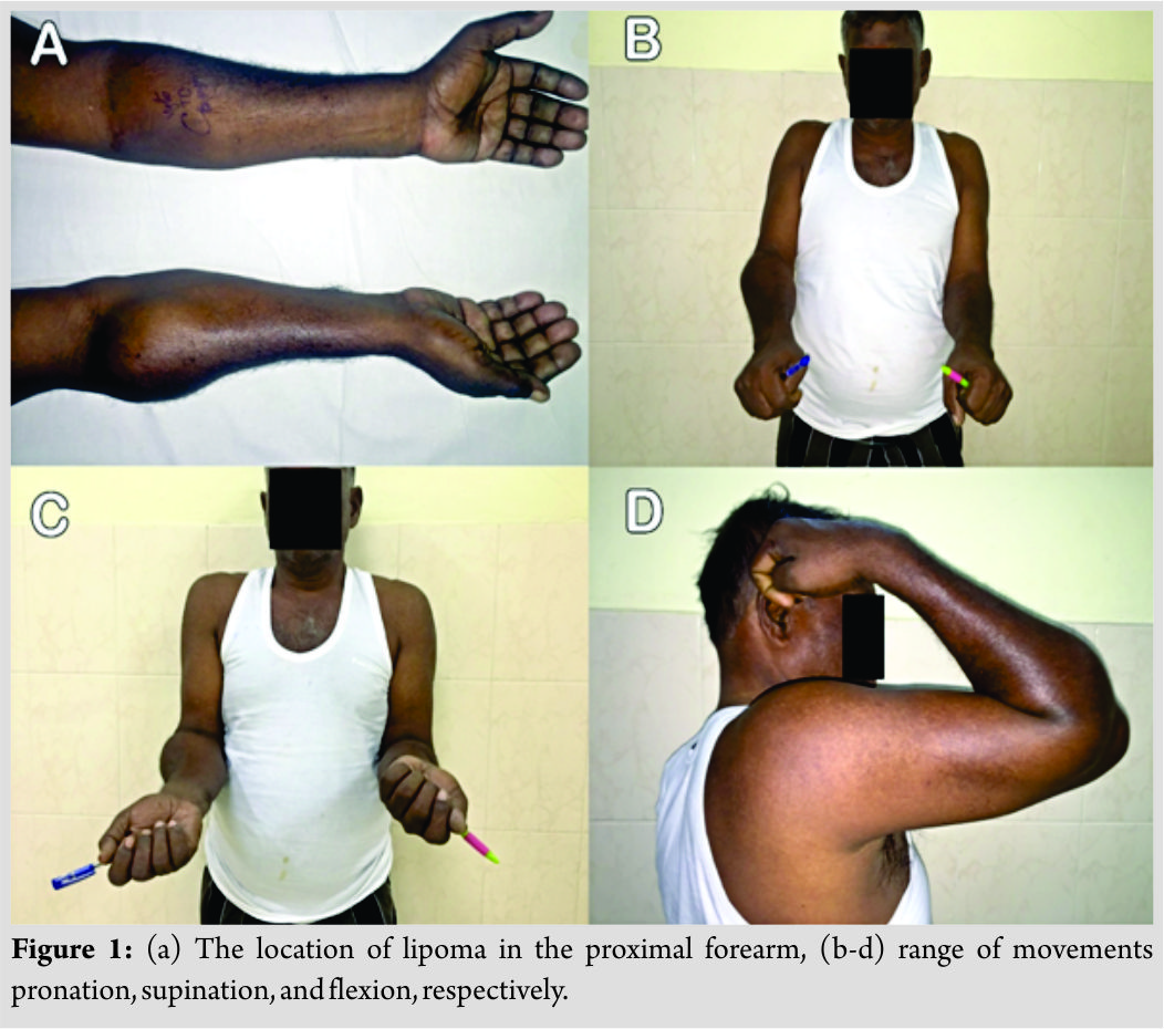
Discussion
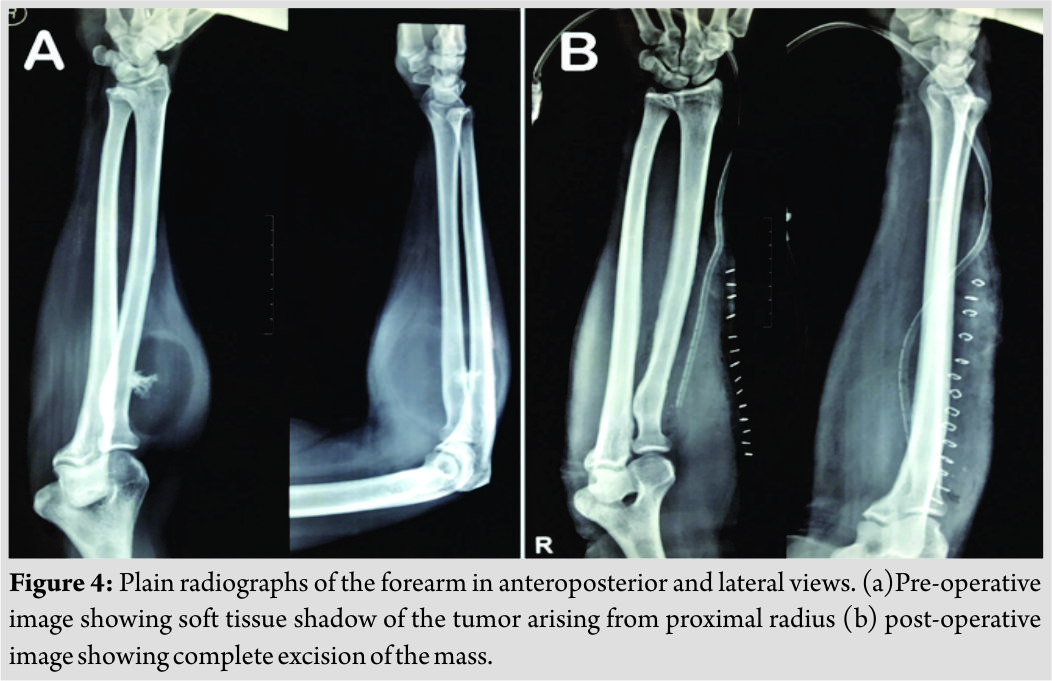
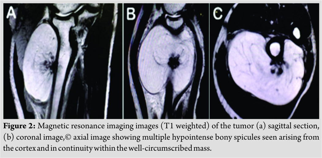
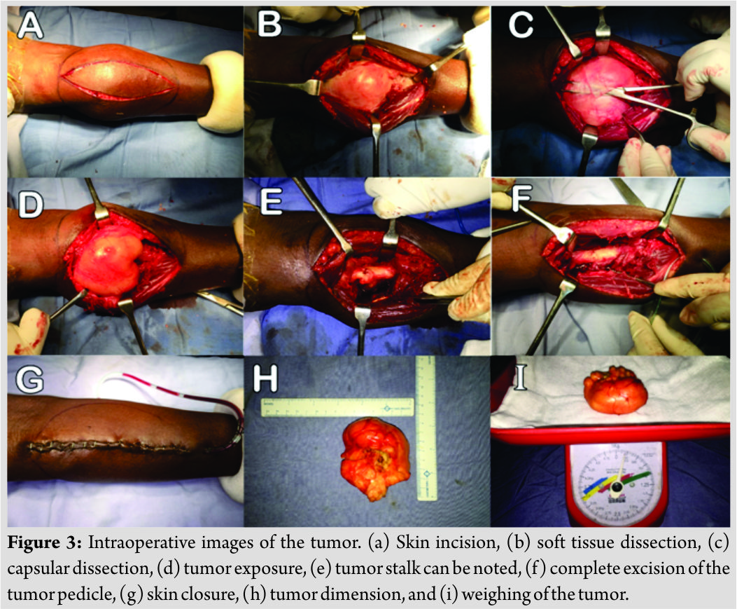
Parosteal lipomas are benign adipose tissue tumors arising from the bone cortex, particularly the periosteum. They have been described by a number of synonyms, such as “periosteallipomas,” “chondrolipomas of soft tissue,” and “lipomas of nerves.” Frequent association with chondroid and/ or osseous tissue has allowed classifying them into four distinct variants: (I) No ossification; (II) pedunculated exostosis; (III) sessile exostosis; and (IV) patchy chondro-osseousmodulation [2]. Parosteal lipoma presents as an immobile, non-tender,and slow growing mass over bones that are not fixed to the skin [5]. On plain radiographs, a parosteal lipoma is a well-defined area of lucency located adjacent to the cortex of a long bone. Very rarely they can cause bony alterations in the form of hyperostotic reactive changes [6]. Superior imaging modalities such as computed tomography (CT) and MRI show a homogeneous lobulated appearance adherent to the surface of the adjacent bone [7, 8]. Sites which are commonly involved are the proximal forearm and the sciatic nerve. MRI is considered better as compared to CT for evaluation of parosteal lipoma. These tumors on MRI appear as ajuxtacortical mass with signal intensity identical to that of subcutaneous fat and heterogeneity in these lesions are invariably present corresponding to the pathologic components in the lesion. On the one hand, T1-weighted images show intermediate signal intensity; on the other hand,T2-weighted images display high signal intensity which representsthe cartilaginous components in parosteal lipoma. Parosteal lipomas are frequently found in the extremities in contrast to the subcutaneous lipomas which are commonly located in femur, radius, tibia, and humerus [9, 10]. They have also been reported in scapula, clavicle, ribs, pelvis, metacarpals, metatarsals, and mandible [6]. The clinical presentation of these ossifying lipomas in the radius varies. They are usually slow-growing tumors with an indolent course. Very often they present as large painless lesions over a long period of time [11]. Symptoms caused by nerve compression are unusual; however, those occurring adjacent to the proximal radius may lead to compression of posterior interosseous nerve[12]. Avramand Hynes [13] in his literature review up to 2004 found out only 18 such reported cases presenting neurological involvement. Tzeng et al. [14] found superficial radial nerve involvement with proximal radius parosteal lipoma which is very rare as the superficial sensory radial nerve has more superficial and medial course than the posterior interosseous nerve. In our case, the patient presented with a mechanical block while eating rather than sensory-motor symptoms. Neurological symptoms have been well documented in the literature, but to the best of our knowledge, there were no reports described with a mechanical block. In addition to this, the duration of growth of the mass was around 30 years which are unusual. Apart from sensory-motor symptoms caused by these benign tumors, mechanical block, especially in a dominant upper limb, may be considered as a relative indication for surgery.
Conclusion
An early diagnosis of such tumors is very vital because it not only reduces the chances of speculated neoplastic changes but also improves the quality of life.
Clinical Message
Parosteal lipoma should be considered as a differential diagnosis in a proximal elbow swelling case. This clinical entity can be diagnosed on MRI scans.
References
1. Myint ZW, Chow RD, Wang L, Chou PM. Ossifying parosteal lipoma of the thoracic spine: A case report and review of literature. J Community Hosp Intern Med Perspect 2015;5:26013.
2. Miller MD, Ragsdale BD, Sweet DE. Parosteal lipomas: A new perspective. Pathology 1992;24:132-9.
3. Kapukaya A, Subasi M, Dabak N, Ozkul E. Osseous lipoma: Eleven new cases and review of the literature. Acta Orthop Belg 2006;72:603-14.
4. Kameyama K, Akasaka Y, Miyazaki H, Hata J. Ossifying lipoma independent of bone tissue. ORL J Otorhinolaryngol Relat Spec 2000;62:170-2.
5. Pompili G, Tarico MS, Scrimali L, Scilletta A, Tamburino S, Curreri SW, et al. A big parosteal lipoma of the thigh. Chirurgia 2011;24:107-10.
6. Fleming RJ, Alpert M, Garcia A. Parosteal lipoma. AJR Am Roentgenol 1962;87:1075-84.
7. Murphey MD, Johnson DL, Bhatia PS, Neff JR, Rosenthal HG, Walker CW, et al. Parosteal lipoma: MR imaging characteristics. AJR Am J Roentgenol 1994;162:105-10.
8. Yu JS, Weis L, Becker W. MR imaging of a parosteal lipoma. Clin Imaging 2000;24:15-8.
9. Schwartz K, Chevrier L. Des lipomesosteoperiostiques. Rev Chir Orthop 1906;33:76.
10. Bohm W. Ueber ‘‘periostale’’ lipome. Beitr Klin Chir 1918;111:440.
11. Piattelli A, Fioroni M, Iezzi G, Rubini C. Osteolipoma of the tongue. Oral Oncol 2001;37:468-70.
12. Nishida J, Shimamura T, Ehara S, Shiraishi H, Sato T, Abe M. Posterior interosseous nerve palsy caused by parosteal lipoma of proximal radius. Skelet Rad 1998;27:375-9.
13. Avram R, Hynes NM. Posterior interosseous nerve compression secondary to a parosteal lipoma: Case report and literature review. Can J Plast Surg 2004;12:69-72.
14. Tzeng CY, Lee TS, Chen IC. Superficial radial nerve compression caused by a parosteal lipoma of proximal radius: A case report. Hand Surg 2005;10:293-6.
 |
 |
 |
 |
| Dr. Shishir Murugharaj | Dr. Shuaib Ahmed | Dr. Harsh Abhay | Dr. Syed Najimudeen |
| How to Cite This Article: Murugharaj S, Ahmed S, Abhay H, Najimudeen S. Parosteal Lipoma of Proximal Radius: A Case Report of an Unusual Swelling and Review of Literature. Journal of Orthopaedic Case Reports 2019 May-June; 9(3): 46-48. |
[Full Text HTML] [Full Text PDF] [XML]
[rate_this_page]
Dear Reader, We are very excited about New Features in JOCR. Please do let us know what you think by Clicking on the Sliding “Feedback Form” button on the <<< left of the page or sending a mail to us at editor.jocr@gmail.com

