[box type=”bio”] Learning Point of the Article: [/box]
By accessing medial and lateral elbow separately by two separate incisions, the morbidity and wound complications of an extensile posterior approach can be reduced and also it has similar, if not better, functional results when compared to a single posterior approach.
Case Report | Volume 9 | Issue 5 | JOCR September – October 2019 | Page 78-81 | Shaligram Purohit, B Sai Gautham, Nandan Marathe, Aditya Anand Dahapute, Swapneel Shah. DOI: 10.13107/jocr.2019.v09i05.1544
Authors: Shaligram Purohit[1], B Sai Gautham[1], Nandan Marathe[1], Aditya Anand Dahapute[1], Swapneel Shah[1]
[1]Department of Orthopedics, Seth Gordhandas Sunderdas Medical College and King Edward Memorial Hospital, Mumbai, Maharashtra, India.
Address of Correspondence:
Dr. B Sai Gautham,
Department of Orthopaedics, Seth Gordhandas Sunderdas Medical College and King Edward Memorial Hospital, Mumbai, Maharashtra, India.
E-mail: saigautham90@gmail.com
Abstract
Introduction: Chronic elbow dislocation is a highly disabling condition to be treated and to provide a successful functional outcome. Surgical treatment of such conditions might result in persisting instability or stiffness of the elbow joint due to associated shortening and contracture of the soft tissues and articular incongruity. Most of the described open reduction techniques are through an extensile posterior approach which might result in increased post-operative stiffness. We report the treatment of such a case with separate medial and lateral incisions with the excellent functional outcome at 1-year follow-up.
Case Report: A 45-year-old lady with 2-month-old elbow dislocation was planned for open reduction of the joint through two separate incisions, medial and lateral. Surgical details and difficulties faced will be analyzed in this paper. The patient currently has 30–140°flexion with complete pronation-supination movements at 1-year follow-up.
Conclusion: Chronic dislocation of the elbow is a highly disabling condition and has a very unpredictable outcome. By combining an understanding in the anatomy and biomechanics of the elbow with a proper surgical technique tailored to the individual patient, it is possible to achieve a functional and painless elbow in the majority of cases. By accessing medial and lateral elbow separately, the morbidity and wound complications of an extensile posterior approach can be reduced and also it has similar, if not better, functional results.
Keywords: Elbow dislocation, medial and lateral incisions.
Introduction
Dislocation of the elbow is not an uncommon orthopedic injury in the Indian subcontinent with an incidence of approximately 20% of all articular dislocations [1]. Posterior or posterolateral is the most common type contributing 80–90% of the total dislocation. Early reduction of the elbow is very simple and has historically produced good results. Tan et al. [2] defined chronic ulnohumeral dislocation as the late unreduced dislocation that has become irreducible by closed manipulation over a period of time. It almost always requires open reduction of the articular surface with the release of associated contracted ligaments and muscles. Such chronic unreduced elbow dislocation usually results from inadequate reduction primarily, inadequate mobilization, or recurrent instability. In the Indian subcontinent, it most commonly occurs due to the initial reduction attempts by traditional bone setters and non-medical treatments. The main goals of therapy are to produce a stable functional joint with satisfactory range of motion before arthritic changes develop in the joint. Treatment of such neglected unreduced elbows is challenging as it requires extensive dissection and release of ligaments both medially and laterally before the joint can be reduced.
Case Report
A 45-year-old lady came to the outpatient department, 2 months after a history of fall on an outstretched arm and injury to the elbow and diagnosed with having a simple elbow dislocation. She was treated with an above elbow slab after the reduction maneuver by a traditional bonesetter for 6 weeks. The patient came to us after the initial treatment as she noted a deformity and severe restriction in movement of the elbow. X-ray showed a persistent elbow dislocation with no associated fractures. (Fig. 1) shows the radiographs of the left elbow joint in anteroposterior and lateral views at the time of the admission. The patient was planned for open reduction of elbow joint through separate medial and lateral incisions with the patient in lateral position.
Surgical procedure
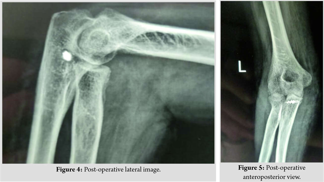
The first step was to identify and preserve the ulnar nerve through a direct medial approach. (Fig. 2) shows the scar site of the medial incision taken. In situ neurolysis of the ulnar nerve was performed between the Osborne fascia and arcade of Struthers. The contacted posterior capsule was excised and the fibrous tissue between the ulnohumeral joint which was blocking the reduction was excised completely, taking care not to damage the cartilage of the joint. Medial collateral ligament (MCL) complex and the flexor mass were found to be completely torn and contracted. Triceps muscle shortening was dealt with complete release and pie-crusting of the muscle subperiosteally. The wound was kept open to allow observation of tension in the ulnar nerve during open reduction and distraction. Next, Kocher’s interval was accessed to enter the lateral part of the joint through a separate lateral incision. (Fig. 3) shows the scar site of the lateral incision through which the Kocher’s interval was accessed. The annular ligament was found to be torn and contracted. The anterior capsule was released and the fibrous tissue was removed. The lateral collateral ligament (LCL) was found to be intact, released subperiosteally, and arthrolysis was done laterally. Open reduction of the elbow joint was done and the elbow stability was assessed. The annular ligament was repaired with triceps fascia (Bell-Tawse procedure) with adequate tension and checked intraoperatively for restriction of pronosupination. MCL was reconstructed with triceps fascia turndown from the medial side and fixed with 2.7mm suture anchors on sublime tubercle after forming a posterior to anterior drill hole tunnel. The sutures along with the fascia were fixed back to the medial epicondyle, thus forming a triangular-shaped stable reconstruction of the MCL. The ulnar nerve was left in cubital tunnel with an adequate cover around it. The post-operative radiographs are shown in (Fig. 4 and 5).
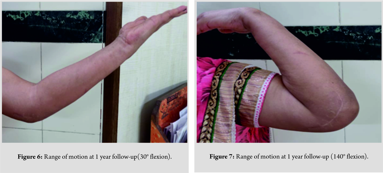
After the medial and lateral reconstruction of the ligaments, the elbow was found to be stable and intraoperative range of motion was checked. Postoperatively, the patient was kept in an above-elbow cast in complete supination for 6 weeks. The patient was regularly followed up and the range of motion was started at the end of 6 weeks. The patient had no instability for daily day to day activities and range of motion was initially from 40 to 100° flexion and from complete supination to 20°pronation. It improved gradually and the patient has 30–140° flexion and complete supination-pronation movements currently at 1 year follow-up. (Fig. 6 and 7) show the patient’s flexion from 30 to 140°.
Discussion
Treatment of chronic elbow dislocation is a demanding surgery to be performed even by the experienced trauma surgeon due to the challenges imposed intraoperatively and during rehabilitation. The surgeon has to deal with the antagonistic goals of treatment, which is to bring about concentric reduction and deal with the instability intraoperatively and to achieve a functional range of motion for the patient postoperatively. Chronic dislocation of elbow invariably leads to contracture and fibrosis of the ligaments and joint capsule with shortening of the surrounding muscles, leading to fixed dislocation [3]. Morrey [4] found consistent surgical findings in all patients having dislocation of the elbow. These include:
1. Contracted triceps
2. Collateral ligament and capsular contracture
3. Variable ulnar nerve involvement
4. Fibrous membrane covering the articular surface.
All these issues have to be addressed during open reduction for successful rehabilitation. However, van der Ley et al. [5] showed no significant difference and better results with nonoperative treatment of simple elbow dislocations. The major soft tissue to be addressed is the LCL complex(circle of Hori) as it is the major structure involved in recurrent instability [6]. The MCL if it prevents relocation might need to be released and reconstructed after successful reduction. Jupiter and Ring [7], in their study, found that all the three cases in their series had LCL rupture and MCL, flexor-pronator mass to be intact. In our case, MCL was ruptured beyond repair and hence the need for reconstruction of the medial dynamic stabilizers with a suture anchor, where as the LCL was found to be intact with extensive adhesions. Silva [8] described the pathologic anatomy of old unreduced elbow dislocation. His observations include shortening of the triceps muscle and both collateral ligaments and fibrosis around the ulnar nerve. The ulnar nerve neurolysis with or without anterior transposition should be done in all cases undergoing open reduction to prevent excessive traction to the nerve. In situ neurolysis was preferred by Ivo et al. [3] as it was found to be experimentally better than the anterior transposition of the nerve. Management of triceps fibrosis and shortening is challenging and has contrasting views. Mahaisavariya et al. [9] showed that leaving the triceps intact resulted in an increased motion of 115°, compared with 89° in those who underwent triceps release. The flexion contracture was markedly greater (by 70°) in those with tricepsplasty. In our case, we found that the reduction can be achieved only after subperiosteal release and pie crusting of the triceps aponeurosis. Mansat et al. [10] described indications and techniques for separate medial and lateral incisions for treating elbow stiffness as posterior based incision might result in increased post-surgical stiffness due to the adhesions and contractures involving the posterior capsule and the muscle. Finally, after reconstruction of collateral ligaments and achievement of anatomical reduction, the elbow joint has to be complemented with adequate immobilization methods for the soft tissue to heal. Arafiles [11] reported a unique technique where he used palmaris longus to reconstruct MCL and used the same graft to form an intraarticular cruciate ligament. The hinged external fixator is an excellent device for the healing of ligament reconstruction and for simultaneous mobilization [7]. The fixator keeps the joint distracted to prevent cartilage attenuation and also, the capsules and muscles can be maintained to an adequate length to prevent stiffness of elbow. Ivo et al. [3] studied biomechanical forces involved in dislocation and came to the conclusion that ligament reconstruction is not required as long as the joint distraction of 15mm by hinged fixator is maintained. This leads to modulation and reestablishment of biomechanically stable ligament complex [4]. Sheps et al. [12] advocated the use of external fixator, only if the joint was unstable. If the joint was stable he mobilized with hinged external brace. In our case, the joint was found to be stable postoperatively and we used the simple method of immobilization by above elbow casting in complete supination with close monitoring of rehabilitation protocol after its removal. The long term follow-up of our patient has satisfactory results with good functional outcome. However, further studies have to be done to compare posterior based and separate incisions and for a proper management protocol which can be followed universally in majority of the patients.
Conclusion
Chronic dislocation of the elbow is a highly disabling condition and has a very unpredictable outcome. By combining an understanding in the anatomy and biomechanics of the elbow with a proper surgical technique tailored to individual patients, it is possible to achieve a functional and painless elbow in the majority of cases. Attaining and maintaining a concentric reduction until the patient mobilizes is the key to a successful outcome.
Clinical Message
Most of the current literature suggests the open reduction of the joint through a single posterior incision and accessing both lateral and medial windows by raising appropriate flaps. In our patient, we have used separate medial and lateral incisions for complete exposure of the joint. By accessing medial and lateral elbow separately, the morbidity and wound complications of an extensile posterior approach can be reduced and also it has similar, if not better, functional results when compared to a posterior approach.
References
1. Jupiter JB. Trauma to the adult elbow fractures of the distal humerus. In: Browner BD, Levine AM, Trafton PG, editors. Skeletal Trauma. Vol. 2. Philadelphia, PA: Saunders; 1992. p. 1141.
2. Tan V, Daluiski A, Capo J, Hotchkiss R. Hinged elbow external fixators: Indications and uses. J Am AcadOrthop Surg 2005;13:503-14.
3. Ivo R, Mader K, Dargel J, Pennig D. Treatment of chronically unreduced complex dislocations of the elbow. Strategies Trauma Limb Reconstr2009;4:49-55.
4. Morrey BF, editor. Chronic unreduced elbow dislocations. In: The Elbow and its Disorders. Philadelphia, PA: Saunders; 2000. p. 431-6.
5. van der Ley J, van Niekerk JL, Binnendijk B. Conservative treatment of elbow dislocations in adults. Neth J Surg 1987;39:167-9.
6. O’Driscoll SW, Morrey BF, Korinek S, An KN. Elbow subluxation and dislocation. A spectrum of instability. Clin OrthopRelat Res 1992;280:186-97.
7. Jupiter JB, Ring D. Treatment of unreduced elbow dislocations with hinged external fixation. J Bone Joint Surg Am 2002;84:1630-5.
8. Silva JF. Old dislocations of the elbow. Ann R Coll Surg Engl1958;22:363-81.
9. Mahaisavariya B, Laupattarakasem W, Supachutikul A, Taesiri H, Sujaritbudhungkoon S. Late reduction of dislocated elbow. Need triceps be lengthened? J Bone Joint Surg Br 1993;75:426-8.
10. Mansat P, Bonnevialle N, Werner B. Indications and technique of combined medial and lateral column procedures in severe extrinsic elbow contractures. Orthopade2011;40:307-15.
11. Arafiles RP. Neglected posterior dislocation of the elbow. A reconstruction operation. J Bone Joint Surg Br 1987;69:199-202.
12. Sheps DM, Hildebrand KA, Boorman RS. Simple dislocations of the elbow: Evaluation and treatment. Hand Clin 2004;20:389-404.
 |
 |
 |
 |
 |
| Dr. Shaligram Purohit | Dr. B Sai Gautham | Dr. Nandan Marathe | Dr. Aditya Anand Dahapute | Dr. Swapneel Shah |
| How to Cite This Article: Purohit S, Gautham B S, Marathe N, Dahapute A A, Shah S. Treatment of Chronic Simple Elbow Dislocation by Two Separate Incisions – A Case Report. Journal of Orthopaedic Case Reports 2019 Sep-Oct; 9(5): 78-81. |
[Full Text HTML] [Full Text PDF] [XML]
[rate_this_page]
Dear Reader, We are very excited about New Features in JOCR. Please do let us know what you think by Clicking on the Sliding “Feedback Form” button on the <<< left of the page or sending a mail to us at editor.jocr@gmail.com


