[box type=”bio”] Learning Point of the Article: [/box]
Arthroscopic approach is a good alternative to open arthrotomy for the treatment of synovial chondromatosis of the ankle.
Case Report | Volume 10 | Issue 6 | JOCR September 2020 | Page 54-59 | Vikaesh Moorthy, Kae Sian Tay, Kevin Koo. DOI: 10.13107/jocr.2020.v10.i06.1874
Authors: Vikaesh Moorthy[1], Kae Sian Tay[2], Kevin Koo[2]
[1]Yong Loo Lin School of Medicine, National University of Singapore, Singapore.
[2]Department of Orthopaedic Surgery, Singapore General Hospital, Singapore.
Address of Correspondence:
Dr. Vikaesh Moorthy,
Yong Loo Lin School of Medicine, National University of Singapore, Singapore.
E-mail: vikaeshmoorthy@yahoo.com
Abstract
Introduction: Primary synovial chondromatosis is a rare disease characterized by the presence of metaplastic cartilaginous nodules arising from the synovia. Synovial chondromatosis has been widely described in the large joints, including elbow, hip, and knee joints, but very rarely in the foot or ankle. Data on the arthroscopic management of this condition in the ankle are also limited.
Case Report: A 50-year-old woman of Asian-Indian origin presented with the right lateral ankle pain of 1-month duration, associated with swelling and numbness of the joint. Magnetic resonance imaging revealed multiple loose bodies (at least 8) within the anterior ankle joint recess intracapsularly. She subsequently underwent right ankle arthroscopic debridement, synovectomy, removal of loose bodies, and microfracture with good post-operative recovery.
Conclusion: We report a rare case of ankle synovial chondromatosis with multiple loose bodies managed arthroscopically. Arthroscopic approach is a good alternative to open arthrotomy for the treatment of synovial chondromatosis of the ankle.
Keywords: Primary synovial chondromatosis, Ankle joint, Arthroscopy, Tumor, Case report
Introduction
Synovial chondromatosis is a disease of unknown etiology, originating from synovia and characterized by the presence of metaplastic cartilaginous nodules in the synovial cavities, bursa, or tendon sheaths [1]. The disease is commonly seen in middle-aged men between 30 and 50 years of age and the number of loose bodies can vary from 1 to 2 in the temporomandibular joint to more than 30 in the ankle joint [2]. Previous reports of synovial chondromatosis mostly involve large joints, including elbow, hip, and knee joints [1, 3, 4, 5, 6]. Synovial chondromatosis is very rarely found in the foot or ankle, although there have been previous reports of primary synovial chondromatosis in the subtalar, calcaneocuboid, naviculocuneiform, and metatarsaophalangeal joints [7, 8]. The rarity of this diagnosis in the literature spurred us to report our case of synovial chondromatosis of the ankle joint. Recently, the arthroscopic approach has gained popularity and preference in the treatment of ankle pathologies. The main advantages of the arthroscopic approach are decreased morbidity and the synchronous visualization and treatment of other articular pathologies [9]. However, data on the arthroscopic management of synovial chondromatosis of the ankle are limited. Herein, we present the case of an adult patient with primary synovial chondromatosis of the right ankle who was managed arthroscopically and discuss the potential benefits of such arthroscopic management.
Case Report
We present a 50-year-old woman of Asian-Indian origin with no significant medical history who developed right lateral ankle pain of 1-month duration, associated with swelling and numbness of the joint. There was no direct trauma or twisting injury of note, and no other joints were involved. On clinical examination, lumps were noted over the medial aspect of the midfoot and tenderness was elicited over the calcaneofibular ligament, anterior talofibular ligament (ATFL), and joint line of the right ankle. The remainder of the foot and ankle examination was unremarkable with good range of motion of the ankles bilaterally and negative anterior impingement. Preoperatively, her ankle-hindfoot American Orthopedic Foot and Ankle Score (AOFAS) was 38 (out of a maximum score of 100), and physical function score of Short Form 36 (SF-36) was 25 (out of a maximum score of 100), indicating a relatively poor pre-operative functional level. Weight-bearing ankle radiographs demonstrated loose bodies at the anterior ankle joint line, with normal alignment and preserved joint spaces noted (Fig. 1).
Magnetic resonance imaging revealed multiple loose bodies (at least 8) within the anterior ankle joint recess intracapsularly but superficial to the ATFL and AITFL, suggestive of synovial chondromatosis (Fig. 2). A 3 mm full-thickness chondral defect on medial talar shoulder and thickened and frayed ATFL was also noted. The patient initially declined surgery and was thus managed conservatively with ankle brace and symptomatic treatment. However, on follow-up 3 months later, there was no improvement in her ankle pain and the patient was keen to undergo surgery. She subsequently underwent right ankle arthroscopic debridement, synovectomy, removal of loose bodies, and microfracture, with intraoperative fluoroscopy (Fig. 3). This was performed through standard anteromedial and anterolateral arthroscopic portals under spinal anesthesia and tourniquet application. Intraoperatively, the right ankle synovial chondromatosis with multiple loose bodies in anterior ankle joint was confirmed. In addition, significant synovitis and osteoarthritis of tibiotalar joint with generalized cartilage thinning were noted.
Postoperatively, a below-knee Backslab was applied and the patient was not allowed weight-bearing for 2 weeks. On follow-up 2 weeks after surgery, wounds were well healed, and stitches were removed. Thereafter, the patient was on an Aircast ankle brace and she was allowed partial weight-bearing with crutches for 4 weeks. Subsequently, the Aircast ankle brace was removed and she was allowed full weight-bearing as tolerated. Her ankle was stable, and she was able to return to activities within 5 months after surgery without pain or limitations. At 6 months after surgery, both her AOFAS ankle-hindfoot score and physical function score had also improved to 71 and 55, respectively, indicating a significant improvement in functional level after the procedure. She was followed-up at 8 months postoperatively, with no complications or recurrence of signs and symptoms.
Discussion
Synovial chondromatosis is a disease composed of benign synovial metaplasia and multiple loose bodies [3]. Primary synovial chondromatosis is characterized by idiopathic stem cell proliferation of the stratum synoviale [10], while secondary synovial chondromatosis is often caused by trauma, degenerating joint diseases, osteochondritis dissecans, rheumatoid arthritis, and tuberculosis arthritis [11]. Our case presented here was evaluated as primary synovial chondromatosis due to the absence of previous trauma or history of inflammatory pathologies. Patients commonly present with symptoms of pain, swelling, and limited motion of the joint [9, 12, 13, 14, 15, 16]. In addition, effusion, diffuse tenderness, and crepitus can be found on clinical examination [17]. In the early stage of the disease, only active synovitis is present, and radiographs appear normal, while in the late stage, loose bodies are present and can be detected on radiographs [18]. Grossly, these loose bodies are consistent with ossified nodules. Milgram [12] classified the disease into three distinct stages:
1. Early stage with intrasynovial metaplasia (and islands of cartilage) without loose bodies
2. Transitional stage with both active and intrasynovial proliferation of cartilaginous nodules with loose bodies (and calcification beginning in the center of these masses) and
3. Late stage with only multiple loose bodies and slight inflammation but no evidence of synovial metaplasia.
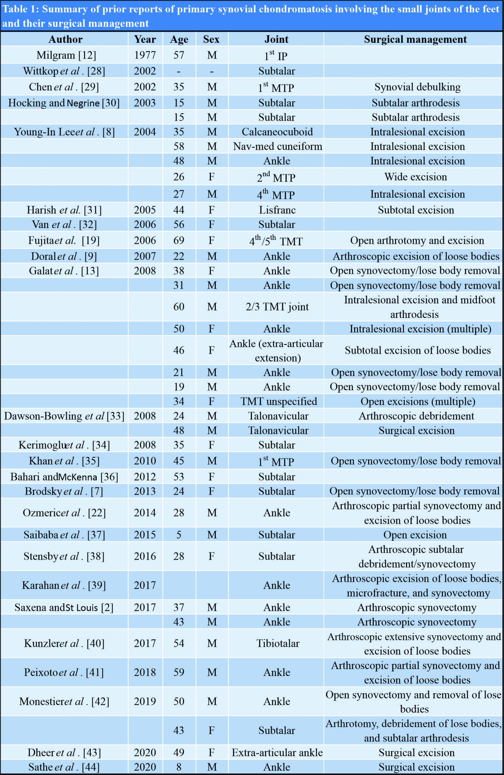
The aim of the treatment consists of decreasing pain and limiting the development of early osteoarthritis. While the treatment decision is made according to the patient’s age, symptoms, and the disease stage, surgery is often the treatment of choice. However, the surgical approach can vary based on the stage of disease – synovectomy for Stage 1, synovectomy and removal of intra-articular bodies for Stage 2, and removal of loose bodies for Stage 3 [12, 19]. Other authors report that the removal of loose bodies alone achieves similar results to synovectomy and removal of loose bodies [20, 21]. A summary of previous case reports of synovial chondromatosis of the foot and ankle is presented in Table 1.While extensive loose body removal and synovectomy have traditionally been achieved with open arthrotomy, the present case report demonstrates that minimally invasive arthroscopic procedures also provide excellent outcomes in the management of synovial chondromatosis [22]. Classically, the treatment of synovial chondromatosis of the ankle consists of arthrotomy with excision of the loose bodies and synovectomy. Post-operative immobilization in a cast for a varying period of time is usually necessary after such surgical treatment [23, 24, 25]. Up to 2006, it has been common to see surgeons managing ankle synovial chondromatosis through open synovectomy, intralesional excision, and even arthrodesis for the more severe cases (Table 1). However, from 2007 onward, this has largely been replaced by arthroscopic synovectomy and excision of loose bodies (Table 1), with excellent post-operative outcomes on follow-up. Due to the recent advances in ankle arthroscopy, therapy can now be performed in this non-invasive manner, with low morbidity and early rehabilitation [7, 9]. Minimally invasive surgery offers a multitude of advantages, including less pain, swelling, infection, and increased patient satisfaction [9]. Recovery after arthroscopic debridement and loose body removal is also much shorter than with open techniques, and there is no need of immobilization postoperatively. The patient can walk without pain, and there have been no reports of recurrence or osteoarthritis of the ankle joint in the follow-up period. However, with arthroscopy, there is also the possibility of limited synovectomy and residual loose bodies, which can increase the risk of recurrence and malignant transformation [22]. While local recurrence may follow incomplete synovectomy, malignant transformation is also a rare possibility. A recent article suggested 5% risk of malignant transformation in patients with primary synovial chondromatosis, which is much higher than that of other well-recognized bone diseases [26]. A single case of malignant transformation has been reported in primary synovial chondromatosis presenting in the foot [13]. Multiple local recurrences, rapid increase in size, cortical destruction, and marrow invasion, may be features of malignant transformation [27] and patients should be followed up after surgery for extended periods of time to allow for early detection of such complications.
Conclusion
We report a rare case of ankle synovial chondromatosis managed with arthroscopic debridement, synovectomy, removal of loose bodies, and microfracture. This case demonstrates that an arthroscopic approach for managing ankle synovial chondromatosis can provide excellent outcomes while maintaining the superior risk profile inherent to arthroscopic procedures. Arthroscopic approach is a good alternative to open arthrotomy for the treatment of synovial chondromatosis of the ankle.
Clinical Message
This case demonstrates that an arthroscopic approach for managing ankle synovial chondromatosis can provide excellent outcomes while maintaining the superior risk profile inherent to arthroscopic procedures. Arthroscopic approach is a good alternative to open arthrotomy for the treatment of synovial chondromatosis of the ankle.
References
1. Wiedemann NA, Friederichs J, Richter U, Buhren V. Secondary synovial chondromatosis of the ankle joint. Orthopade 2011;40:807-11.
2. Saxena A, St Louis M. Synovial chondromatosis of the ankle: Report of two cases with 23 and 126 loose bodies. J Foot Ankle Surg 2017;56:182-6.
3. Shearer H, Stern P, Brubacher A, Pringle T. A case report of bilateral synovial chondromatosis of the ankle. Chiropr Osteopat 2007;15:18.
4. Bojanic I, Bergovec M, Smoljanovic T. Combined anterior and posterior arthroscopic portals for loose body removal and synovectomy for synovial chondromatosis. Foot Ankle Int 2009;30:1120-3.
5. Murphey MD, Vidal JA, Fanburg-Smith JC, Gajewski DA. Imaging of synovial chondromatosis with radiologic-pathologic correlation. Radiographics 2007;27:1465-88.
6. Aydogan NH, Kocadal O, Ozmeric A, Aktekin CN. Arthroscopic treatment of a case with concomitant subacromial and subdeltoid synovial chondromatosis and labrum tear. Case Rep Orthop 2013;2013:636747.
7. Brodsky JW, Jung KS, Tenenbaum S. Primary synovial chondromatosis of the subtalar joint presenting as ankle instability. Foot Ankle Int 2013;34:1447-50.
8. Young-In Lee F, Hornicek FJ, Dick HM, Mankin HJ. Synovial chondromatosis of the foot. Clin Orthop Relat Res 2004;423:186-90.
9. Doral MN, Uzumcugil A, Bozkurt M, Atay OA, Cil A. Arthroscopic treatment of synovial chondromatosis of the ankle. J Foot Ankle Surg 2007;46:192-5.
10. Leu JZ, Matsubara T, Hirohata K. Ultrastructural morphology of early cellular changes in the synovium of primary synovial chondromatosis. Clin Orthop Relat Res 1992;276:299-306.
11. Chillemi C, Marinelli M, de Cupis V. Primary synovial chondromatosis of the shoulder: Clinical, arthroscopic and histopathological aspects. Knee Surg Sports Traumatol Arthrosc 2005;13:483-8.
12. Milgram JW. Synovial osteochondromatosis: A histopathological study of thirty cases. J Bone Joint Surg Am 1977;59:792-801.
13. Galat DD, Ackerman DB, Spoon D, Turner NS, Shives TC. Synovial chondromatosis of the foot and ankle. Foot Ankle Int 2008;29:312-7.
14. Pau M, Bicsak A, Reinbacher KE, Feichtinger M, Karcher H. Surgical treatment of synovial chondromatosis of the temporomandibular joint with erosion of the skull base: A case report and review of the literature. Int J Oral Maxillofac Surg 2014;43:600-5.
15. Robinson D, Hasharoni A, Evron Z, Segal M, Nevo Z. Synovial chondromatosis: The possible role of FGF 9 and FGF receptor 3 in its pathology. Int J Exp Pathol 2000;81:183-9.
16. Maurice H, Crone M, Watt I. Synovial chondromatosis. J Bone Joint Surg Br 1988;70:807-11.
17. Adelani MA, Wupperman RM, Holt GE. Benign synovial disorders. J Am Acad Orthop Surg 2008;16:268-75.
18. Dworak DP, McGuire MH. Primary synovial osteochondromatosis in the ankle: A case report. Am J Orthop (Belle Mead NJ) 2011;40:E96-8.
19. Fujita I, Matsumoto K, Maeda M, Kizaki T, Okada Y, Yamamoto T. Synovial osteochondromatosis of the lisfranc joint: A case report. J Foot Ankle Surg 2006;45:47-51.
20. Dorfmann H, De Bie B, Bonvarlet JP, Boyer T. Arthroscopic treatment of synovial chondromatosis of the knee. Arthroscopy 1989;5:48-51.
21. Shpitzer T, Ganel A, Engelberg S. Surgery for synovial chondromatosis. 26 cases followed up for 6 years. Acta Orthop Scand 1990;61:567-9.
22. Ozmeric A, Aydogan NH, Kocadal O, Kara T, Pepe M, Gozel S. Arthroscopic treatment of synovial chondromatosis in the ankle joint. Int J Surg Case Rep 2014;5:1010-3.
23. Holm CL. Primary synovial chondromatosis of the ankle. A case report. J Bone Joint Surg Am 1976;58:878-80.
24. Sekosky M, Lefkowitz H, Steiner I. Osteochondromatosis of the ankle. J Foot Surg 1990;29:330-3.
25. Wagner S, Bennek J, Grafe G, Schmidt F, Thiele J, Wittekind C, et al. Chondromatosis of the ankle joint (reichel syndrome). Pediatr Surg Int 1999;15:437-9.
26. Davis RI, Hamilton A, Biggart JD. Primary synovial chondromatosis: A clinicopathologic review and assessment of malignant potential. Hum Pathol 1998;29:683-8.
27. McCarthy C, Anderson WJ, Vlychou M, Inagaki Y, Whitwell D, Gibbons CL, et al. Primary synovial chondromatosis: A reassessment of malignant potential in 155 cases. Skeletal Radiol 2016;45:755-62.
28. Wittkop B, Davies AM, Mangham DC. Primary synovial chondromatosis and synovial chondrosarcoma: A pictorial review. Eur Radiol 2002;12:2112-9.
29. Chen A, Shih SL, Chen BF, Sheu CY. Primary synovial osteochondromatosis of the first metatarsophalangeal joint. Skeletal Radiol 2002;31:122-4.
30. Hocking R, Negrine J. Primary synovial chondromatosis of the subtalar joint affecting two brothers. Foot Ankle Int 2003;24:865-7.
31. Harish S, Saifuddin A, Cannon SR, Flanagan AM. Synovial chondromatosis of the foot presenting with lisfranc dislocation. Skeletal Radiol 2005;34:736-9.
32. Van P, Wilusz PM, Ungar DS, Pupp GR. Synovial chondromatosis of the subtalar joint and tenosynovial chondromatosis of the posterior ankle. J Am Podiatr Med Assoc 2006;96:59-62.
33. Dawson-Bowling S, Hamilton P, Bismil Q, Gaddipati M, Bendall S. Synovial osteochrondomatosis of the talonavicular joint: A case report. Foot Ankle Int 2008;29:241-4.
34. Kerimoglu S, Aynaci O, Saracoglu M, Cobanoglu U. Synovial chondromatosis of the subtalar joint: A case report and review of the literature. J Am Podiatr Med Assoc 2008;98:318-21.
35. Khan Z, Yousri T, Chakrabarti D, Awasthi R, Ashok N. Primary synovial osteochondromatosis of the first metatarsophalangeal joint, literature review of a rare presentation and a case report. Foot Ankle Surg 2010;16:e1-3.
36. Bahari S, McKenna J. Subtalar and extra articular synovial chondromatosis. J Case Rep 2012;2:120-4.
37. Saibaba B, Sudesh P, Govindan G, Prakash M. Pediatric subtalar joint synovial chondromatosis report of a case and an up-to-date review. J Am Podiatr Med Assoc 2015;105:435-9.
38. Stensby JD, Fox MG, Kwon MS, Caycedo FJ, Rahimi A. Primary synovial chondromatosis of the subtalar joint: Case report and review of the literature. Skeletal Radiol 2018;47:391-6.
39. Karahan HG, Cetin O, Kurtulmus A, Kayali C. Arthroscopic management and treatment of synovial chondromatosis and talus osteochondral defect in the ankle joint. A case study. Ortop Traumatol Rehabil 2017;19:293-6.
40. Kunzler DR, Safavi PS, Warren BJ, Janney CF, Panchbhavi V. Arthroscopic treatment of synovial chondromatosis in the ankle joint. Cureus 2017;9:e1983.
41. Peixoto D, Gomes M, Torres A, Miranda A. Arthroscopic treatment of synovial chondromatosis of the ankle. Rev Bras Ortop 2018;53:622-5.
42. Monestier L, Riva G, Stissi P, Latiff M, Surace MF. Synovial chondromatosis of the foot: Two case reports and literature review. World J Orthop 2019;10:404-15.
43. Dheer S, Sullivan PE, Schick F, Karanjia H, Taweel N, Abraham J, et al. Extra-articular synovial chondromatosis of the ankle: Unusual case with radiologic-pathologic correlation. Radiol Case Rep 2020;15:445-9.
44. Sathe P, Agnihotri M, Vinchu C. Synovial chondromatosis of ankle in a child: A rare presentation. J Postgrad Med 2020;66:112-3.
 |
 |
 |
| Dr. Vikaesh Moorthy | Dr. Kae Sian Tay | Dr. Kevin Koo |
| How to Cite This Article: Moorthy V, Tay KS, Koo K. Arthroscopic Treatment of Primary Synovial Chondromatosis of the Ankle: A Case Report and Review of Literature. Journal of Orthopaedic Case Reports 2020 September;10(6): 54-59. |
[Full Text HTML] [Full Text PDF] [XML]
[rate_this_page]
Dear Reader, We are very excited about New Features in JOCR. Please do let us know what you think by Clicking on the Sliding “Feedback Form” button on the <<< left of the page or sending a mail to us at editor.jocr@gmail.com


