[box type=”bio”] Learning Point of the Article: [/box]
Early identification of injury and timely intervention with CRPP for distal radius fracture and the same line of management for radial shaft, neck fracture, and ulnar fracture using elastic TENS nail gives better results in this kind of unusual combination fractures of the forearm.
Case Report | Volume 10 | Issue 6 | JOCR September 2020 | Page 86-89 | Shanti Lal Sankhla, Piyush Joshi, Devendra Singh. DOI: 10.13107/jocr.2020.v10.i06.1888
Authors: Shanti Lal Sankhla[1], Piyush Joshi[1], Devendra Singh[1]
[1]Department of Orthopaedics, Dr. S.N. Medical College, Jodhpur. India.
Address of Correspondence:
Dr. Shanti Lal Sankhla,
Department of Orthopaedics, Dr. S.N. Medical College, H-15, Shastri Nagar, Jodhpur – 342 003. India.
E-mail: sankhla.shantilal@yahoo.com
Abstract
Introduction: We report an extremely rare combination of Monteggia equivalent Type 1 lesion (diaphyseal ulna and radial neck fractures without dislocation) with ipsilateral radius shaft and distal radius fractures in a 13-year-old boy. There are only a few cases of Monteggia or Monteggia equivalent injury with ipsilateral forearm fractures in children, and injury pattern being reported by us is not only rare but also the only case reported, thus far to the best of our knowledge.
Case Report: A 13-year-old, right-hand dominant boy presented in casualty with a history of fall 1 day back with pain, swelling and deformity in the left forearm with bleeding from the left forearm, and restriction of movement of fingers and thumb of the left hand. On examination, there was a wound of size 1.5 cm on the upper third-forearm over the ulnar aspect. No neurovascular deficit was present. X-rays were performed, which suggested Type I Monteggia fracture equivalent lesion (diaphyseal ulna and radial neck fractures without dislocation) with ipsilateral distal radius and radial shaft fractures. The patient was operated with toileting, debridement, and close reduction of proximal ulnar fracture with titanium elastic nail (TENS) Distal radius was managed by percutaneous fixation with two K-wires under the guidance of image intensifier, while the shaft of radius fracture was managed by close reduction and internal fixation with elastic TENS nail with a lateral entry point and radial neck fracture was managed by the Metaizeau technique. Follow-up of the patient showed subsequent union of all fractures with good functional outcome.
Conclusion: We have highlighted an extremely rare combination of injuries. Early recognition and prompt surgical intervention can lead to a satisfactory outcome, even in these complex injuries.
Keywords: Monteggia equivalent, radius fractures, child.
Introduction
Monteggia fracture-dislocation is an uncommon injury in children comprising only 0.4% of all forearm fractures [1] This fracture was first described by Giovanni Batista Monteggia in 1814. It was not until 1967 that Bado renamed this fracture as the Monteggia lesion and classified the adult injury into four types depending on the direction of radial head dislocation [2] According to the current classification, Type 1 variety is the most common (59%) followed by Type 3 variety (26%). He described two Monteggia equivalent injuries. Subsequently, Types 3 and 4 Monteggia equivalent lesions were described. Despite the increased understanding of Monteggia lesions, the injury continues to represent a challenge for the orthopedic surgeon. In 1943, Sir Watson-Jones wrote that “no fracture presents so many problems; no injury is beset with greater difficulty; and no treatment characterized by more general failure’’ [1]. There are few reports in the literature regarding the association of Monteggia lesion with ipsilateral limb fractures. Association of Type 1 Monteggia equivalent injury with ipsilateral radius shaft and distal radius fractures in children is very rare [3] and injury pattern described by us reported not once before in the literature.
Case Report
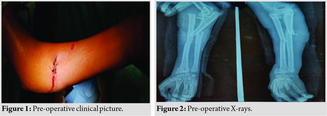
A 13-year-old right-hand dominant boy presented in casualty with a history of fall 1 day back with pain, swelling, and deformity in the left forearm with bleeding from the left forearm and restriction of movement of fingers and thumb of the left hand. On examination, there was a wound of size 1.5 cm on upper third-forearm over the volar and ulnar aspect and a wound of the size of 1 cm on mid-forearm over radial aspect (Fig. 1). No neurovascular deficit was present. X-rays were performed, which suggested Type I Monteggia fracture equivalent lesion (diaphyseal ulna and radial neck fractures without dislocation) with ipsilateral distal radius and radial shaft fractures (Fig. 2). Intravenous antibiotics were started, and splintage was given, and the patient kept for surgery the following morning.
Surgery was performed without a tourniquet. The open wound was thoroughly debrided and irrigated. The ulnar fracture was managed with close reduction and internal fixation with titanium elastic nail (TENS). Distal radius was managed by percutaneous fixation with two K-wires under the guidance of image intensifier, while the shaft of radius fracture was managed by close reduction and internal fixation with elastic TENS nail with a lateral entry point [Fig. 3]. 

Discussion
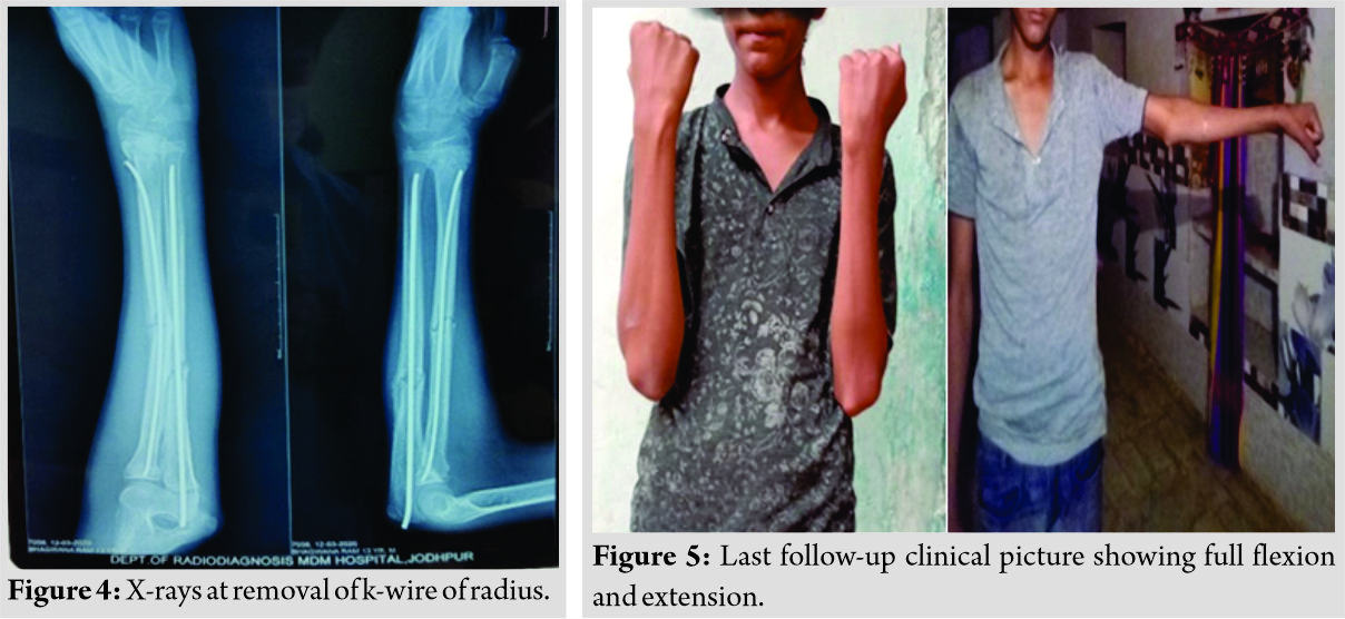
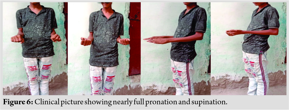
Monteggia fracture-dislocation is a rare injury in children. Three mechanisms of injury have been proposed, which are direct trauma, hyperpronation, and hyperextension [ 1]. Monteggia proposed the direct blow theory, in which fracture occurs with a direct blow to the forearm first produces fracture through the ulna. Then, by continued deformation or direct pressure, the radial head is forced anteriorly with respect to the capitellum, causing radial head dislocation. According to hyperpronation theory, hyperpronation forcibly rotates the radius over the middle of the ulna resulting in either anterior dislocation of radial head or fracture of proximal one-third of radius, along with a fracture of shaft of the ulna as elicited by Evans during cadaveric dissections. Tompkins analyzed both the above theories and proposed, these injuries were caused by a combination of static and dynamic forces. His study postulated three steps in fracture mechanism: Hyperextension, followed by radial head dislocation due to the pull of biceps. Subsequently, the weight of the body is transferred to the ulna, leading to the fracture in tension. Radial head dislocation is a more common than radial neck fracture as the annular ligament is more lax. In addition, a combination of Monteggia lesion along with ipsilateral distal forearm fracture is a very rare injury. Such injuries are also called bipolar fractures of the forearm, as described by Odena [ 4]. The previous injury patterns reported in the literature were mostly in combination with radial head dislocation [ 5-9], and only six had Monteggia equivalent injuries [ 3,10-13]. Our case has Type 1 Monteggia equivalent injury with ipsilateral forearm fracture with distal radius fracture with radial neck fracture in a child, being the first case of its kind. This type of lesion is rare in children probably because the annular ligament is relatively lax and the radial head fracture and dislocates more easily anteriorly, rather than a radial neck. Sood et al. described a case of Type 1 Monteggia equivalent injury with epiphyseal injury of distal radius and ulna. They had fixed the ulna using a dynamic compression plate, which would need the second surgery for removal. They had reduced the radial neck under direct vision. They performed closed manipulation under the image intensifier for the distal forearm. However, their patient developed signs of median nerve compression and needed carpal tunnel decompression and fixation of the distal radius with K-wire [ 10]. We used elastic TENS nail for fixation of proximal ulna shaft fracture instead to avoid second surgery for implant removal, along with close reduction and two K wires fixation for distal end radius fracture and elastic TENS nail for shaft radius fracture and radial neck with same TENS nail used for the reduction of the radial shaft with Metaizeau technique under image intensifier. Our case had an uneventful post-operative period. Osada et al. also described injury pattern nearly the same as us where initially closed reduction was attempted with no success, and subsequently, three out of four fractures were treated with open reduction and internal fixation with K-wires. They had a follow-up up to 30 months with proximal radius epiphysis partially closed, and ulnar diaphysis and the radial neck were posteriorly convex 20° and 18°, respectively. The various complications associated with such injuries include posterior interosseous nerve palsy, compartment syndrome, median nerve involvement, myositis ossificans, and elbow stiffness in the previous cases due to their different approach of management [ 1]. No similar complications found in our case in a follow-up of 6 months with full range of motion and good functional outcome.
Conclusion
Radial head dislocates more easily in a child due to the elasticity of annular ligament. Hence, the occurrence of radial neck, shaft radius, and distal radius injury, as seen in our case, is a very rare occurrence in Monteggia injuries. In our case, the same line of management was used for radial shaft and neck fracture and ulna fracture using elastic TENS nail. For distal radius fracture, close reduction and two K wires fixation under image, guidance was done. In our case, approaches such as Kocher and Boyd should be avoided to reduce post-operative complications such as revision surgery for removal of implant, neurovascular injury, and myositis ossificans. Anatomic reduction of all fractures should be performed. The child should be monitored for growth disturbances in the future. These precautions prevent any untoward complication and may lead to overall good functional and radiological outcome.
Clinical Message
Monteggia fracture-dislocation in a child is rare, and Monteggia equivalent injuries still rarer as children have lax annular ligament. This report highlights an extremely rare injury occurring in combination with ipsilateral radial shaft and distal radius fracture. The awareness of this possible injury combination is important for early recognition and prompts surgical intervention in these complex injuries with a good result.
References
1. Shah AS, Waters PM. Monteggia-fracture dislocation in children. In: Flynn JM, Skaggs DL, Waters PM, editors. Rockwood and Wilkins’ Fractures in Children. Philadelphia, PA: Wolters Kluwer; 2015. p. 527-63.
2. Bado JL. The Monteggia lesion. Clin Orthop Relat Res 1967;50:71-86.
3. Faundez AA, Ceroni D, Kaelin A. An unusual Monteggia Type-I equivalent fracture in a child. J Bone Joint Surg Br 2003;85:584-6.
4. Odena IC. Bipolar fracture-dislocation of the forearm. J Bone Joint Surg Am 1952;34A:968-76.
5. Zrig M, Mnif H, Koubaa M, Bannour S, Amara K, Abid A. An unusual Monteggia Type I equivalent fracture: A case report. Arch Orthop Trauma Surg 2011;131:973-5.
6. Soin B, Hunt N, Hollingdale J. An unusual forearm fracture in a child suggesting a mechanism for the Monteggia injury. Injury 1995;26:407-8.
7. Peter N, Myint S. Type I Monteggia lesion and associated fracture of the distal radius and ulna metaphysis in a child. CJEM 2007;9:383-6.
8. Gupta V, Kundu ZS, Kaur M, Kamboj P, Gawande J. Ipsilateral dislocation of the radial head associated with fracture of distal end of the radius: A case report and review of the literature. Chin J Traumatol 2013;16:182-5.
9. Bhandari N, Jindal P. Monteggia lesion in a child: Variant of a Bado Type-IV lesion. A case report. J Bone Joint Surg Am 1996;78:1252-5.
10. Biyani A. Ipsilateral Monteggia equivalent injury and distal radial and ulna fracture in a child. J Orthop Trauma 1994;8:431-3.
11. Sood A, Khan O, Bagga T. Simultaneous Monteggia Type I fracture equivalent with ipsilateral fracture of the distal radius and ulna in a child: A case report. J Med Case Rep 2008;2:190.
12. Osada D, Tamai K, Kuramochi T, Saotome K. Three epiphyseal fractures (distal radius and ulna and proximal radius) and a diaphyseal ulnar fracture in a seven-year-old child’s forearm. J Orthop Trauma 2001;15:375-7.
Song KS, Bae KC. Type III Monteggia equivalent fracture with ipsilateral distal radial epiphyseal and ulnar metaphyseal fracture in a child: Case report. J Korean Orthop Assoc 2004;39:563-5.
 |
 |
 |
| Dr. Shanti Lal Sankhla | Dr. Piyush Joshi | Dr. Devendra Singh |
| How to Cite This Article: Sankhla SL, Joshi P, Singh D. An Extremely Rare Combination of Monteggia Equivalent Type 1 Lesion (Diaphyseal Ulna and Radial Neck Fractures Without Dislocation) with Ipsilateral Radius Shaft and Distal Radius Fractures in a Child. Journal of Orthopaedic Case Reports 2020 September;10(6): 86-89. |
[Full Text HTML] [Full Text PDF] [XML]
[rate_this_page]
Dear Reader, We are very excited about New Features in JOCR. Please do let us know what you think by Clicking on the Sliding “Feedback Form” button on the <<< left of the page or sending a mail to us at editor.jocr@gmail.com


