 [box type=”bio”] Learning Point of the Article: [/box]
Displaced pediatric calcaneal tuberosity avulsion fractures are rare, but when you see one, you must fix it fast, and fix it well.
Case Report | Volume 11 | Issue 3 | JOCR March 2021 | Page 25-28 | Ehab S Saleh, Ahmed Elabd . DOI: 10.13107/jocr.2021.v11.i03.2072
Authors: Ehab S Saleh[1], Ahmed Elabd[2]
[1]Department of Orthopedic Surgery, Oakland University William Beaumont School of Medicine, Rochester, Michigan,
[2]Department of Orthopedic Surgery and Rehabilitation, Texas Children’s Hospital, Texas.
Address of Correspondence:
Dr. Ehab S Saleh,
Department of Orthopedic Surgery, Pediatric Orthopedic Surgeon, William Beaumont Hospital, Royal Oak, Michigan.
E-mail: ehab.saleh3@beaumont.org
Abstract
Introduction: Calcaneus fractures are rare in the pediatric population, and avulsion fracture of the calcaneal tuberosity is even less common. In adults, those fractures are usually associated with poor bone quality, however, this is not the case in children. It is a fracture that requires emergent intervention to prevent devastating skin and soft-tissue-related complications.
Case Report: We report a case of a 9-year-old female who had a displaced calcaneal tuberosity fracture with heel skin impending compromise, after a fall at an indoor gymnastic facility. The child had a history of acute lymphoblastic leukemia, diagnosed at age 4, she was in remission at the time of injury. In the present report, besides reporting a rare injury among the pediatric population, we also describe the operative management, the post-operative course, and we review the literature.
Conclusion: Pediatric calcaneal tuberosity fractures, although rare, can lead to devastating complications if not addressed promptly, and should be treated in an expedited fashion.
Keywords: Pediatric, calcaneal tuberosity, skin compromise.
Introduction
Pediatric calcaneal tuberosity avulsion fractures are rare, reports of this injury are sporadic [1, 2, 3]. In the adult population, this injury is usually associated with poor bone quality, and fixation with screws or Kirschner wires only can result in treatment failure, even when combined with post-operative cast immobilization [4]. Fixation with cannulated screws or Kirschner wires is usually enough in the pediatric population, and unlike the situation in the adult population, fixation augmentation with tension band wires, small lateral plates, and suture anchors are usually not necessary. Furthermore, some adult and pediatric patients with associated gastrocnemius tightness will require a gastrocnemius soleus lengthening to decrease the deforming forces on the fixation construct [1, 5].
Case Report
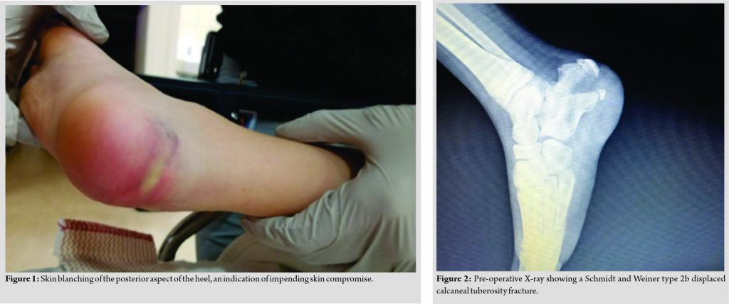
A 9-year-old female presented to our hospital in February 2018, with a right heel injury. She was at an indoor gymnastic facility, when she jumped into a foam pit, landing at the bottom of the pit. She immediately noticed right heel pain and swelling. She was initially seen at an urgent care; where she was evaluated with an X-ray and then was referred to our institution for definitive management. The patient had a medical history of acute lymphoblastic leukemia diagnosed at age 4 years, she was treated with vincristine, intrathecal methotrexate injection, and oral dexamethasone until she achieved remission, she was then kept on maintenance therapy until early 2015. In our emergency room, her evaluation did show right heel swelling and skin blanching of the posterior aspect of the heel, an indication of impending skin compromise (Fig. 1). The X-rays were positive for a Schmidt and Weiner type 2b displaced calcaneal tuberosity fracture [6] (Fig. 2). She was splinted in plantar flexion to decrease the pressure on the heel skin and was taken to the operating room emergently.
Operative technique
In the operating room, the heel skin was still blanched, without skin necrosis. Under general anesthesia, the patient was positioned prone and a tourniquet was placed on the right thigh. The approach was through an incision parallel to the lateral aspect of the Achilles tendon. The short saphenous vein and the sural nerve were protected. A small incision was made on the plantar aspect of the heel, to apply one limb of a pointed reduction clamp, the other limb of the pointed reduction clamp was applied through the surgical wound. Reduction was confirmed using the image intensifier on the lateral and axial views. Two guide wires for the 5.0 cannulated screws were inserted; then, the two screws were inserted with washers in lag fashion, making sure to engage the plantar cortex of the calcaneus (Fig. 3). Wound closure was done in a standard fashion.
Rehabilitation protocol
Postoperatively, a short leg cast was applied with the ankle in plantar flexion. At 4 weeks, the cast was replaced by another short leg cast in neutral ankle flexion for 2 more weeks. The patient was non-weight-bearing for 6 weeks. At her 6 weeks follow-up visit, the cast was replaced with a cam boot, and the patient was instructed to ambulate weight-bearing as tolerated with the boot for 2 more weeks, and a home exercise program was initiated at that point. At 8 weeks, the cam boot was discontinued, and the patient was instructed to continue the home exercise program and to resume all activities except for contact sports. At her 12 weeks follow-up, she was pain free and regained full motion and strength and was able to return to sports activities. The child was followed for 2 years; she fully healed the fracture and resumed all daily life and sports activities (Fig 4). Because of her medical history, she was evaluated with a bone density scan postoperatively, which showed normal bone density.
Discussion
Fractures of the calcaneus are common in adults, accounting for 1–2% of all adult fractures [6, 7]. However, in children, it is less common, with an incidence of only 1 in 100.000 fractures [8, 9]. They constitute one-third of pediatric tarsal fractures [10]. Historically, this low incident has been attributed to the pediatric calcaneus having a larger, more resilient cartilage component, and to the relative strength of the bone in relation to the body weight of the child [10, 11]. Non-displaced calcaneus fractures in young children tend to be missed, which is another contributing reason for the reported low incidence of this fracture in children [12]. Schmidt and Weiner [6] proposed a classification system for pediatric calcaneal fractures in which all calcaneal tuberosity fractures were classified as Type 2 ([1] beak fracture; [2] avulsion fracture of insertion of Achilles tendon). On the other hand, Walling et al. [3] considered calcaneal tuberosity fractures different from other calcaneal fractures because they involve an apophysis, hence, they could be classified into four types, analogous to the Salter-Harris classification. In this case report, the fracture described can be classified as Schmidt Type 2b or Salter-Harris type 4 fracture. There are few case reports on pediatric calcaneal tuberosity fractures in the literature. Alvarez Fernandez et al. [1] described a 10-year-old girl that initially presented with non-traumatic heel pain, and minimal changes on the initial radiographs, managed initially with rest and analgesia, however, there was a recurrence of symptoms 6 months later with heel deformity and ankle equines, at which time radiographs showed a displaced calcaneal tuberosity separation which the author attributed to repetitive microtrauma leading to epiphysiolysis. The fracture was treated with open reduction and internal fixation with screws combined with Z lengthening of the calcaneal tendon, and the result was good. Walling et al. [3] reported on 11 patients who had calcaneal apophyseal avulsion fractures, six patients had open fractures from lawn mowers injury (SH type 1 and 3) who were treated surgically, all had growth disturbances of the posterior calcaneus, and problems with shoe wear, heel pain, and scar pain. Two adolescent patients had a slipped apophysis (Salter-Harris type 1) that resembled a slipped capital femoral epiphysis, both were treated conservatively, and had posterior calcaneal symptomatic growth deformities and intermittent heel pain. Three adolescent patients had Salter-Harris type 4 split apophyses, all were treated by closed reduction and casting initially, but one patient continued to be symptomatic and was treated with fixation with a staple, all three eventually were asymptomatic at follow-up. In 1995, Cole et al. [2] reported the treatment of this fracture in four children, three of them had surgery with fixation using cannulated screws or Kirschner wires, while one was treated with a cast. All patients returned to full activity, with good functional results. Inokuchi et al. [12] in his series of 20 pediatric calcaneal fractures reported on a 9 years old with an avulsion fracture of the calcaneus tuberosity that was treated with open reduction and fixation with Kirschner wires, with good result. The other patient in his report that required surgery was a 13-year-old male with bilateral calcaneal intra-articular fractures, where one side was treated with open reduction and Kirschner wire fixation and the other side treated with closed reduction and casting. All other fractures in his report were managed with casting only.
Conclusion
We present a rare pediatric fracture that can compromise the local soft tissue and potentially cause devastating complications, but at the same time can have excellent result once treated properly. Most pediatric calcaneal fractures can be treated conservatively, with expected good result, but displaced calcaneal tuberosity fractures and intra-articular calcaneal fractures are an indication for operative treatment [11, 12].
Clinical Message
Fixing pediatric calcaneal tuberosity fractures with screws, or Kirschner wires, supplemented with a cast usually gives good stability, with no reported fracture displacement or implant failure, even in the case of our patient, with her medical history of leukemia, and treatment with chemotherapy and corticosteroids.
References
1. Alvarez Fernandez J, Vaquero MV, Cimiano JG. Epiphysiolysis of the great tuberosity of the calcaneum: Brief report. J Bone Joint Surg Br 1989;71:321.
2. Cole RJ, Brown HP, Stein RE, Pearce RG. Avulsion fracture of the tuberosity of the calcaneus in children. A report of four cases and review of the literature. J Bone Joint Surg Am 1995;77:1568-71.
3. Walling AK, Grogan DP, Carty CT, Ogden JA. Fractures of the calcaneal apophysis. J Orthop Trauma 1990;4:349-55.
4. Rauer T, Twerenbold R, Flückiger R, Neuhaus V. Avulsion fracture of the calcaneal tuberosity: Case report and literature review. J Foot Ankle Surg 2018;57:191-5.
5. Azar FM BJ, Canale ST. Campbell’s Operative Orthopedics. 13th ed. Netherlands: Elsevier, Inc.; 2017.
6. Schmidt TL, Weiner DS. Calcaneal fractures in children. An evaluation of the nature of the injury in 56 children. Clin Orthop Relat Res 1982;171:150-5.
7. Brunet J. Calcaneal fractures in children: Long-term results of treatment. J Bone Joint Surg Br 2000;82:211-6.
8. Bernard L. Nonoperative treatment of fractures of the calcaneus. J Bone Joint Surg Am 1963;45:865-7.
9. Wiley J, Profitt A. Fractures of the os calcis in children. Clin Orthop Relat Res 1984;188:131-8.
10. Ishikawa SN. Conditions of the calcaneus in skeletally immature patients. Foot Ankle Clin 2005;10:503-13.
11. Abdelgawad AA, Kanlic E. Minimally invasive (sinus tarsi) approach for open reduction and internal fixation of intra-articular calcaneus fractures in children: Surgical technique and case report of two patients. J Foot Ankle Surg 2015;54:135-9.
12. Inokuchi S, Usami N, Hiraishi EH. Calcaneal fractures in children. J Pediatr Orthop 1998;18:469-74.
 |
 |
| Dr. Ehab S Saleh | Dr. Ahmed Elabd |
| How to Cite This Article: Saleh ES, Elabd A. Pediatric Calcaneal Tuberosity Avulsion Fracture: A Case Report and Literature Review. Journal of Orthopaedic Case Reports 2021 March;11(3): 25-28. |
[Full Text HTML] [Full Text PDF] [XML]
[rate_this_page]
Dear Reader, We are very excited about New Features in JOCR. Please do let us know what you think by Clicking on the Sliding “Feedback Form” button on the <<< left of the page or sending a mail to us at editor.jocr@gmail.com


