Symptomatic isolated congenital ACLD should be treated by correction of bony deformity first to restore anatomic alignment and consider ACL reconstruction only if instability persists.
Dr. Chloe Xiaoyun Chan,
National University Hospital, Singapore; Department of Orthopaedic Surgery, 5 Lower Kent Ridge Road, Singapore 119074.
E-mail: chloe_xiaoyun_chan@nuhs.edu.sg
Introduction: Isolated congenital ACLD is a rare condition with limited literature on the optimal management approach. At present, patients with instability symptoms have been managed with ACL reconstruction in case reports. We present a case report of symptomatic isolated congenital anterior cruciate ligament deficiency (ACLD) managed effectively with gradual correction of biplanar proximal tibial deformity alone.
Case Presentation: This was a case of bilateral isolated congenital ACLD in a 15-year-old girl with chronic bilateral knee instability, bilateral mild genu valgum, and positive Lachman’s tests. Biplanar tibial deformity was evident with a 50 proximal tibia valgus and a posterior tibial slope angle of 260 on the more symptomatic right knee. This was treated with a proximal tibial osteotomy and gradual correction with a hexapod frame using the CORA method. The right knee alignment was restored to normal. At 2-year post-surgery, her symptoms of instability had resolved, and there was a soft end point on the Lachman’s test.
Conclusion: We recommend that symptomatic isolated congenital ACLD be treated by correction of any existing bony deformities first, keeping in view of ACL reconstruction if instability persists thereafter. To date, there are no reports on correction of proximal tibial deformities as the first-line treatment in isolated congenital ACLD before consideration of ACL reconstruction. To the best of our knowledge, this is the first report of symptomatic isolated congenital ACLD managed with correction of the biplanar deformity of the proximal tibia alone. Our management strategy proved to be effective in the treatment of this patient’s instability, with good post-operative outcomes.
Keywords: Congenital anterior cruciate ligament deficiency, hexapod frame, deformity correction, knee instability, case report.
Congenital anterior cruciate ligament deficiency (ACLD) is a very rare pathology that occurs approximately 0.017 per 1000 live births [1]. It can either occur unilaterally or bilaterally, in isolation or in association with other limb deformities or syndromes. These include congenitally short femur, proximal focal femoral deficiency, fibular hemimelia, abnormalities of the menisci (congenital absence of menisci, ring meniscus), congenital knee dislocation, osteochondritis dissecans, thrombocytopenia-absent radius syndrome, and Larsen syndrome. In the earlier case reports of patients with obvious skeletal deformities mentioned above and concomitant congenital ACLD, management had been targeted at realignment osteotomies and limb lengthening procedures [2, 3, 4, 5, 6]. The restoration of anatomical alignment and adaptation of soft-tissue structures, such as the hypertrophy of ligament of Humphrey, had often provided sufficient stability to the knee [7]. However, in cases where instability persisted, subsequent ACL reconstruction has proven useful [7, 8]. The indication for ACL reconstruction has since been extended to treat cases of isolated congenital ACLD [9, 10, 11, 12]. The reconstruction of the ACL in congenital ACLD patients is technically challenging. A narrow femoral notch and dysplastic or absent tibial eminence make instrumentation and proper tunnel positioning very challenging [7, 8, 12]. The presence of open physes will also be a concern in very young patients. There is also a risk of ACL graft failure if coexisting bony deformities are not addressed. Furthermore, there is the absence of long-term post-operative outcomes in this group of patients managed with just ACL reconstruction alone. To date, there are no reports on analysis and correction of proximal tibial deformities as the first-line treatment in isolated congenital ACLD. Given the lack of substantial evidence guiding the treatment of isolated congenital ACLD, we present a case of symptomatic isolated congenital ACLD managed with correction of the biplanar deformity of the proximal tibia alone. Our management strategy proved to be effective in the treatment of this patient’s instability, with good post-operative outcomes.
Clinical presentation
Our patient is a 15-year-old girl, who presented with chronic bilateral knee instability resulting in recurrent falls. There was no prior history of trauma. Clinical examination revealed bilateral mild genu valgum. Both knees demonstrated positive Lachman test with no end point. There was no limb length discrepancy. There was full range of movement of the knee.
Pre-operative radiographical findings
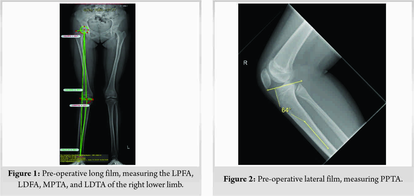
Radiographical measurements on long leg films revealed the following measurements on the more symptomatic right lower limb (Figs. 1, 2):
• Lateral proximal femoral angle (LPFA) of 820
• Lateral distal femoral angle (LDFA) of 850
• Medial proximal tibial angle (MPTA) of 950
• Posterior proximal tibial angle (PPTA) of 640
• Lateral distal tibial angle (LDTA) of 900
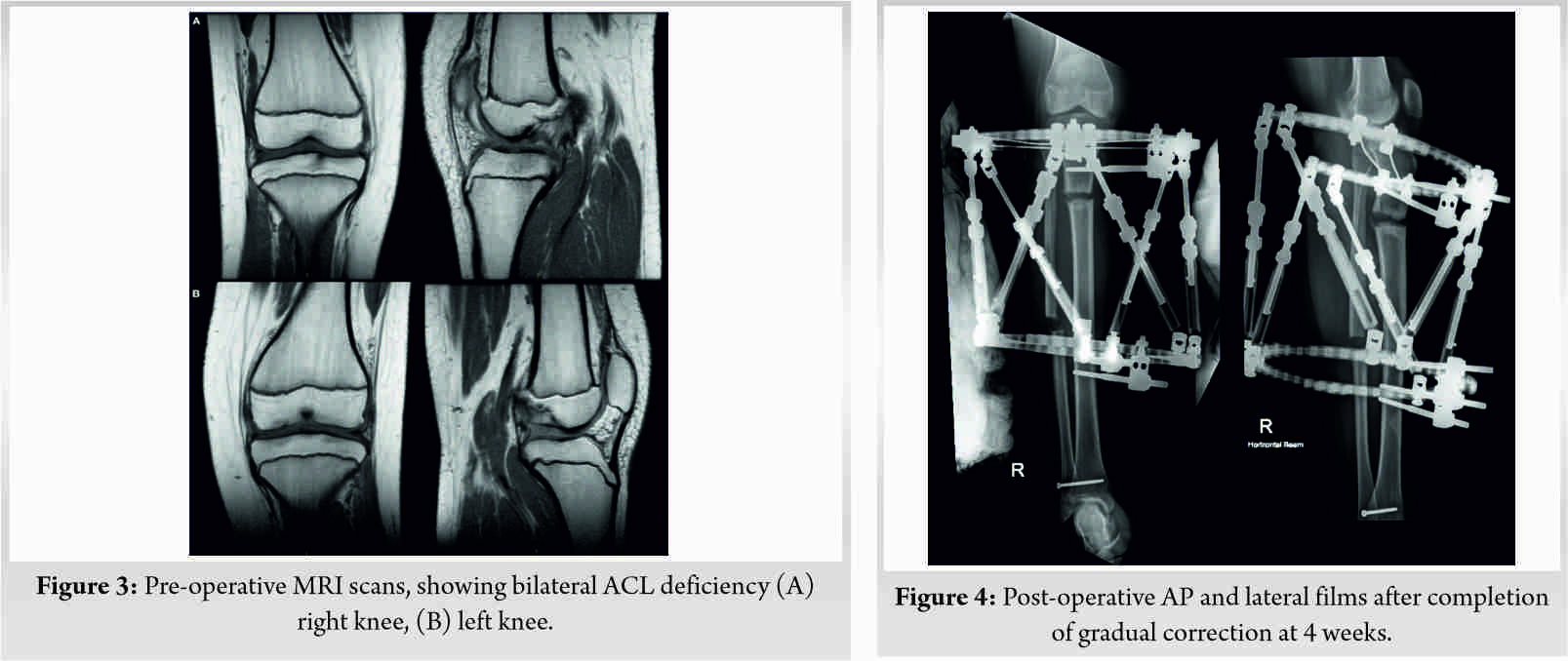
The fibula was normal. Magnetic resonance imaging (MRI) of both knees demonstrated absence of ACL and hypoplastic tibial eminences (Fig. 3). The PCL and both meniscus were normal.

Treatment – deformity analysis, planning, and execution
Based on the deformity analysis, the proximal tibia was the main contribution of deformity, with minimal contribution from the distal femur. Deformity correction of the proximal tibia was performed based on the center of rotation of angulation (CORA) method. The CORA lies approximately 5 cm distal to the joint line in the coronal plane and approximately 3 cm distal to the joint line in the sagittal plane of the proximal tibia. The proximal tibial osteotomy was performed using the CORA of the coronal plane. To enable correction of the high posterior tibial slope, a resultant posterior translation of the proximal fragment was necessary. Gradual correction with a hexapod frame was chosen over an acute opening wedge biplanar correction as the latter is technically difficult with higher risk of peroneal nerve injury [13]. The gradual correction was based on a proximal tibial Taylor Spatial Frame (TSF) module with the proximal ring as the reference ring. The following parameters were used: Proximal ring (2/3 ring, 180 mm), distal ring (full ring, 155 mm), and struts (FastFX medium size). A concomitant 8 mm of lengthening was programmed during the simultaneous valgus and posterior slope correction to prevent fragment impingement during the correction process. A distraction rate of 1 mm/day was utilized. The distraction was carried out over 20 days.
Post-operative rehabilitation protocol
Immediate postoperatively, the patient was kept on non-weight-bearing of the operated limb with use of crutches or wheelchair mobilization. On post-operative day 7, adjustments of struts were initiated and distraction confirmed on radiographs. She was kept on non-weight-bearing until gradual correction was completed and was allowed touch down weight-bearing. Progression to full weight-bearing was allowed when there was radiographic evidence of adequate bridging callus on three cortices. Pin sites were dressed regularly with antiseptic dressings. No strut change was required.
Post-operative radiographic and clinical findings
(Fig. 4) demonstrates the final correction at 4 weeks post-surgery. The mechanical axis of the right lower limb was corrected to lie between zone 1 and 2. The post-operative MPTA was 89 degrees and PPTA was 770 (Fig. 5, 6). There was a soft end point on the Lachman’s test. Her International Knee Documentation Committee (IKDC) score improved from 48 preoperatively to 79 postoperatively. At 2 years post-surgery, her symptoms of instability had resolved without the need for an ACL reconstruction. She is planned for a similar surgery on the contralateral left side.
The management of this case was directed at correcting the proximal tibial deformities in the coronal and sagittal planes to restore anatomical alignment. While ACL reconstruction has been proven to be useful in similar case studies, our management demonstrates that achieving anatomical alignment alone can restore sufficient functional anterior stability of the knee. The earliest report of ACL reconstruction in symptomatic congenital ACLD was by Katz et al. [14] in 1967 on five patients with congenital knee dislocation. Subsequently, Gabos et al. [7] and Chahla et al. [8] reported a combination of six cases performed in patients with the classical associated limb deformities (fibular hemimelia and congenital short femur) who had previously undergone deformity correction, with decent outcomes. The management algorithm in this group of patients is clear – performing both bony correction and soft tissue (ACL) reconstruction. However, with regard to the treatment of isolated congenital ACLDs, literature thus far have supported direct ACL reconstruction. To date, there are no reports on correction of proximal tibial deformities as the first-line treatment in isolated congenital ACLD. In 2019, Sachleben et al. [9] reported the post-ACL reconstruction outcomes of 14 knees, 4 of which were isolated congenital ACLDs. The mean follow-up duration of the four patients was 3.7 years. Three of four knees had improved Lachman and pivot shift tests to IKDC Grade A (the remaining knee did not have available data). Davanzo et al. [10] compared the outcomes between conservative and operative treatment of two siblings with symptomatic bilateral isolated congenital ACLD. A 6-year-old male had bilateral sequential ACL reconstructions performed. At the 5-year follow-up, he had significant laxity of both knees with Grade III Lachman test, positive pivot shift tests, and a modified Lysholm score of 53 and 49 on the right and left sides, respectively. This outcome paled in comparison to his 15-year-old sister who was treated with physical therapy and proprioceptive exercises. At the 2-year follow-up, both knees had Grade I Lachman test, negative pivot shift tests, and a modified Lysholm score of 91 bilaterally. There are several other case reports of isolated congenital ACLD managed with ACL reconstruction by Sonn and Caltoum [15] (two knees in monozygotic twins with 32 months follow-up, using bone-patella-bone autograft), Bedoya et al. [16] (a 13-year-old girl with 2 years follow-up, using posterior tibial tendon allograft), Lee et al. [12] (a 21-year-old girl with 6 months follow-up, using anterior tibialis tendon allograft), and Knorr et al. [11] (a 5-year-old boy with 5 years follow-up, using patellar autograft). All of which had reported positive outcomes. On the contrary, Steckel et al. [17] had a patient who developed post-operative fixed posterior subluxation of the tibia, manifesting as extension problems and severe patellofemoral pain. While these cases were classified as isolated ACLDs in view of the lack of classical associated limb deformities, there was no mention of altered anatomy and mechanical axis of the knee. It has been established that increasing anterior-posterior tibial slope causes an anterior shift in tibial resting position that is accentuated under axial loads. Giffin et al. [18] performed a cadaveric study which demonstrated up to 4.4 mm of anterior tibial translation on increasing tibial slope from 8.80 to 13.20 . The study suggests that a decreasing slope may be protective in an ACL-deficient knee. In our case, in view of the significant posterior tibial slope of 260, it was essential to correct the bony deformity before addressing any form of ligamentous deficiency. Correction of the biplanar deformity alone has proven to provide sufficient stability, without the need to perform an ACL reconstruction which can be technically challenging in this rare knee condition.
We recommend that symptomatic isolated congenital ACLD be treated by correction of any bony deformity first to restore anatomic alignment, keeping in view a possible ACL reconstruction if instability persists thereafter.
To date, there have only been case reports of treatment of symptomatic isolated congenital ACLD with ACL reconstruction. There has yet to be analysis of proximal tibial deformities in such cases, with correction of the deformity as the first-line treatment before consideration of ACL reconstruction. To the best of our knowledge, this is the first report of symptomatic isolated congenital ACLD managed with correction of the biplanar deformity of the proximal tibia alone. Our management strategy proved to be effective in the treatment of this patient’s instability, with good post-operative outcomes.
References
- 1.Tachdjian MO. Pediatric Orthopedics. 2nd ed. Philadelphia, PA: Saunders; 1990. p. 609-18, 2277-80. [Google Scholar]
- 2.Johansson E, Aparisi T. Missing cruciate ligament in congenital short femur. J Bone Joint Surg Am 1983;65:1109-15. [Google Scholar]
- 3.Thomas NP, Jackson AM, Aichroth PM. Congenital absence of the anterior cruciate ligament: A common component of knee dysplasia. J Bone Joint Surg Br 1985;67:572-5. [Google Scholar]
- 4.Cuervo M, Albinana J, Cebrian J, Juarez C. Congenital hypoplasia of the fibula: Clinical manifestations. J Pediatr Orthop B 1996;5:35-8. [Google Scholar]
- 5.Hall JG, Lenin J, Kuhn JP, Ottenheimer EJ, van Berkum KA, Mckusick VA. Thrombocytopenia with absent radius. Medicine (Baltimore) 1969;48:411-39. [Google Scholar]
- 6.Kaelin A, Hulin PH, Carlioz H. Congenital aplasia of the cruciate ligaments. A report of six cases. J Bone Joint Surg Br 1986;68:827-8. [Google Scholar]
- 7.Gabos PG, El Rassi G, Pahys J. Knee reconstruction in syndromes with congenital absence of the anterior cruciate ligament. J Pediatr Orthop 2005;25:210-4. [Google Scholar]
- 8.Chahla J, Pascual-Garrido C, Rodeo SA. Ligament reconstruction in congenital absence of the anterior cruciate ligament: A report of two cases. HSS J 2015;11:177-81. [Google Scholar]
- 9.Sachleben BC, Nosreddine AY, Nepple JJ, Tepolt FA, Kasser JR, Kocher MS. Reconstruction of symptomatic congenital anterior cruciate ligament insufficiency. J Pediatr Orthop 2019;39:59-64. [Google Scholar]
- 10.Davanzo D, Fornaciari P, Barbier G, Maniglio M, Petek D. Review and long-term outcomes of cruciate ligament reconstruction versus conservative treatment in siblings with congenital anterior cruciate ligament aplasia. Case Rep Orthop 2017;2017:1636578. [Google Scholar]
- 11.Knorr J, Accadbled F, Cassard X, Ayel JE, de Gauzy JS. Isolated congenital aplasia of the anterior cruciate ligament treated by reconstruction in a 5-year-old boy. Rev Chir Orthop Reparatrice Appar Mot 2006;92:803-8. [Google Scholar]
- 12.Lee JJ, Oh WT, Shin KY, Ko MS, Choi CH. Ligament reconstruction in congenital absence of the anterior cruciate ligament: A case report. Knee Surg Relat Res 2011;23:240-3. [Google Scholar]
- 13.Paley D. Problems, obstacles, and complications of limb lengthening by the Ilizarov technique. Clin Orthop Relat Res 1990;250:81-104. [Google Scholar]
- 14.Katz MP, Grogono BJ, Soper KC. The etiology and treatment of congenital dislocation of the knee. J Bone Joint Surg Br 1967;49:112-20. [Google Scholar]
- 15.Sonn KA, Caltoum CB. Congenital absence of the anterior cruciate ligament in monozygotic twins. Int J Sports Med 2014;35:1130-3. [Google Scholar]
- 16.Bedoya MA, McGraw MH, Wells L, Jaramillo D. Bilateral agenesis of the anterior cruciate ligament: MRI evaluation. Pediatr Radiol 2014;44:1179-83. [Google Scholar]
- 17.Steckel H, Klinger HM, Baums MH, Schultz W. Cruciate ligament reconstruction in knees with congenital cruciate ligament aplasia. Sportverletz Sportschaden 2005;19:130-3. [Google Scholar]
- 18.Giffin JR, Vogrin TM, Zantop T, Woo SL, Harner CD. Effects of increasing tibial slope on the biomechanics of the knee. Am J Sports Med 2004;32:376-82. [Google Scholar]


