Continuous compression implants can be used to provisionally reduce posterior column with posterior wall acetabular fractures and stabilize small pelvic bone fragments that may be difficult to hold with lag screws.
Dr. Rachel B. Sotsky, Department of Orthopaedic Surgery, Johns Hopkins Hospital, 4940 Eastern Ave, Building A Suite 667, Baltimore, MD 21224, United States. E-mail: rsotsky1@jhmi.edu
Introduction: Continuous compression implants (CCI) are a fixation device formed from nitinol, a shape memory alloy. This alloy is durable enough to augment fixation and combined with its small footprint, versatile enough to insert into areas that are too small for K wires or lag screws to hold a provisional fixation.
Case Report: We used CCIs to successfully stabilize the transverse segments in three posterior column with posterior wall fractures.
Conclusion: CCIs can be used to provisionally reduce posterior column with posterior wall acetabular fractures and stabilize small pelvic bone fragments that may be difficult to hold with lag screws. These cases highlight a novel augmentation of the surgical treatment of posterior column with posterior wall fractures.
Keywords: Posterior column acetabular fracture, continuous compression staples, shape memory alloy.
The posterior column acetabular fracture was originally coined by Letournel in the 1900 s. It is categorized by the separation of the whole posterior column [1]. Such fracture patterns are uncommon in adults younger than 60 years, in contrast to posterior wall fractures which are the third most common acetabular fractures in adults younger than 60 years and the fourth most common for adults older than 60 years [2, 3]. The posterior column fracture is commonly associated with impaction of the femoral head into the acetabulum [2], similar to the mechanism of posterior wall fractures. Posterior wall fractures can also lead to an unstable acetabulum that depends on the percent of the posterior acetabular wall involved. Posterior column with posterior wall acetabular fractures is typically fixed with open reduction internal fixation (ORIF) through the classic Kocher-Langenbeck approach [1]. Fixation is achieved using any combination of plates in bridge, buttress, or neutralization mode, spring plates, or lag screws [1]. However, achieving compression in small unstable posterior column fractures with plates and screws in the pelvis is difficult and can place important neurovascular structures at risk. For example, it is challenging to position lag screws or rim plates through a small fracture fragment while avoiding joint penetration [3]. Because of difficulties with using larger fixation devices on small fractures of the posterior acetabular column, we postulated that continuous compression implants (CCIs) can be used to augment traditional fixation devices in areas where larger implants may be too cumbersome. CCIs are formed from nitinol, a shape memoryalloy ( S M A ), 16–32 times more elastic than popular metals in orthopedics [4]. The CCI staple is manufactured with the distal limbs of the staple pointed toward the midline of the bridge (Fig. 1). However, when it is inserted into the patient, the holding device keeps these distal limbs straight. As the staple is ejected, the distal limbs regain their manufactured position and once again point toward the midline. Due to this shape memory property, CCIs can provide continuous interfragmentary compression at the fracture site [4]. In addition, with their small footprint, CCIs can be inserted into small areas where lag screws may be too large to hold a provisional fixation. We present three cases of patients followed for 1 year at our institution where CCIs were used to augment the fixation of posterior column with posterior wall fractures.
Case 1
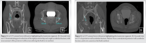
A 17-year-old male, without pertinent past medical history, sustained a right posterior column with posterior wall acetabulum fracture after a single-motor vehicle collision as a front-seat passenger. On arrival, he was neurovascularly intact. Radiographs demonstrated a right posterior wall acetabular fracture with a posterior hip dislocation, involving 21% of the posterior wall, measured by the Keith et al. method (Fig. 2) [5, 6]. The operative treatment for the acetabular fracture is described below.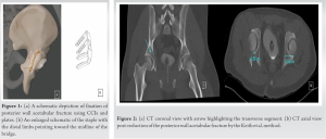
Case 2
A 23-year-old male, without pertinent past medical history, sustained a right posterior column with posterior wall acetabular fracture with ipsilateral posterior hip dislocation and knee traumatic arthrotomy from a motor vehicle collision as a back-seat passenger. The CT scan demonstrated disruption involving 29% of the posterior wall as measured the Keith et al. method (Fig. 3). The operative treatment for the acetabular fracture is described.
Case 3
A 40-year-old man presented as an unrestrained front seat passenger from a high-speed, vehicle motor vehicle collision, sustaining a right posterior column with posterior wall acetabulum fracture and left posterior wall acetabulum fracture (Fig. 4). He was neurovascularly intact on presentation. Anteroposterior (AP) view of the pelvis demonstrated a right transverse posterior wall acetabular fracture/dislocation and a left posterior wall acetabular fracture. The operative treatment for the right acetabular fracture, which utilized a CCI, is described.
Operative treatment of the hip using compression staples
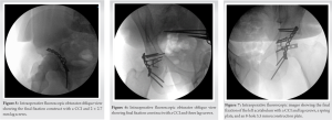
After placing the patient in prone position on the operating table, a Kocher-Langenbeck approach was used to access the posterior wall of the acetabulum. Provisional reduction was achieved using K-wires in all three cases. Definitive reduction of the posterior wall was obtained with 2 × 2.7 mm lag screws in Cases 1 and 2. Lag screws along with a 3-hole spring plate and an 8-hole 3.5 mm reconstruction plate were used for the third case. Reduction of the transverse segments was achieved using BME ELITE CCI, Depuy Synthes Companies, (BME ELITE Continuous Compression Implants, DePuy Synthes, Raynham, MA). Using the BME ELITE Drilling template kit, the correct implant bridge size and configuration was selected. Drill holes were accurately made across the intended fusion site using a drilling template. A BME ELITE Implant insertion tool, with the staple loaded, is used to inset the staples as far as possible into the predrilled holes. Fluoroscopy was used to ensure proper placement (Fig. 5-7). (Fig. 1) shows a schematic view of the reduction.
Post-operative management
Postoperatively, all patients were toe touch weight bearing for 12 weeks. Patient 3 additionally required an ORIF of the left acetabulum, which was fractured without hip dislocation. Physical therapy was initiated postoperatively for all patients, and all were progressed to weight bearing as tolerated at the 3-month visit. On the last clinic visit at 12 months, radiographs revealed maintained alignment of the posterior wall acetabular fractures. There was no evidence of the staples or other fixation device failure or loosening (Fig. 8-10 corresponding to patients 1, 2, and 3, respectively). There were no complications and all patients reported minimal pain. All patients returned to ambulating without a limp and required no assistive devices.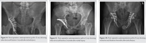
In this report, we described three novel cases where CCIs have been successfully used as reduction aids to augment the final fixation of posterior column with posterior wall acetabular fractures. All three patients went on to bony union with maintained functional status and no evidence of malunion or hardware failure. Acetabular fractures commonly occur due to high-energy trauma, such as motor vehicle accidents or falls from a height [1]. Unfortunately, 30% of outcomes after ORIF are unsatisfactory [7]. Some factors contribute to poor fixation outcomes such as obesity, avascular necrosis, and advanced age [7]. Quality of the anatomical reduction of the fracture remains the most important factor, especially for optimal prognosis and functional outcome [7]. However, it is sometimes hard to achieve anatomic reduction because of difficulty with using lag screws to stabilize small fracture fragments. Hence, we recommend CCIs which have a small footprint and are durable enough to achieve provisional fixation. CCIs are made of nitinol, an alloy of nickel and titanium. The new generation of this alloy has super elasticity and retains its properties at body temperature [4, 8]. This contrasts with static compressive fixation methods, such as lag screws which may lose the initial compression generated over time [9]. SMA staples are often utilized clinically as fixation devices for osteotomies, arthrodesis, and fracture repairs especially of short bones, facial bones, and the distal extremities [9]. There have been other successful usage of CCI as fixation. For example, staples were described as a preliminary fixation method for an inferior glenoid fracture [4]. Yang et al. also reviewed 51 cases, including two hip arthrodesis, that used CCIs and reported patients with robust bone reunion, no inflammation, and no tissue reaction on follow-up [10]. Schnabel et al. found that CCIs had an advantage of a smaller surgical approach, ease of instrumentation, and decreased fracture site displacement in transverse patella fractures compared to tension band wiring in cadaveric specimens [8]. Lavelle et al. also described the use of nitinol vertebral body stapling for prevention of curve progression in idiopathic scoliosis [11]. However, to the authors knowledge, the use of CCIs has not been previously described for augmenting posterior column with posterior wall pelvic fixation. Here, we demonstrate three cases of posterior column with posterior wall acetabular fractures successfully treated with novel augmentation with CCIs. The advantages of CCIs makes them a favorable fixation method for transverse segments present in posterior acetabular fractures [4, 9]. With its durable alloy and small footprint, CCIs can provide strong compression in small areas, provide provisional reduction, reinforce fixation constructs, and reduce the amount of hardware required for fixation. Despite its small footprint, CCIs supply enough force to encourage bone healing [4, 10]. BME ELITE CCIs, described in our case series, supply increased compression force of about 150 N compared to the SPEED or SPEEDTITAN implants [12]. Constructs using the BME ELITE implants were found to provide significantly more compression than a locking titanium midfoot compression plate with and without 4.0 mm lag screws [13]. CCIs dynamically supply compression in contrast to mechanical static compression staples [9, 14]. Single staple implants can average about 18.7 ± 2.5 N to 27.6 N ± 10.7 N in force [15], with bending stiffness of 45 N/mm. Due to the small footprint, double-staple constructs can be considered and can achieve 237% greater compressive force, 34% greater bending stiffness, and 104% greater bending strength than constructs with a single implant [15]. When placed in an orthogonal configuration, double staples can supply 2.16 times the compressive force compared to a single-staple application [15]. There are several studies corroborating the strength of the implant [4, 12]. The specifications of the BME ELITE implant can range from 15 to 30 mm in bridge size, 15–20 mm in leg length, and 2–4 legs per staple [13]. Compared to lag screws which have only two main points of contact with bone, at the head and at the tip of the screw, four-legged staples allow more even load distribution through the four tips of the staple [9, 15]. With such a small size, there is an increased versatility in providing fixation in areas constrained by neighboring soft tissue, where other larger fixation tools may be limited [4]. However, care must be exercised to avoid inserting staples into the joints when fixing lateral fracture pieces with bone depths <15 mm. Similar challenges are faced with bone fragments from highly comminuted fractures and osteoporotic bone [4]. In our series, all patients were young adults, so this concern was attenuated. There is also a concern for galvanic corrosion when using different combinations of metals [4]. However, nitinol is a near-equiatomic nickel-titanium alloy that is biologically safe [9]. Another limitation is the high cost of the device [4].
We used CCIs to successfully stabilize the transverse segments in three posterior column with posterior wall fractures. On the last clinic visit at 12 months, radiographs revealed maintained alignment of the posterior wall acetabular fractures. There was no evidence of the staples or other fixation device failure or loosening. There were no complications and all patients reported minimal pain. All patients returned to ambulating without a limp and required no assistive devices.
We present a series of three patients whose posterior column with posterior wall fractures went on to bony union with augmentation using CCIs. Our series demonstrates the viability of utilizing CCIs to augment fixation of posterior column with posterior wall acetabular fractures. This technique has value to surgeons, as the device has a small anatomic footprint, is technically simple, and provides excellent compression.
Large fixation devices may compromise the quality of anatomical reduction of small fractures of the posterior acetabular column. CCIs may present a novel and simple method of addressing this obstacle due to its small footprint and continuous integumentary force provision.
References
- 1.Letournel E. Acetabulum fractures: Classification and management. Clin Orthop Relat Res 1980;151:81-106 [Google Scholar]
- 2.Ferguson TA, Patel R, Bhandari M, Matta JM. Fractures of the cetabulum in patients aged 60 years and older: An epidemiological and radiological study. J Bone Joint Surg Br 2010;92:250-7. [Google Scholar]
- 3.Pease F, Ward A, Stevenson A, Cunningham J, Sabri O, Acharya M, et al. Posterior wall acetabular fracture fixation: A mechanical analysis of fixation methods. J Orthop Surg (Hong Kong) 2019;27:2309499019859838. [Google Scholar]
- 4.Wu JC, Mills A, Grant KD, Waiter PJ. Fracture fixation using shape-memory (Ninitol) staples. Orthop Clin North Am 2019;50:367-74. [Google Scholar]
- 5.Keith J, Brashear H, Guilford W. Stability of posterior fracture-dislocations of the hip. Quantitative assessment using computed tomography. J Bone Joint Surg Am 1988;70:711-4. [Google Scholar]
- 6.Firoozabadi R, Spitler C, Schlepp C, Hamilton B, Agel J, Routt M, et al. Determining stability in posterior wall acetabular fractures. J Orthop Trauma 2015;29:465-9. [Google Scholar]
- 7.Long HT, Deng ZH, Zou M, Lin ZY, Zhu JX, Zhu Y. Effects of the cetabular fracture index and other factors of posterior wall acetabular fracture on functional outcome. J Int Med Res 2017;45:1394-405. [Google Scholar]
- 8.Schnabel B, Scharf M, Schwieger K, Windolf M, van der Pol B, Braunstein V, et al. Biomechanical comparison of a new staple technique with tension band wiring for transverse patella fractures. Clin Biomech (Bristol, Avon) 2009;24:855-9. [Google Scholar]
- 9.Schipper ON, Ellington JK. Nitinol compression staples in foot and ankle surgery. Orthop Clin North Am 2019;50:391-9. [Google Scholar]
- 10.Yang PJ, Zhang YF, Ge MZ, Cai TD, Tao JC, Yang HP. Internal fixation with Ni-Ti shape memory alloy compressive staples in orthopedic surgery. A review of 51 cases. Chin Med J (Engl) 1987;100:712-4. [Google Scholar]
- 11.Lavelle WF, Samdani AF, Cahill PJ, Betz RR. Clinical outcomes of nitinol staples for preventing curve progression in idiopathic scoliosis AIS supplement. J Pediatr Orthop 2011;31:S107-13. [Google Scholar]
- 12.DePuy Synthes. Continuous Compression Implants. Ray nham, MA: DePuy Sy nthe s. Available from: https://www.jnjmedtech.com/sites/default/files/2021- 12/CCI%20Trauma%20Indication%20Brochure%20103162- 210602.pdf [Last accessed on 2021]. [Google Scholar]
- 13.DePuy Synthes. Product Overview BME ELITE TM Continuous Compression Implant. Raynham, MA: DePuy Synthes. Available from: https://www.jnjmedtech.com/sites/default/files/2021- 12/BME%20ELITE%20Brochure%20127248-210527.pdf [Last accessed on 2021]. [Google Scholar]
- 14.Farr D, Karim A, Lutz M, Calder J. A biomechanical comparison of shape memory compression staples and mechanical compression staples: Compression or distraction? Knee Surg Sports Traumatol Arthrosc 2010;18:212-7. [Google Scholar]
- 15.Gaston GR, Lee SK. Effects of Using Single vs Double Nitinol Implants on Construct Compression and Stability. Available from: https://www.jnjmedtech.com/sites/default/files/2021- 07/%28Two%20Parallel%20BME%20ELITE%20vs.%20Plat e%20%26%20Screw%20White%20Paper%29175335- 210428_EffectsUsingSinglevDoubleNitinolImplants%20%28 1%29%20%281%29%20%281%29.pdf [Last accessed on 2021]. [Google Scholar]