Floating hip with hip dislocation is difficult to manage but can reduce the hip dislocation with knee spanning external fixator and manage in stages shall reduce the complications.
Dr. Nabin Kumar Sahu, Department of Orthopaedics, IMS and SUM Hospital, Kalinga Nagar, Bhubaneswar, Odisha, India. E-mail: nabinsahu08@gmail.com
Introduction: Floating hip with hip dislocation is a very high-energy, devastating, and rare injury whose treatment is very challenging, and the outcome is usually poor.
Case Report: A 35-year-old man presented posterior wall fracture acetabulum and dislocation of the hip with ipsilateral distal third shaft femur fracture with intra-articular extension fracture and un-displaced patella fracture. We achieved a reduction of hip dislocation by a knee-spanning external fixator followed by open reduction and internal fixation with anatomical locking plate for distal third femur fracture with intra-articular extension followed by open reduction and internal fixation for posterior wall of acetabulum with recon plate in Kocher-Langenbeck approach in stages. The patient was able to partial weight bear after 12 weeks of the injury and mobilized independently without any support after 5 months.
Conclusion: Floating hip with hip dislocation is difficult to manage but reducing the hip dislocation with knee spanning external fixator and management in stages will reduce the complications and better outcome.
Keywords: Floating hip, hip dislocation, knee spanning external fixator.
A floating hip is a very high-energy, devastating, and rare injury for which treatment is very challenging and the outcome is poor. A floating hip with hip dislocation is a more devastating injury than a floating hip. A floating hip is a rare case with an incidence rate of one in ten thousand fracture patients. A floating joint is a skeletal discontinuity proximal and distal to that joint [1]. The floating hip term can be delineated as a fracture of the pelvis or acetabulum with an ipsilateral femur fracture. Floating hips have been classified into three types by Mueller. Type A is a combination of acetabulum and femur fractures; type B is a combination of pelvic and femur bone fractures; and type C is a combination of pelvic, acetabulum, and femur fractures [2]. Leibergall is classified into two types: groups A and B. Group A included a femur fracture with an ipsilateral pelvic bone fracture, either an open book fracture or an unstable shear fracture of the pelvis, whereas group B included a femur fracture with an ipsilateral acetabulum fracture [3]. Nathan classified type A as a femur fracture with pelvic ring fracture and type B as a femur fracture with acetabulum fracture [4]. Nathan’s irreducible posterior hip dislocation with a floating hip was reduced by open reduction by the Kocher-Langenbeck approach [4]. We are reporting a case of irreducible hip dislocation with floating hip and distal femur fracture, which is a very rare case, and reporting the method, stages of treatment, and outcome of the case.
We present a case of a 35-year-old man who came to our emergency with a history of road traffic accidents after sustaining multiple fractures and irreducible hip dislocation with type A floating hip as per the Mueller classification of floating hip and type C1 distal femur intercondylar fracture with shaft femur fracture in the lower limb. The patient had no head injury with Glasgow Coma Scale 15/15 without any abdominal, pelvis, or chest injury.
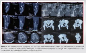
X-ray and computed tomography (CT) scan imaging of the pelvis and knee joint which are shown in Fig. 1 and 2 revealed a posterosuperior hip dislocation with a posterior wall fracture of the acetabulum (Thompson and Epstein classification type II) and an ipsilateral shaft femur with a distal third femur fracture with intra-articular of the right lower limb. He had an associated injury, a segmental fracture of the clavicle with ipsilateral upper limb dysfunction with hand power 2/5, which was a partial incomplete brachial plexus injury.
His D-dimer value was 1045 which is very high. We planned for emergency hip dislocation reduction by the close reduction method with the help of Schanz pins and an external fixator. We applied traction to the proximal femur with 3 Schanz pins and a fixator and did the reduction maneuver by the Allis method with knee flexion and internal rotation but couldn’t reduce the hip dislocation picture [5]. After the failure of the Allis reduction maneuver, hip dislocation was reduced by the closed method after completing the knee-spanning external fixator while maintaining the limb length of the fractured femur which is shown in intraoperative C-arm pictures in Fig. 3 and 4 and X-ray and CT scan of pelvis with both hips in Fig. 3.
On the same anesthesia, intercondylar femur fracture fixation was done by close reduction and internal fixation with two 6.5 mm CC screws to congruent the articular surface. After 3 days, fixators were removed, and the distal third femur fracture with intra-articular extension was fixed with a distal femoral locking plate and two interfragmentary screws and traction was applied to the remaining tibial fixator. On the same anesthesia, the segmental clavicle was fixed with an anatomical plate. After 7 days of injury, the acetabulum was fixed in the Kocher-Langenbeck approach in the left lateral position by a 3.5 mm, 9-hole recon plate, and two interfragmentary screws with the help of a trochanter osteotomy, which was fixed with two 6.5 mm CC screws. Immediate post-operative day 2 X-rays were done which is shown in Fig. 5. Immediate post-operative after 15 days 30–45° by continuous passive movement of the knee joint, and hip flexion were started till 6 weeks. After 6 weeks, knee bending was increased to 90°–120° till 12 weeks by a physiotherapist. Gait training was started after 12 weeks with support from a physiotherapist followed by weight-bearing mobilization with the help of a stick till 5 months.
We followed up on this patient for 6 weeks, 12 weeks, 6 months, and 1 year and found a satisfactory X-ray with a satisfactory clinical outcome. The patient started weight bearing after 12 weeks with support and started squatting and sitting cross-leg after 5 months.
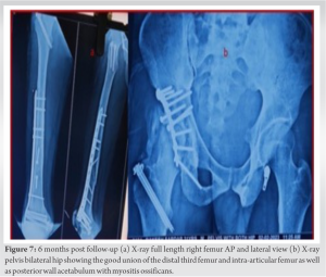
He achieved full knee flexion and extension, HIP flexion 0–110°, abduction and adduction 0–30° with external and internal rotation 45° after 5 months of surgery as shown in Fig. 6 and found satisfactory union in 6 months of post-operative X-ray which is shown in Fig. 7.
He was independently walking after 6 months of surgery. In X-ray myositis ossificans developed in the hip abductors after 6 weeks and was started on indomethacin.
Floating hip dislocation is a high-velocity trauma that involves multiple injuries and polytrauma. This is a very rare case and a big challenge to manage trauma surgeons, which needs surgical management in the correct sequence at the right time. Radiologic evaluation by multiple views of X-rays and CT scans is very important to avoid missing diagnoses because hip dislocation is missed by regular X-rays by 50% in floating hip cases [6]. Floating hip dislocation is very difficult to reduce, and it is an orthopedic emergency to avoid avascular necrosis as a later complication. Many articles describe many techniques for close reduction of hip dislocation by using the Steinmann pin and traction method [3, 7-9]. N.C. Tiedeken et al. describe the open reduction of hip dislocation and the fixation of the acetabulum in irreducible floating hip dislocation [4]. We achieved floating hip dislocation reduction by applying a knee-spanning external fixator by alignment of the limb length, which was failed by the Steinmann pin and Allis maneuver by longitudinal traction and internal rotation [5]. Many complications can be faced during and after treatment of this type of injury [10]. It helps to decrease the chance of avascular necrosis of the head of the femur and improves the general condition of the patient.
Hip dislocation with a floating hip is a very rare case and a very high-energy trauma that is difficult to manage hemodynamically as well as fracture and dislocation. We achieved a favorable outcome for this patient outcome as we hemodynamically optimized immediately and hip dislocation was reduced immediately within 12 h of injury by a knee-spanning external fixator, which failed to reduce several attempts of reduction technique, which reduced the chance of avascular necrosis of the head and arthritis of the hip joint. Subsequent femur and acetabulum fracture fixation was done in stages, which reduced the chance of fat embolism syndrome and other complications. The surgical line of management of floating hips has been up for debate for decades. No definitive guidelines are available for the order of fixation for these fractures. This is our endeavor to present an organized structural planning for the floating hip.
Floating hip with hip dislocation is a complex trauma that can be simplified the treatment by reducing the hip dislocation with the help of knee spanning external fixator by maintaining the limb length alignment and fixation of fractures can be done by stages will be better options to decrease the chance of complication in such type of polytrauma case.
References
- 1.Simpson NS, Jupiter JB. Complex fracture patterns of the upper extremity. Clin Orthop Relat Res 1995;318:43-53. [Google Scholar]
- 2.Muller EJ, Siebenrock K, Ekkernkamp A, Ganz R, Muhr G. Ipsilateral fractures of the pelvis and the femur-floating hip? A retrospective analysis of 42 cases. Arch Orthop Trauma Surg 1999;119:179-82. [Google Scholar]
- 3.Liebergall M, Mosheiff R, Safran O, Peyser A, Segal D. The floating hip injury: Patterns of injury. Injury 2002;33:717-22. [Google Scholar]
- 4.Tiedeken NC, Saldanha V, Handal J, Raphael J. The irreducible floating hip: A unique presentation of a rare injury. J Surg Case Rep 2013;2013:rjt075. [Google Scholar]
- 5.Allis OH. An Inquiry into the Difficulties Encountered in the Reduction of Dislocations of the Hip. Philadelphia, PA: Dornan; 1896. [Google Scholar]
- 6.Helal B, Skevis X. Unrecognised dislocation of the hip in fractures of the femoral shaft. J Bone Joint Surgery. Br Vol 1967;49:293-300. [Google Scholar]
- 7.Chi-Chuan W, Chun-Hsuing S, Lih-Huei C. Femoral shaft fractures complicated by fracture-dislocations of the ipsilateral hip. J Trauma 1993;34:70-5. [Google Scholar]
- 8.Harper M. Traumatic dislocation of the hip with ipsilateral femoral shaft fracture: A method of treatment. Injury 1982;18:391-4. [Google Scholar]
- 9.Suzuki T, Shindo M, Soma K. The floating hip injury: Which should we fix first? Eur J Orthop Surg Traumatol 2006;16:214-8. [Google Scholar]
- 10.Cannada LK, Justin MH, Preston JB, Heidi I, Hassan M, Jason H, et al. Treatment and complications of patients with ipsilateral acetabular and femur fractures. J Orthop Trauma 2017;31:650-6. [Google Scholar]


