Osteochondroma can rarely develop in flat bones and management with excision provides good results with minimal recurrence.
Dr. Danish Jameel Khan, Department of Trauma and Orthopedics, Queen Elizabeth Hospital, Birmingham, United Kingdom. E-mail: drdanishkhannew.540@gmail.com
Introduction: Osteochondromas are benign bone tumors common in metaphyseal ends of long bones like distal femur and are relatively uncommon in flat bones such as scapula. Patients usually present with either a visual deformity requiring treatment for cosmetic reason or present with mechanical symptoms hindering activities of daily living. The tumor is mostly benign and malignant transformation is rarely seen. Treatment usually involves surgical excision of the lesion with minimal chances of recurrence if complete excision of the lesion is done.
Case Report: Here, we present the case of a 12-year-old boy presenting with a symptomatic dorsal scapular osteochondroma who underwent successful surgical excision without any recurrence.
Conclusion: Osteochondroma can be seen on flat bones and should be kept in the differential. Treatment is by excision and usually has good long-term outcomes.
Keywords: Osteochondroma, dorsal scapula, pediatric.
Osteochondromas or exostoses are benign bone tumors, which arise from normal, expanding epiphyseal cartilage located in sites away from the growth plate, resulting in the typical asymmetrical mass of enchondral bone with a cartilaginous cap [1]. The presence of medullary and cortical bone with the continuity of the tumor is pathognomonic for osteochondroma and aids in establishing the diagnosis [2]. Common sites of osteochondromas include the metaphyseal ends of long bones such as distal femur, proximal tibia, and proximal humerus. Scapular involvement is rare and seen in 3–4.6% of the cases [3]. They remain mostly asymptomatic, patients present either for cosmetic reasons as it leads to a bony hard swellings causing visual deformity or for functional reasons as it may lead to reduced movements and hamper activities of daily living [4]. Osteochondromas usually grow with skeletal growth and cease to grow after skeletal maturity and a growth continuing into adulthood should raise suspicion of malignant transformation [5]. Few cases of osteochondroma on flat bone have been previously described in the literature. [6]. Here, we report a case of 12-year-old boy who underwent successful surgical excision of a symptomatic dorsal scapular osteochondroma.
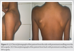
A 12-year-old boy presented to the outpatient department with a history of pain and gradual onset progressive swelling in the left upper back (Fig. 1a and b) for the past 2 years. The swelling led to a visual deformity and impaired the activities of daily living as the patient had difficulty lying supine while sleeping. On clinical examination, a bony swelling was observed along the dorsal aspect of the left scapula measuring approximately 2 cm × 2 cm. The swelling was non-tender and was fixed to the underlying bone and moved with the scapula. Complete active and passive ranges of motion were observed without any restriction in thoracoscapular movements. No bony swellings in other parts of the body were noted.
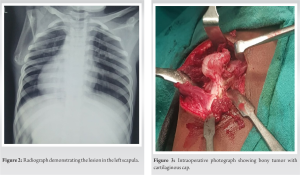
Radiographic evaluation of the scapula showed that the swelling originated from the lateral border of scapula (Fig. 2). The lesion caused significant distress and hampered activities of daily living therefore decision for surgical excision of swelling was taken.
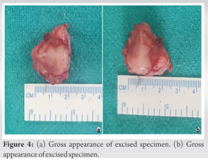
The swelling was exposed through a dorsal scapular incision and a bony lesion with stalk and cartilaginous covering was observed (Fig. 3). Extra periosteal resection was performed, and the sample was sent for histopathological examination which confirmed the diagnosis of osteochondroma without evidence of malignant changes (Fig. 4a and 4b).
Postoperatively the patient’s deformity completely disappeared with complete scapulothoracic range of motion. At 3 month follow-up, the patient had completely recovered and was able to sleep supine without any discomfort. After 1 year of follow-up, there was no recurrence of symptoms or swelling.
Scapular involvement by osteochondroma is seen in 3.0–4.6% of all reported cases and comprises 14.4% of all tumors of the scapula. The ventral surface is involved most commonly, presenting as an isolated unilateral lesion [4]. They are usually asymptomatic, and patients usually present for cosmetic reasons or mechanical restriction to shoulder movements. Pain may occur secondary to fracture through the stalk of a pedunculated osteochondroma or due to malignant transformation [7]. Our patient had mechanical symptoms leading to difficulty in sleeping in supine position. Osteochondroma radiographs show cortex and medulla of swelling continuous with the parent bone [1]. Few cases of solitary osteochondroma of scapula have been reported in literature with most of these describing osteochondroma on the volar surface leading to pseudo-winging of scapula. Literature for dorsal scapular osteochondroma is relatively less available. Jadhav et al. presented case of dorsal scapular osteochondroma in 2016 presenting with difficulty in sleeping in supine position [7]. Yadkikar and Yadkikar also presented a similar case in 2013 with dorsal scapular osteochondroma [8]. Salgia et al. in 2013 also treated patient with similar presentation but the patient was from a higher age group and was predominantly concerned about cosmesis [9]. In our case, the patient symptoms improved symptomatically after complete excision of lesion, and at 1 year follow-up, there was no evidence of recurrence of lesion. Thus, the overall prognosis following excision is good, provided that excision is complete, with incomplete excision leading to recurrence [10].
Osteochondromas though rare may present in flat bones such as scapula and differential diagnosis of osteochondroma should be kept in mind while working up such cases. Complete surgical excision provides excellent clinical outcome with complete resolution of deformity as well as the mechanical symptoms associated with the lesion as evidenced in this case.
Osteochondroma can be seen on flat bones and should be kept in the differential. Treatment is by excision and usually has good long-term outcomes provided complete excision has been carried out.
References
- 1.Fukunaga S, Futani H, Yoshiya S. Endoscopically assisted resection of a scapular osteochondroma causing snapping scapula syndrome. World J Surg Oncol 2007;5:37. [Google Scholar]
- 2.Alabdullrahman LW, Byerly DW. Osteochondroma. In: StatPearls. Treasure Island, FL: StatPearls Publishing; 2024. [Google Scholar]
- 3.Esenkaya I. Pseudowinging of the scapula due to subscapular osteochondroma. Orthopedics 2005;28:171-2. [Google Scholar]
- 4.Puri A, Agarwal MG. Current concepts in bone and soft tissue tumours. In: Textbook of Orthopaedic Oncology. Hyderabad: Paras Medical Books Pvt. Ltd.; 2007. [Google Scholar]
- 5.Kumar N, Ramakrishnan V, Johnson GV, Southern S. Endoscopically-assisted excision of scapular osteochondroma. Acta Orthop Scand 1999;70:394-6. [Google Scholar]
- 6.Aalderink K, Wolf B. Scapular osteochondroma treated with arthroscopic excision using prone positioning. Am J Orthop (Belle Mead NJ) 2010;39:E11-4. [Google Scholar]
- 7.Jadhav PU, Banshelkikar SN, Seth BA, Goregaonkar AB. Osteochondromas at unusual sites-case series with review of literature. J Orthop Case Rep 2016;6:52-4. [Google Scholar]
- 8.Yadkikar SV, Yadkikar VS. Osteochondroma on dorsal surface of the scapula in 11 years old child-A case report. Int J Med Res Health Sci 2013;2:305. [Google Scholar]
- 9.Salgia A, Biswas S, Agarwal T, Sanghi S. A rare case presentaion of osteochondroma of scapula. Med J Dr DY Patil Univ 2013;6:338. [Google Scholar]
- 10.Krieg JC, Buckwalter JA, Peterson KK, El-Khoury GY, Robinson RA. Extensive growth of an osteochondroma in a skeletally mature patient. A case report. J Bone Joint Surg Am 1995;77:269-73. [Google Scholar]


