Hereditary hemochromatosis may be a predisposing factor for Kienböck’s disease, which in turn may aid in the early detection and treatment of these conditions.
Dr. Alexander C R Top, Department of Orthopaedics, AZ Delta, Deltalaan 1, 8800 Roeselare, Belgium. E-mail: alexander.top1995@gmail.com
Introduction: Avascular necrosis of the lunate bone has been extensively researched, although the etiology of the condition remains controversial. Even though many treatments for the disease exist, a better understanding of the pathophysiology can improve our decision-making between preventive and therapeutic measures. Various hematological disorders have been found to predispose for Kienböck’s disease. On the other hand, there has not yet been any reference in literature to a relationship between this condition and hereditary hemochromatosis (HH).
Case Report: We present two cases of Kienböck’s disease in two patients who are third-degree relatives and diagnosed with HH. A 61-year-old Caucasian female patient with type 1 HH presented with symptomatic Kienböck’s disease on the left side. The patient is a third-degree relative of a 51-year-old male Caucasian patient with Kienbock’s disease on the right side, known as having the same hereditary hematological condition.
Conclusion: Our findings suggest a potential correlation between the aforementioned conditions. The prevalence of these coexisting pathologies should be studied further.
Keywords: Kienböck, lunate, osteonecrosis, hemochromatosis.
Kienböck’s disease is a condition characterized by avascular necrosis (AVN) of the lunate bone. It was first described in 1910 by the Austrian radiologist Robert Kienböck [1]. Symptoms can include wrist pain, decreased grip strength, and range of motion of the affected joint. However, incidental determination of the condition is common [2]. Other than the patient’s perceived symptoms, there are little other findings that guide the choice of therapy. Although distinct degrees of lunar collapse could evoke outspoken symptoms in some, numerous studies found no clear link between these radiological findings and the symptomatology [2-4]. Treatment is therefore predominantly based on patient complaints. Just as the factors predisposing to symptoms are not clearly defined, the etiology of the condition remains dubious. In general, it affects 20–40-year-old men regularly performing physical labor or having a history of wrist trauma. More recent literature describes equal involvement in terms of gender with the onset of the disease usually later in women [3]. Other low evidence predisposing factors include smoking, excessive alcohol consumption, ulnar variance, and specific comorbidities such as diabetes, peripheral vascular, and certain genetic conditions [4]. Factors largely associated with AVN of the hip as well. Despite inconclusive data, one can assume that the vascularization of the lunate plays an important role in the onset of the disease. Lamas et al. mapped the arterial supply of this carpal bone by studying 27 wrists using latex injections and the Spalteholz technique, finding palmar nutrient vessels entering the proximal end of the os lunatum through the radioscaphocapitate ligament [4]. Degeneration of this ligament through blunt force and repetitive microtrauma could therefore be a cause of the vascularization of the bone. Vessels entering the dorsal aspect of the lunate could anastomose with the volar ones, preventing ischemia. Considering this delicate vascularization, not only the blood supply but also the vital blood-carried minerals and nutrients could impact the health of this bone. We refer in this context to one specific condition. Hereditary hemochromatosis (HH), a common autosomal recessive disorder in Caucasians, is characterized by excessive iron absorption. A C282Y mutation of the HFE gene is found in most cases [5, 6]. Men and women are affected equally in regard to gene inheritance. In vitro studies have proven the excess of iron to impact different cascades, altering osteoclastogenesis, resulting in bone loss in HH-impaired patients [7]. These studies help us better understand the impact on bone integrity but currently fail to adequately emulate these mostly animal-based and vitreous findings in clinical cases. Little evidence can be found on the correlation between HH and lunar osteomalacia. We present two affiliated patients with unilateral Kienböck’s disease and HH who received treatment in our center.
Case 1
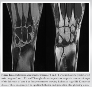
A 61-year-old female patient, housewife, right hand dominant with type 1 HH and homozygous for the Cys282Tyr mutation of the HFE gene presented with increasing mechanical left wrist tenderness and concomitant functional deficit without any history of wrist-involved injuries, professional or recreational activities associated with excessive stress on the wrist joint. The patient is an active smoker. Physical examination showed 45° palmar flexion (contralateral 80°) and dorsiflexion (contralateral 70°). Prosupination was full and symmetric. Posteroanterior and lateral radiographs displayed lunate collapse with a revised carpal height ratio of 1.26 (measured using the Nattrass Method), differing significantly from the mean value of the revised carpal height ratio in the general population of 1.57 ± 0.05 [8] (Fig. 1).
Case 2
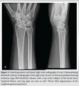
A 51-year-old male patient, driver and third-degree relative of our female study participant, also known with type 1 HH and homozygous for the Cys282Tyr mutation in the HFE gene is presented. He quit smoking at age 35 and is known for psoriasis. He is right-hand dominant and presented with 14-year history of atraumatic right wrist complaints. The physical examination showed 30° palmar flexion (contralateral 75°), 80° dorsiflexion (contralateral 80°) and Grip strength of 30 kg on the right, and 50 kg on the left, measured with the Jamar dynamometer. X-rays were obtained showing lunar collapse with a revised carpal height ratio of 1.31 (Fig. 3) and stage IIIb Kienböck’s disease in accordance with the Lichtman classification. Additional therapy was consensually waived given the current unstated clinical implications.
Since its discovery in 1910, several pathogenetic pathways have been proposed for Kienböck’s disease with many well-defined risk factors playing a key role. These can be further categorized into different groups, including traumatic events ranging from repetitive microtrauma to acute wrist injuries, deprived vascularization, rheumatological conditions, and certain medical conditions, associated with impaired osseous blood flow [10]. Concerning Kienböck’s disease, HH is not commonly mentioned in the literature and its role is probably underestimated. After all, most current research largely focuses on morphological predisposing factors, rather than hematological ones. This iron overload disorder can lead to osteoporosis, chondrocalcinosis, and arthropathy. A few studies linked the condition to AVN of the femur [11, 12]. Both patients had successful monthly phlebotomy treatments for their iron excess. Both relatives presented with clear lunate collapse. One chose skillful neglect, and the other one underwent an operative treatment with good relief of pain and fine recovery of function.
We suggest a close pathological relationship between HH and AVN of the lunate (Kienböck’s disease) based on these two case reports in a male and female relative, which have never been published in the literature. Awareness of this possible cause of wrist pain with this hereditary condition is mandatory.
In patients with Kienböck’s disease, possible predisposing conditions such as HH should not be overlooked. Identification of both pathologies may help us better understand the pathophysiology and possibly future treatment of lunatomalacia.
References
- 1.Wagner JP, Chung KC. A historical report on Robert Kienböck (1871-1953) and Kienböck’s disease. J Hand Surg Am 2005;30:1117-21. [Google Scholar]
- 2.Van Leeuwen WF, Janssen SJ, Ter Meulen DP, Ring D. What is the radiographic prevalence of incidental kienböck disease? Clin Orthop Relat Res 2016;474:808-13. [Google Scholar]
- 3.Lamas C, Carrera A, Proubasta I, Llusà M, Majó J, Mir X. The anatomy and vascularity of the lunate: Considerations applied to Kienböck’s disease. Chir Main 2007;26:13-20. [Google Scholar]
- 4.Golay SK, Rust P, Ring D. The radiological prevalence of incidental kienböck disease. Arch Bone Jt Surg 2016;4:220-3. [Google Scholar]
- 5.Beaton MD, Adams PC. The myths and realities of hemochromatosis. Can J Gastroenterol 2007;21:101-4. [Google Scholar]
- 6.Pietrangelo A. Hereditary hemochromatosis--a new look at an old disease. N Engl J Med 2004;350:2383-97. [Google Scholar]
- 7.Jandl NM, Rolvien T, Schmidt T, Mussawy H, Nielsen P, Oheim R, et al. Impaired bone microarchitecture in patients with hereditary hemochromatosis and skeletal complications. Calcif Tissue Int 2020;106:465-75. [Google Scholar]
- 8.Nattrass GR, King GJ, McMurtry RY, Brant RF. An alternative method for determination of the carpal height ratio. J Bone Joint Surg Am 1994;76:88-94. [Google Scholar]
- 9.Lichtman DM, Mack GR, MacDonald RI, Gunther SF, Wilson JN. Kienböck’s disease: The role of silicone replacement arthroplasty. J Bone Joint Surg Am 1977;59:899-908. [Google Scholar]
- 10.Irisarri C. Aetiology of Kienbőck’s disease. J Hand Surg 2004;29:279-85. [Google Scholar]
- 11.Rollot F, Wechsler B, Du Boutin le TH, De Gennes C, Amoura Z, Hachulla E, et al. Hemochromatosis and femoral head aseptic osteonecrosis: A nonfortuitous association? J Rheumatol 2005;32:376-8. [Google Scholar]
- 12.Albers CE, Albers J, Sapra A, Bhandari P, Ranjit E. Double whammy: A case of concurrent alcohol use and hereditary hemochromatosis leading to avascular necrosis of the femur. Cureus 2021;13:e18067. [Google Scholar]


