Early recognition and prompt surgical intervention are crucial in managing heterotopic ossification of the Achilles tendon, particularly in patients with underlying risk factors such as diabetes mellitus, and the presented surgical approach and postoperative management contribute to evolving strategies for addressing this rare condition.
Dr. Krishna Amith Kumar, Department of Orthopaedics, Aster Hospital Mankhool, Al Mankhool, Dubai. E-mail: dr_kamit@yahoo.com
Introduction: Heterotopic ossification of the Achilles tendon (HOTA) is a rare but consequential complication to an inflammatory event, often presenting challenges in diagnosis and management.
Case Report: We report a case of a 42-year-old male with Type II diabetes mellitus, managed with Metformin, who presented with acute pain and swelling in the right ankle following a running activity. Clinical examination revealed tenderness and a visible swelling proximal to the calcaneum attachment of the tendon which was hard in consistency, and a positive Thompson test indicative of a potential Achilles tendon injury. Imaging, including X-ray and magnetic resonance imaging, confirmed a complete avulsion tear with cranial retraction of the torn tendon and heterotopic ossification seen proximal to the torn end of the tendon. Surgical intervention, employing a posterior paramedian approach under tourniquet control, involved tendon approximation using the Speed bridge technique, incision over the proximal tendon and removal of the ossified tendon, and repair of the incised tendon sheath. Postoperatively, the patient received a tailored medication regimen and was advised strict non-weight-bearing measures, wound management, and limb elevation. The patient was discharged in a stable condition.
Conclusion: This case underscores the importance of early recognition and prompt surgical intervention in managing HOTA, particularly in patients with underlying risk factors. The presented surgical approach and postoperative management contribute to the evolving strategies for addressing this rare clinically significant condition.
Keywords: Heterotopic ossification, diabetes mellitus, surgical intervention, speed bridge technique.
The Achilles tendon, recognized as one of the vital and adaptable tendons in the body, is situated in the posterior aspect of the lower leg [1]. It distinguishes itself as the thickest tendon in the human body, capable of withstanding significant tensile forces [2]. Despite being strongest, it is prone to various injuries due to its continuous functional demands during activities such as walking and running, compounded by its limited blood supply. Prolonged exposure to heavy loads on the Achilles tendon can result in chronic inflammation, degeneration, and various forms of fractures [3]. Heterotopic ossification (HO), characterized by the abnormal formation of bone within soft tissues, is frequently associated with inflammatory events [4]. Achilles tendon ossification represents an atypical clinical phenomenon, with occasional reports appearing in the medical literature since its initial description in 1908. It is distinguished by the presence of an ossified mass within the fibrocartilaginous structure of the tendon, located either within the tendon body or at its attachment to the calcaneus. Typically, it manifests as a firm, tender mass, although it can also be entirely asymptomatic [5]. Several cases manifest acutely, revealing a previously asymptomatic region with established ossification within a fractured tendon. In contrast, there are instances where symptoms gradually develop, characterized by the onset of pain and swelling [6]. HO of the Achilles Tendon (HOTA) shows a male predilection. In some instances, the ossified mass may fracture, leading to associated partial or complete rupture of the tendon. Conditions linked to HOTA encompass metabolic diseases such as diabetes mellitus and Wilson’s disease, as well as infectious diseases, including chronic osteomyelitis [7]. HOTA is usually managed through conventional preventive measures targeting the occurrence of HO, which involves prophylactic non-steroidal anti-inflammatory drugs (NSAIDs) and low-dose radiotherapy. In cases where intervention is necessary (in cases of failed conservative treatment), surgical resection of the ossified mass is the most common treatment approach [8]. As the understanding of HO continues to evolve, particularly in the context of specific anatomical structures like the Achilles tendon, the presented case contributes valuable insights to the existing body of knowledge. This report serves not only as a documentation of a rare clinical entity but also as a resource for clinicians involved in the management of similar cases, shedding light on the intricacies of diagnosis, surgical intervention, and post-operative care in the context of Achilles tendon injuries complicated by HO.
A 42-year-old male, with a history of Type II diabetes mellitus managed with Metformin, presented with pain and swelling in the right ankle persisting for 7 days. The onset of symptoms followed a running activity, causing difficulty in movement and walking. Notably, there were no known drug allergies, and the patient had no familial history of medical illnesses.
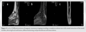
On clinical examination, the right ankle exhibited tenderness and visible swelling. The positive Thompson test indicated a potential Achilles tendon injury. A hard palpable mass was felt proximal to the torn end of the tendon. Palpation revealed a gap in the Tendon Achilles, accompanied by pain during ankle movement. Neurovascular assessments showed no distal deficits. The X-ray report highlighted marked soft tissue swelling, multiple irregular osseous fragments in the thickened soft tissue on the posterior aspect of the distal leg, and a noted calcaneal spur, suggesting a likely diagnosis of heterotrophic ossification and a potential Achilles tendon tear (Fig. 1). Magnetic resonance imaging (MRI) findings revealed a complete full-thickness avulsion tear involving the tendon Achilles insertion with cranial retraction of the torn tendon. The distance between the retracted tendon and the insertion site was about 28 mm. Diffuse thickening of the retracted tendon-Achilles was seen. Focal ossification was seen at the torn edge of the tendon and in the substance of the mid tendon. Diffuse peritendinous soft- tissue edema and fluid collection was seen (Fig. 2).
The patient was diagnosed with a laceration of the right Achilles tendon along with HO of tendo achilles, and surgical intervention was recommended.
Surgical intervention
Under spinal anesthesia, the patient was positioned prone, and a posterior paramedian incision, 3 cm above the palpable rupture, exposed the severed tendon ends after incising the paratenon. The sural nerve was identified, isolated, and protected. Following freshening and debridement of the tendon end, a successful approximation was achieved with the ankle in a plantar-flexed position. and by utilizing the speed bridge technique, the tendon was sutured with Krakow locking loop technique with a suture tape , was secured at the avulsed site in a calcaneum with two anchors. The skin incision was extended proximally up to the solid mass felt inside the tendon, incision was made in the mass of the tendon and the ossified mass was removed from within the substance of the tendon keeping the fibers intact. The remaining tendon was sutured and sealed keeping the tendon intact. Closure involved suturing superficial tear fibers with non-absorbable sutures, proximal tendon exploration, and removal of calcified bone (Fig. 3 and 4).
The limb was immobilized with a below-knee slab in plantar flexion, and the patient was transferred to the recovery room. On discharge, the patient was in stable condition and received a personalized medication plan aimed at pain management, infection prevention, and facilitating healing. Strict non-weight-bearing measures were advised for 8 weeks, accompanied by wound management, limb elevation, and a scheduled outpatient review after 3 days for dressing.
The presented case highlights a rare clinical entity, HOTA, characterized by the abnormal formation of bone within the fibrocartilaginous substance of the Achilles tendon. While the literature on this condition remains limited, it is crucial to explore the etiology, clinical presentation, and management strategies, as demonstrated in this case. The common occurrence of calcific deposits within the intra-tendinous tissue is a well-recognized pathology affecting tendons. However, it is crucial to differentiate tendon ossification from the mere accumulation of calcific deposits. While the process of calcification in tendinous tissue is familiar, the origin of cells involved in tendon ossification remains poorly understood. Several hypotheses have been suggested to elucidate this unusual and not yet fully understood process that results in ossification within the tendon substance. However, none of the proposed hypotheses has been conclusively proven to date [5]. The multifaceted etiology of HOTA involves significant contributions from trauma, including repetitive incidents such as falls, sports injuries, and surgical interventions. Metabolic conditions, especially diabetes mellitus, prominently feature in the literature as key factors associated with HOTA [8]. The presented case, showcasing HOTA in a diabetic patient, aligns with this understanding, highlighting the pivotal role of metabolic factors in the pathogenesis of this condition. Clinical manifestations of HOTA can vary, ranging from an asymptomatic ossified mass to acute presentations with fractures in previously asymptomatic areas [6]. In this case, the patient, a 42-year-old male with Type II diabetes, presented with pain and swelling following a running activity, ultimately leading to the diagnosis of a laceration of the Achilles tendon associated with HO. The clinical examination and the Thompson test in this case raised Achilles tendon injury suspicion. X-ray and MRI confirmed an avulsion tear with heterotopic calcification. The surgical intervention used the posterior paramedian approach and the speed bridge technique for secure fixation, aiming for early mobilization and reduced re-rupture risk. Proximal tendon exploration addressed potential irritation sources. Postoperatively, a below-knee slab in plantar flexion maintained tendon approximation, and medication focused on pain management, infection prevention, and healing. Due to the patient’s diabetic status, meticulous wound care and an 8-week strict non-weight-bearing protocol were crucial for minimizing complications and promoting healing. There is generally no standardized treatment exists for HO. It has been proposed that NSAIDs, localized low-dose irradiation, retinoids, BMP inhibitors, and antagonists may have a prophylactic effect on HO formation. While these options are effective in preventing HO, their efficacy diminishes after fibroproliferation and cartilage formation. Surgical resection remains the sole treatment option once bone tissue formation is completed [9]. The documented case of a 42-year-old male with HOTA presents a unique intersection with previous reports on diabetes mellitus as a risk factor for Achilles tendon pathology. While diabetes has been recognized as a risk factor for Achilles tendon rupture, prior instances involving diabetic individuals are notably scarce. Sobel et al. reported a case involving a 61-year-old diabetic female with complete ossification of a ruptured Achilles tendon, potentially accompanied by a fracture of the ossified mass [10]. Notably, the present case aligns with this limited pool of reports, further emphasizing the association between diabetes mellitus and Achilles tendon ossification. The rarity of documented cases involving both diabetes mellitus and Achilles tendon ossification suggests the need for heightened clinical awareness in managing diabetic patients presenting with symptoms of tendon pathology. Diabetes, with its known impact on tissue healing and repair, may contribute to the unique manifestation of HO in the Achilles tendon. The interplay of diabetes as a metabolic factor and trauma, such as repetitive incidents and sports injuries, underscores the complexity of this case. The presence of ossification in the Achilles tendon in both cases raises questions about the underlying mechanisms and cellular processes involved in the ossification of tendinous tissue in diabetic individuals. While the literature lacks a comprehensive understanding of these mechanisms, the convergence of these cases highlights the need for further research to elucidate the specific pathways and interactions contributing to Achilles tendon ossification in the diabetic population.
The case reported underscores the importance of a comprehensive approach in managing Achilles tendon injuries with HO, especially in patients with comorbid conditions such as diabetes. The multidisciplinary collaboration, accurate diagnostic imaging, and the chosen surgical technique collectively contribute to a favorable outcome in this challenging clinical scenario. Further research into the interplay between diabetes and Achilles tendon pathology is warranted to enhance our understanding and refine treatment strategies for similar cases in the future.
This case underscores the critical significance of early identification and timely surgical intervention in addressing HOTA, particularly in patients with underlying risk factors such as diabetes mellitus. The presented surgical approach and post-operative management contribute valuable insights to the evolving strategies for managing this rare and clinically significant condition.
References
- 1.Egger AC, Berkowitz MJ. Achilles tendon injuries. Curr Rev Musculoskelet Med 2017;10:72-80. [Google Scholar]
- 2.Wong M, Jardaly AH, Kiel J. Anatomy, bony pelvis and lower limb: Achilles tendon. In: StatPearls. Treasure Island, FL: StatPearls Publishing; 2023. Available from: https://www.ncbi.nlm.nih.gov/books/nbk499917 [Last accessed date on 2023]. [Google Scholar]
- 3.Manfreda F, Ceccarini P, Corzani M, Petruccelli R, Antinolfi P, Rinonapoli G, et al. A silent massive ossification of Achilles tendon as a suspected rare late effect of surgery for club foot. SAGE Open Med Case Rep 2018;6:2050313X18775587. [Google Scholar]
- 4.Magnusson SP, Agergaard AS, Couppé C, Svensson RB, Warming S, Krogsgaard MR, et al. Heterotopic ossification after an Achilles tendon rupture cannot be prevented by early functional rehabilitation: A cohort study. Clin Orthop Relat Res 2020;478:1101-8. [Google Scholar]
- 5.Cortbaoui C, Matta J, Elkattah R. Could ossification of the Achilles tendon have a hereditary component? Case Rep Orthop 2013;2013:539740. [Google Scholar]
- 6.Harris PC, Denton JS. Heterotopic ossification of the Achilles tendon following ankle fracture: A case report. Foot Ankle Surg 2000;6:275-9. [Google Scholar]
- 7.Vaishya R, Maduka CO, Agarwal AK, Vijay V, Vaish A. Heterotopic ossification of tendo Achilles: An uncommon clinical entity. J Orthop Case Rep 2019;9:45-7. [Google Scholar]
- 8.Su L, Arshi A, Beck JJ. Extensive atraumatic heterotopic ossification of the Achilles tendon in an adolescent with metabolic syndrome: A case report. JBJS Case Connect 2020;10:e0394. [Google Scholar]
- 9.Chang SH, Matsumoto T, Okajima K, Naito M, Hirose J, Tanaka S. Heterotopic ossification of the peroneus longus tendon in the retromalleolar portion with the peroneus quartus muscle: A case report. Case Rep Orthop 2018;2018:7978369. [Google Scholar]
- 10.Sobel E, Giorgini R, Hilfer J, Rostkowski T. Ossification of a ruptured Achilles tendon: A case report in a diabetic patient. J Foot Ankle Surg 2002;41:330-4. [Google Scholar]


