The purpose of this article is to emphasize the uncommon occurrence of angiolipoma and how to manage it.
Dr. Yogadeepan Dhakshinamurthi, Department of Orthopedics, Dr. Muthus Hospital, Near Jai Shanthi Theatre, Trichy Road, Singanallur, Coimbatore, Tamil Nadu, India. E-mail: yogadeepan5992@gmail.com
Introduction: Angiolipomas are benign small soft tissue rubbery tumors common between the age group of 20 and 30. They are very common in the extremities and neck. It is composed of adipose tissues and blood vessel components.
Case Report: We report a case of 82-year-old lady with complaints of right foot pain. She was diagnosed to have a sclerotic lesion over the cuboid. Biopsy revealed angiolipoma which is a very rare tumor in bone. The patient underwent intralesional curettage along with cortico-cancellous bone grafting.
Conclusion: Although angiolipoma is very rare in bone, its occurrence should not be excluded. It can rarely occur in old age and it has to be carefully evaluated and managed to prevent recurrence.
Keywords: Angiolipma, curettage, cuboid, cortico-cancellous graft.
Angiolipomas are subcutaneous benign tumors. More common in the extremities, neck, and trunk. It has adipose and vascular components. Peak incidence is second and third decades. Usually, the lesion is well-circumscribed and encapsulated. Symptoms depend on the site of the lesion. Microscopically, angiolipomas have a mature adipocytic proliferation variably associated with a vascular component. The blood vessels contain fibrin microthrombi. Differential diagnosis includes angioleiomyomas, schwannomas, glomus tumors, hemangiomas, and giant cell granuloma. Histopathology and immunohistochemistry are crucial for diagnosis. S 100 is expressed in the adipocytic component whereas CD31 and CD34 are expressed in capillary network. Treatment is excisional curettage and recurrence is rare.
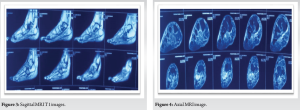
An 82-year-old lady came with complaints of right foot pain over the past 2 months. No history of any recent trauma or injury. Pain was dull aching, gradually increasing. No diurnal variation and no constitutional symptoms. Clinical examination shows tenderness over the lateral aspect of mid foot. Minimal warmth present. Neurovascular status normal. X-ray shows a sclerotic lesion over the cuboid. No breach in the cortex. No adjacent bone involvement (Fig. 1). MRI shows a hyperintense lesion in T2 weighted images confined to the cuboid and hypointense lesion in T1 images (Fig. 2-4).
An open biopsy was performed and the result revealed intraosseous angiolipoma of cuboid (Fig. 5 and 6). Histology revealed adipocytic tissue with thin blood vessels and spindle cells in a myxoid stroma.
The patient was planned for intralesional curettage and bone grafting with or without fixation depending on stability after grafting. Under spinal anesthesia patient’s right foot was draped, right iliac crest also prepared for bone graft harvesting. Across the dorsilinear incision over the dorsum of foot, the cuboid was exposed. Upon exposing the cuboid, the lateral cortex was found to be breached (Fig. 7). Extended curettage was done with a bone scoop and hydrogen peroxide (Fig. 8 and 9). A tricortical graft was harvested from the iliac crest and placed over the defect. The remaining void filled with a cancellous graft harvested from the iliac crest (Fig. 10-12).

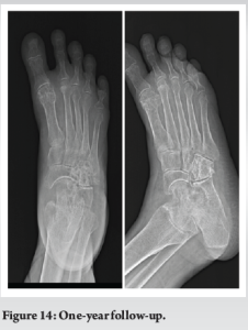
After grafting, cuboid was very stable on all foot movements. Hence, fixation was not done. Below knee pop slab was applied. Post-operative period was uneventful (Fig. 13). The patient was kept on the slab for 6 weeks followed by partial weight bearing. Complete weight bearing started around 3 months. She had one-year follow-up and her pain completely settled and she was doing her all day-to-day activities (Fig. 14).
Lipomas are mesenchymal neoplasm in which angiolipoma is a variant that has proliferating capillaries interspersed with adipose tissues. Lipomas have infiltrating and non-infiltrating types. Angiolipoma was first described by Brown in the year 1912. However, it was described as a separate entity by Howard and Helwig in 1960 [1]. Even though it is rare, many familial cases of autosomal dominance traits have been reported [2]. Brault in 1868 first described intraosseous lipoma involving shaft of the femur. Not many cases of intraosseous angiolipomas are reported. Usually, it arises from long bones, such as humerus, radius, facial bones, and flat bones, such as ribs and pelvis. Cuboidal origin is very rare. A review of the literature shows around twenty cases of intraosseous angiolipomas have been reported so far and no cases have been reported in cuboid. Angiolipomas included 5–17% of lipomas [3]. They are slow-growing tumors. Pain may be due to the mass effect. Chromosome 13 mutations are reported in some cases [4]. Some cases show PIK3CA mutations [5]. The etiology of intraosseous angiolipoma is not very clear. Embryonic sequestration of multipotent mesenchymal cells, which gets activated by hormones after puberty. Some studies state trauma as a possible cause. However, Hart et al. study demonstrated trauma as a less likely cause for this lesion [6-9]. Shea et al. study demonstrated the association of angiolipomas with mast cells in which there are average 10 times more mast cells in angiolipomas than in classical lipomas [10]. Mast cells secrete VEGF which promotes angiogenesis and TGF alpha and beta which produce inflammation and proliferation of endothelial cells, respectively [11]. The histological study is needed to diagnose these rare variants and adequate sample preparation intraoperatively is necessary for appropriate diagnosis as these tumors exhibit non-specific clinical characteristics and mode of presentation. Surgical excision is the primary treatment option. Chances of malignant transformation are rare. Recurrence after thorough excision is rare. Other options like embolization can be used in some cases [7, 12, 13].
Angiolipomas should also be considered in the case of evaluating an intraosseous tumor even though its occurrence is very rare. It is usually benign and adequate curettage prevents recurrence.
Angiolipomas can present as intraosseous tumors. It should be differentiated from other malignant tumors like angioliposarcoma. It can rarely present in old age. Angiolipoma is usually common in long bones, facial bones, and flat bones, such as pelvis and ribs. Cuboid angiolipoma is very rare and to our knowledge, it is the first case getting reported.
References
- 1.Howard WR, Helwig EB. Angiolipoma. Arch Dermatol 1960;82:924-31. [Google Scholar]
- 2.Garib G, Siegal GP, Andea AA. Autosomal-dominant familial angiolipomatosis. Cutis 2015;95:E26-9. [Google Scholar]
- 3.Arenaz Búa J, Luáces R, Lorenzo Franco F, García-Rozado A, Crespo Escudero JL, Fonseca Capdevila E, et al. Angiolipoma in head and neck: Report of two cases and review of the literature. Int J Oral Maxillofac Surg 2010;39:610-5. [Google Scholar]
- 4.Panagopoulos L, Gorunova L, Andersen K, Lobmaier I, Bjerkehagen B, Heim S. Consistent involvement of chromosome 13 in angiolipoma. Cancer Genomics Proteomics 2018;15:61-5. [Google Scholar]
- 5.Saggini A, Santonja C, Nájera L, Palmedo G, Kutzner H. Frequent activating PIK3CA mutations in sporadic angiolipoma. J Cutan Pathol 2021;48:211-6. [Google Scholar]
- 6.Rastogi N, Rohatgi G. Intraosseous angiolipoma of head of humerus-an extremely rare entity, with review of literature. Asian Pac J Health Sci 2017;4:133-5. [Google Scholar]
- 7.Yu K, Van Dellen J, Idaewor P, Roncaroli F. Intraosseous angiolipoma of the cranium: Case report. Neurosurgery 2009;64:E189-90. [Google Scholar]
- 8.Morgan KM, Hanft S, Xiong Z. Cranial intraosseous angiolipoma: Case report and literature review. Intractable Rare Dis Res 2020;9:175-8. [Google Scholar]
- 9.Lewis DM, Brannon RB, Isaksson B. Intraosseous angiolipoma of the mandible. Oral Surg 1980;50:4. [Google Scholar]
- 10.Shea CR, Prieto VG. Mast cells in angiolipomas and hemangiomas of human skin: Are they important for angiogenesis? J Cutan Pathol 1994;21:247-51. [Google Scholar]
- 11.Ribatti D, Belloni AS, Nico B, Salà G, Longo V, Mangieri D, et al. Tryptase-and leptin-positive mast cells correlate with vascular density in uterine leiomyomas. Am J Obstet Gynecol 2007;196:e1-7. [Google Scholar]
- 12.Amirjamshidi A, Ghasemi B, Abbasioun K. Giant intradiploic angiolipoma of the skull. Report of the first case with MR and histopathological characteristics reported in the literature and a review. Surg Neurol Int 2014;5:50. [Google Scholar]
- 13.Hatae R, Mizoguchi M, Arimura K, Kiyozawa D, Shimogawa T, Sangatsuda Y, et al. Giant cranial angiolipoma with arteriovenous fistula: A case report. Surg Neurol Int 2022;13:314. [Google Scholar]


