Tarsal middle phalangeal bipartition should be kept in mind in the clinical setting.
Dr. Uluman Şişman, School of Medicine, Koç University Hospital, Maltepe Mahallesi, Davutpaşa Caddesi, No:4 Topkapı, 34010 Istanbul, Türkiye. E-mail: usisman17@ku.edu.tr
Introduction: Bipartite bone formation is a congenital variation occurring due to the incomplete ossification of newly forming bones in the body. The patella and sesamoid bones are the most common bipartite bone sites. However, some unusual bones can also have this kind of variation and it is important to diagnose them correctly and avoid unnecessary overtreatment. Such unique conditions are being presented as individual case reports and published. Although distal phalangeal bipartition is reported before, to our knowledge, this is the first and only case to be reported to have bipartition in the tarsal middle phalanges in the literature.
Case Report: In this case report, we are presenting the case of a 10-year-old boy, who presented to the Emergency Department due to a glass cut to the plantar site of the right foot, and bipartition in the 3rd middle phalanges of both feet has been found in the radiographies.
Conclusion: Differentiating bipartite bone formations from fractures is important in the clinical setting. Unique presentations of these bony formations, such as tarsal middle phalanges as in our case, should be considered. By doing so, overtreatment and unnecessary interventions can be prevented.
Keywords: Bipartite, middle phalanx, variation, tarsal phalanges, case report.
Bipartite bone formation is an occurrence that has been seen after the incomplete fusion of the ossification centers during the bone formation process. These variations can be seen in different bones in the body, some of them being more common in the population. For instance, bipartite sesamoid bones (patella, hallux sesamoids, os perineum) are more common than other unusual variations (calcaneus, medial cuneiform, navicular) [1-3]. The importance of recognizing these variations is that these got detected during imaging studies, which are usually taken for a different purpose. During the evaluation of results, these variations can be evaluated as a fractured structure rather than individual bone formations. For differentiation, fractures have irregular margins with cortical disruption, yet bipartite bones have smooth margins with cortical continuation [1]. Therefore, recognizing these variations and differentiating them from fractures are important. Nevertheless, not all of these are diagnosed easily, especially the ones which are located at unusual bones. For instance, bipartition of distal phalanges was reported before in literature [4]. In this case report, we are presenting a 10-year-old boy who has bipartite bone formation in the 3rd middle phalanges of both feet, which is the first reported bipartition in the middle phalanges in literature.
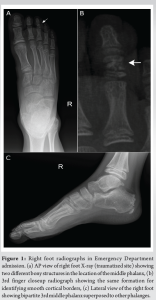
A 10-year-old boy was admitted to the Emergency Department with a cut on the sole of his right foot, which happened while kicking a glass in the same day. In the physical examination, a 5 cm vertical cut has been detected on the plantar side of the right foot. It was located between the 2nd web space and the 3rd middle phalanx. Structures deeper into the cutaneous layer seemed to be unaffected by the cut. All fingers have a full range of motion for flexion. Circulation and sensorineural examination showed no abnormal findings. X-ray imaging was taken for both feet and no foreign bodies in deeper structures were found in the right side. Nevertheless, an incidental bipartite bone formation has been detected in the 3rd middle phalanges in bilateral feet (Fig. 1 and 2). With a detailed radiographical evaluation, the possibility of fracture is ruled out. In further questioning, the patient did not describe any previous symptoms related to these variations. For the treatment, the wound is irrigated and closed with primary suturing. Empirical antibiotics and analgesics are initiated, the tetanus vaccine is delivered, and the patient is discharged on the note of changing the wound dressing for once in 2 days.

Bipartite bones are rare congenital variations that are caused by the incomplete fusion of different ossification centers during the bone formation process. Different bony structures tend to be more susceptible to the occurrence of this variation, which makes them more common in the population. Bipartite patella and sesamoid bones are the most common sites of bipartition variations [5]. Less common presentations, such as scaphoid, medial cuneiform, and navicula bipartition are also getting reported as individual case reports by time [6]. Typically, these variations by themselves usually do not cause symptoms. Hence, they are getting detected incidentally in imaging studies, which are taken for a different reason (such as a trauma to the affected site). In the setting of Emergency Department conditions or Orthopedics clinics, these variations can be misinterpreted as fractured bone structures in the radiographs. This misinterpretation in acute settings can lead to overtreatment and delay the proper treatment of the main reason of the presentation of the patients. Therefore, it is important to differentiate these formations from fractures. In the radiographies, fractures tend to have irregular margins with small fissure-like lines, together with cortical disruption in the case of any displacement. On the other hand, bipartite bone formations have smooth continuous cortical borders without any sharp edges [1, 7]. Some of the most common forms even have their own classifications based on the type of the location of ossification centers, such as Saupe Classification Systems for Bipartite Patella [5, 8]. In addition, hallux sesamoids can be formed as more than 2 bones, which are also known as multipartite hallux sesamoids [9]. These two examples are common bipartite presentations, and they are getting recognized and diagnosed more accurately by virtue of the alertness about the presence of these formations. Nevertheless, for the rare presentations of these variations in unusual locations, misinterpretation of the findings can happen easily. Navicular bone, middle cuneiform, and scaphoid are reported unusual sites for bipartition to occur [1, 10]. In the literature, distal phalangeal bipartition was reported before as a rare case presentation [4]. In our case, tarsal middle phalangeal bipartition is a newly reported site for this variation to occur. Our patient was admitted to the Emergency Department due to a complaint with a glass cut wound due to trauma to the sole of the foot, and radiological evaluation showed 2 separated bony structures in 3rd middle phalanges bilaterally. Combining the results of the physical examination and the radiological suspicion, the patient was diagnosed with bipartition and a treatment plan was formulated around the wound. By doing so, additional unnecessary interventions and treatments have been avoided.
Proper and careful evaluation of the radiographies is important to differentiate between multipartite variations and fractured segments. Considering the medical history of the patient and making a proper physical examination can also aid the diagnostic process for an accurate diagnosis. By doing so, overtreatment and unnecessary interventions can be prevented.
To our knowledge, this is the first case to be reported to have bipartition in the tarsal middle phalanges in the literature.
References
- 1.Chan BY, Markhardt BK, Williams KL, Kanarek AA, Ross AB. Os conundrum: Identifying symptomatic sesamoids and accessory ossicles of the foot. AJR Am J Roentgenol 2019;213:417-26. [Google Scholar]
- 2.Osebold WR, Remondini DJ, Lester EL, Spranger JW, Opitz JM. An autosomal dominant syndrome of short stature with mesomelic shortness of limbs, abnormal carpal and tarsal bones, hypoplastic middle phalanges, and bipartite calcanei. Am J Med Genet 1985;22:791-809. [Google Scholar]
- 3.Pollack D, Diament M, Kotlyarova Y, Gellman Y. The bipartite medial cuneiform. J Am Podiatr Med Assoc 2021;111(6):10.7547/20-025. [Google Scholar]
- 4.Bhatti A, Thirkannad S. Bipartite distal phalanx--watch out for this condition! Hand Surg 2011;16:211-3. [Google Scholar]
- 5.Jennings CM, Tjiattas-Saleski L. Bipartite patella. J Am Osteopath Assoc 2016;116:816. [Google Scholar]
- 6.Keles-Celik N, Kose O, Sekerci R, Aytac G, Turan A, Guler F. Accessory ossicles of the foot and ankle: Disorders and a review of the literature. Cureus 2017;9:e1881. [Google Scholar]
- 7.Candan B, Torun E, Dikici R. The prevalence of accessory ossicles, sesamoid bones, and biphalangism of the foot and ankle: A radiographic study. Foot Ankle Orthop 2022;7:24730114211068792. [Google Scholar]
- 8.Abdelmohsen SM, Hussien MT. Painful knee. Int J Surg Case Rep 2023;114:109165. [Google Scholar]
- 9.Lee SY, Tan TJ, Yan YY. Fracture of a bipartite medial hallux sesamoid masquerading as a tripartite variant: A case report and review of the literature. J Foot Ankle Surg Sep 2019;58:980-3. [Google Scholar]
- 10.Brookes-Fazakerley SD, Jackson GE, Platt SR. An additional middle cuneiform? J Surg Case Rep 2015;2015:rjv076. [Google Scholar]


