This article emphasizes the critical importance of early diagnosis and long-term follow-up in managing cervical spinal enchondromas, ensuring timely intervention and reducing the risk of recurrence or malignant transformation.
Dr. Hitesh Modi, Senior Spine Consultant, Department of Spine Surgery, Zydus Hospitals and Healthcare Research Private Limited, Ahmedabad. Gujarat. India. E-mail: modispine@gmail.com
Introduction: Chondromas are benign cartilaginous tumors classified into periosteal chondromas and enchondromas. While periosteal chondromas grow on the bone surface, enchondromas develop within the medullary cavity. Enchondromas constitute 4–8% of all bone tumors, with spinal enchondromas being exceptionally rare, particularly in the cervical region. Despite their benign nature, spinal enchondromas can cause significant clinical symptoms and have the potential for recurrence or malignant transformation.
Case Report: A 14-year-old female presented with a swelling on the posterior aspect of her neck, accompanied by dull, aching pain radiating into the right upper limb, and muscle weakness assessed at IV/V. Imaging studies, including computed tomography (CT) and magnetic resonance imaging, revealed a lobulated lesion in the right lamina of the C4 vertebra extending to C5, causing spinal cord and nerve root indentation. The patient underwent a C4-C5 laminectomy with complete tumor excision. Histopathological examination confirmed the diagnosis of enchondroma. Follow-up and Outcomes: At 6 months, the patient experienced complete resolution of pain and significant improvement in neurological symptoms. Follow-up CT scans at 3 years and at 10 years did not exhibit any recurrence, and the patient remained symptom-free throughout the follow-up period.
Conclusion: This case highlights the successful long-term outcome following the surgical resection of a cervical spine enchondroma, demonstrating that aggressive surgical intervention can lead to sustained symptom-free outcomes. The 10-year follow-up provides valuable insight into the long-term prognosis of cervical spine enchondromas, emphasizing the importance of early and complete surgical resection along with extended surveillance.
Keywords: Enchondroma of cervical spine, benign cartilaginous tumor, surgical resection, long-term outcome.
Chondromas are benign cartilaginous tumors that can be classified into periosteal chondromas and enchondromas [1]. Periosteal chondromas grow outside the bone cortex, while enchondromas arise within the medullary cavity [2]. Enchondromas account for 4–8% of all bone tumors, with spinal enchondromas comprising only 2% of these cases [3]. Enchondromas rarely transform into malignant chondrosarcomas but can cause significant clinical symptoms, particularly when they occur in the spine. The primary treatment for symptomatic or potentially malignant enchondromas is surgical resection [4]. Despite their benign nature, spinal enchondromas are particularly rare, especially in the cervical region, with only a few cases reported in the literature [5]. Most documented cases have a follow-up period of fewer than 3 years, limiting our understanding of their long-term outcomes [6]. The potential for recurrence and malignant transformation, though low, necessitates thorough and prolonged monitoring [7]. Our case study aims to fill this gap by providing a detailed 10-year follow-up of a cervical spine enchondroma, highlighting the importance of long-term surveillance. The aim of this study is to present a rare case of enchondroma in the C4-C5 vertebrae of a 14-year-old female, treated surgically, and followed for 10 years with no recurrence. This study hypothesizes that aggressive surgical resection of spinal enchondromas, even in early stages, can lead to long-term symptom-free outcomes and prevent recurrence. By documenting this extended follow-up period, we seek to contribute valuable data to the limited body of literature on spinal enchondromas and underscore the importance of long-term follow-up in these patients.
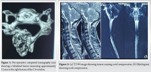
A 14-year-old female presented to our outpatient department with a swelling on the posterior aspect of the right side of her neck, which was progressively associated with dull, aching pain radiating into the right upper limb. Along with the pain, she reported weakness in her right upper limb, with muscle strength assessed at IV/V. The patient had no significant past medical history, nor was there a relevant family history of similar conditions. There was no prior trauma or previous surgeries reported. On physical examination, a prominent swelling was noted on the right posterior aspect of the neck. Neurological assessment confirmed diminished muscle strength in the right upper limb, graded at IV/V. To further investigate the condition, imaging studies were performed. Computed tomography (CT) revealed a lobulated lesion approximately 12 mm in diameter located in the right lamina of the C4 vertebra, extending toward the lamina of the C5 vertebra (Fig. 1). Magnetic resonance imaging demonstrated a predominantly hyperintense and well-defined lesion at the C4-C5 disc space. This lesion caused indentation of the spinal cord and the exiting nerve root, without any observable bone marrow edema (Fig. 2).
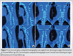
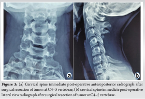
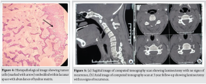
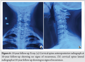
Following the diagnostic imaging, the patient underwent surgical intervention. A C4-C5 laminectomy was carried out to remove the hard, irregular tumor mass completely (Fig. 3). Intraoperative samples were sent for histopathological examination to confirm the diagnosis. Histopathological analysis of the excised tumor revealed multiple fragmented masses of cartilage islands separated by fibro-osseous septa. The tumor cells were organized in small clusters within nodules, consistent with the diagnosis of enchondroma (Fig. 4) At the 6-month follow-up, the patient reported complete resolution of pain and notable improvement in neurological symptoms, with upper limb strength returning to V/V. Repeat CT scans at this stage showed no signs of recurrence. During the 3-year follow-up, CT scans continued to reveal no evidence of recurrence (Fig. 5), and the patient remained asymptomatic. At the 10-year follow-up, the patient maintained an absence of recurrence and did not report any neck pain or neurological deficits (Fig. 6 and 7).
This case underscores the successful long-term outcome following the surgical resection of a cervical spine enchondroma, with a decade-long follow-up demonstrating no recurrence and no neurological impairment.
The aim of this study was to document a rare case of enchondroma in the cervical spine, treated surgically, with a comprehensive 10-year follow-up. Enchondromas are benign cartilaginous tumors that are uncommon in the spine, particularly in the cervical region. Our case involved a 14-year-old female with a C4-C5 enchondroma, who underwent complete surgical excision and demonstrated no recurrence over a decade. This case highlights the importance of long-term monitoring and supports the hypothesis that aggressive early surgical intervention can lead to sustained symptom-free outcomes. Previous studies have documented the occurrence and treatment of spinal enchondromas, but follow-up periods have generally been short. For instance, Fahim et al. reported a case of periosteal chondroma in the pediatric cervical spine with a follow-up period of < 3 years, emphasizing the rarity and surgical challenges associated with these tumors (Fahim et al. 2009) [7]. Similarly, McLoughlin et al. described spinal chondromas, noting their low incidence and potential for recurrence, with follow-ups averaging around 2–3 years (McLoughlin et al. 2008) [8]. Our study extends these findings by providing a significantly longer follow-up, demonstrating that aggressive surgical resection can achieve durable outcomes without recurrence. Willis and Heilbrun (2005) [5] reported a case of enchondroma in the cervical spine with a short-term follow-up, highlighting the rarity of the condition and the need for more extensive data to understand long-term outcomes. In contrast, our case report offers a decade-long follow-up, contributing valuable insights into the long-term prognosis of cervical spine enchondromas. This extended follow-up period is crucial for understanding the full spectrum of potential outcomes and reinforces the importance of long-term surveillance in these patients. Some studies have reported contrasting outcomes, particularly concerning the recurrence and potential malignant transformation of spinal enchondromas. Jing et al. (2017) documented a case of a large thoracic spine enchondroma with a relatively high recurrence rate, suggesting that tumor size and location may influence the likelihood of recurrence (Jing et al. 2017) [9]. In contrast, our case involved a smaller cervical lesion with no recurrence over 10 years, indicating that early and complete surgical resection may mitigate these risks. Another study by Pansuriya et al. (2010) discussed the various subtypes of enchondromatosis and their differing prognoses, noting that while enchondromas generally have a benign course, certain subtypes may exhibit more aggressive behavior (Pansuriya et al. 2010) [10]. Our study provides additional evidence that solitary enchondromas, particularly when surgically resected early, can have an excellent long-term prognosis with minimal risk of recurrence or malignant transformation. While our study provides valuable long-term follow-up data, it is limited by its nature as a single case report. The findings may not be generalizable to all patients with spinal enchondromas due to variations in tumor size, location, and patient demographics. In addition, our study’s retrospective nature and reliance on historical imaging and clinical data may introduce biases. Further research with larger sample sizes and prospective designs is needed to validate our findings and provide more comprehensive insights into the management and prognosis of spinal enchondromas.
Our case report demonstrates that aggressive surgical resection of cervical spine enchondromas can lead to excellent long-term outcomes, with no recurrence observed over a 10-year period. This study adds to the limited body of literature on spinal enchondromas and underscores the importance of long-term follow-up in these patients. Early surgical intervention, coupled with regular monitoring, can effectively manage this rare condition and provide patients with a sustained, symptom-free life.
Prompts recognition and accurate diagnosis of cervical spinal enchondromas are essential for preventing complications and they must be paired with vigilant long-term surveillance to effectively manage the risk of recurrence and malignant transformation.
References
- 1.Robles LA, Mundis GM. Chondromas of the lumbar spine: A systematic review. Global Spine J 2021;11:232-9. [Google Scholar]
- 2.Willis BK, Heilbrun MP. Enchondroma of the cervical spine. Neurosurgery 1986;19:437-40. [Google Scholar]
- 3.Wilpshaar TA, Bovée JV. Bone: Enchondroma. Atlas Genet Cytogenet Oncol Haematol 2018;3(1) DOI:10.4267/2042/70187. [Google Scholar]
- 4.Omlor GW, Lohnherr V, Lange J, Gantz S, Mechtersheimer G, Merle C, et al. Outcome of conservative and surgical treatment of enchondromas and atypical cartilaginous tumors of the long bones: Retrospective analysis of 228 patients. BMC Musculoskelet Disord 2019;20:134. [Google Scholar]
- 5.Willis BK, Heilbrun MP. Enchondroma of the cervical spine. Clin Radiol Extra 2005;60:40-3. [Google Scholar]
- 6.Jeong DM, Paeng SH. Enchondroma of the cervical spine in young women: A rare case report. Asian J Neurosurg 2015;10:334-7. [Google Scholar]
- 7.Fahim DK, Johnson KK, Whitehead WE, Curry DJ, Luerssen TG, Jea A. Periosteal chondroma of the pediatric cervical spine. J Neurosurg Pediatr 2009;3:151-6. [Google Scholar]
- 8.McLoughlin GS, Sciubba DM, Wolinsky JP. Chondroma/chondrosarcoma of the Spine. Neurosurg Clin N Am 2008;19:57-63. [Google Scholar]
- 9.Jing G, Gao J, Guo L, Yin Z, He E. Large enchondroma of the thoracic spine: A rare case report and review of the literature. BMC Musculoskelet Disord 2017;18:155. [Google Scholar]
- 10.Pansuriya TC, Kroon HM, Bovée JV. Enchondromatosis: Insights on the different subtypes. Int J Clin Exp Pathol 2010;3:557-69. [Google Scholar]


