
A radial head fracture involving three or more fragments does not necessarily require a radial head prosthesis or excision. Fixation can lead to favorable outcomes. Repair of the lateral collateral ligament (LCL), fixation of the coronoid, and reinforcement of the anterior capsule contribute to enhanced joint stability in terrible triad elbow injuries.
Dr. Karthik Sangani, Department of Orthopaedics, Narayana Health City, Benguluru, Karnataka, India. E-mail: karthiksang2024@gmail.com
Inroduction: Terrible triad includes posterior dislocation of the elbow, radial head fracture, and coronoid fracture. It renders the elbow joint unstable, creating a need for surgical intervention. The purpose of our study was to analyze the outcomes in such patients who underwent surgery in this tertiary care center.
Case Report: Ten cases were identified in this monocentric single surgeon retrospective study. The cases had radial head tripod fixation with Headless screws, coronoid fixation with transosseous suture tape, and lateral collateral ligament (LCL) repair with suture anchors/ethibond. The assessment was done using the Oxford elbow score and range of motion.
Results: Eight of the ten cases had Mason type 3 radial head fracture pattern, and the remaining had type 2. Six cases had Regan Morey type 1 coronoid fracture, rest had type 2. The mean follow-up period was 38 months (range 4–64 months). The Oxford elbow scores for Pain: A mean of 98.6 (standard deviation [SD] 3.1), Function: A mean of 97.8 (SD 3.6), and Psychosocial domain: A mean of 96 (SD 5.8). The flexion-extension range of motion arc mean is 130° (SD 6.1). The mean Pronation is 69° (SD 2.23). The mean supination is 79° (SD 2.23). Heterotopic ossification was noticed in one patient.
Conclusion: A radial head fracture with three or more fragments should not always indicate a radial head prosthesis or excision. Fixation can also achieve good results. LCL repair, coronoid fixation, and the anterior capsule add to joint stability.
Keywords: Terrible triad, tripod fixation, oxford elbow score, lasso technique of coronoid fixation, lateral collateral ligament repair, level four evidence.
“TERRIBLE TRIAD” was entitled by Robert Hotchkiss in 1996, to describe a spectrum of elbow injuries. Posterolateral dislocation of the elbow accompanied by Radial head and coronoid fractures constituted the triad [1]. Although relatively uncommon, it accounted for 30% of elbow dislocations in a study by van Reit and Morrey [2]. Our study aimed to assess the outcomes in patients with terrible triad elbow injuries who underwent surgery at our hospital.
A total of ten cases of terrible triad who underwent surgical intervention between 2016 and 2022 at our Hospital were identified and their assessment was done retrospectively. Cases with other associated injuries and cases with radial head excision were excluded.
Surgical technique
After pre-operative assessment and radiological imaging, the patient was posted for surgery in a supine position with a sidearm board (Fig. 1). The lateral elbow approach in the Kaplan plain was used (Fig. 2). Dislocation was reduced, if not done already. Sutures were passed around the displaced coronoid fragment along with the anterior capsule using suture tape or Ethibond5. A mini Incision over the dorsal aspect of the proximal ulna was made to create drill holes from the dorsal to the volar aspect near the base of the coronoid. A suture passer was used to shuttle the free ends of the suture tape through one in each of the two drill holes onto the dorsal aspect of the ulna and secured with a knot. The juxtaposition following the transosseous fixation of the coronoid along with the anterior capsule was confirmed under vision through the lateral approach.

The fragments of the radial head were identified and extracted. The fragments were reduced and held with bone clamps. A guide pin was passed across the fracture site. Fixation was done using two 2 mm Headless screws (Fig. 3). This extra corporeally fixed radial head was placed into its position over the radial neck in proper alignment. A 1.2 mm K wire was passed from the periphery of the radial head to the far cortex of the distal fragment. A cannulated drill bit passed over it. After confirming the screw size, a cannulated 2 mm headless screw was passed over the k wire and tightened, providing adequate compression at the fracture site till it engaged the far cortex and proximal end sunk below the chondral surface. This process was repeated for two other screws in different angles of prono-supination, to position the screws on the periphery of the intact radial head toward the radial shaft in a tripod manner [3]. The position of the screws was confirmed on the image intensifier. Lateral collateral ligament (LCL) repair was done using suture anchors, ethibond, or fiber tape. Two Drill holes were made in the lateral epicondyle from the anterolateral and posterolateral aspect in a converging manner in the axial plain. Fiber tape was passed through it for a transosseous fixation of LCL. The stability of the elbow was checked under the c-arm while subjecting it to range of motion (ROM) and valgus-varus stress.

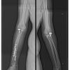
The medial collateral ligament (MCL) was not repaired in any of the patients. The rehabilitation followed a standard protocol. The follow-up assessment was done using the X-rays, ROM in flexion-extension arc and pronation-supination, and the Oxford elbow score. The Oxford elbow scoring system is a type of patient-reported outcome measure [4]. It evaluates the elbow in three independent domains: Elbow function, pain, and social-psychological affect. Fig 4a and b depict the post-operative X-ray images of the same patient.

Out of the ten cases, six were female and four were male. The mean age was 42.2 years. The mode of injury in seven of them was RTA involving a two-wheeler. The mean duration from the time of injury to surgery was 3 days (range 1–6 days). Eight of the ten cases had Mason type 3 radial head fracture pattern, and the remaining had type 2. Six cases had Regan Morey type 1 coronoid fracture, rest had type 2 fracture [5]. The mean follow-up period was 38 months (range 4–64 months). The Oxford elbow scores for Pain: Mean of 98.6 (standard deviation [SD] 3.1), Function: Mean of 97.8 (SD 3.6), and Psychosocial domain: Mean of 96 (SD 5.8). The flexion-extension ROM arc mean was 130° (SD 6.1). The mean Pronation was 69° (SD 2.23). The mean supination was 79° (SD 2.23). None of the cases had redislocation. Grade 1 heterotopic ossification developed in a patient was the only complication among the cases.
The mode of injury in a terrible triad elbow is a fall on an outstretched hand in a supinated forearm leading to a valgus force and axial loading. The failure mechanism of soft tissue constraints around the elbow, as described by O’Driscoll et al., progresses in an orderly fashion from lateral to medial. Starting from the LCL, it involves the anterior and posterior capsule and the MCL [6]. Trochlea causes a shear fracture of the coronoid. LCL, MCL, and coronoid form the primary stabilizers of the joint. These primary stabilizers are disrupted in this injury pattern leading to instability, justifying the injury to be terrible. Most daily activities require an elbow varus, so an incompetent LCL can disable the person. For this reason, even though MCL is a key stabilizer LCL repair warrants priority [7]. Several treatment algorithms have been developed for the management of this injury complex. Pugh et al. proposed a protocol where the coronoid and radial head, LCL injuries are addressed serially with the addition of MCL repair if needed [8]. Controversies still exist over the approach of choice, the need for a dual approach, the need for coronoid fixation, and the management of choice for radial head fractures. Papatheodorou et al. studied the impact of coronoid fixation in terrible triad elbow injuries [9]. Another study from 2019 ascertained that reattachment of the anterior capsule can reduce the risk of elbow arthritis [10]. Garrigues et al. used the suture lasso technique for coronoid fixation. In their multicentric comparative study, the suture lasso technique showed better outcomes with fewer complications compared to internal fixation [11]. The use of headless screws for radial head fixation gives the advantage of placing them beyond the 100° safe zone and also avoids interference with the joint because of its subchondral positioning [3]. Watters et al. compared radial head replacement with fixation and found the overall outcomes to be similar [12]. Wu et al., conducted a study comparing screw fixation, arthroplasty, and plate fixation for 3-part radial head fractures. The study concluded that screw fixation resulted in good early bone union with minimal complications [13]. Implant loosening, capitellar erosion, and forearm pain were associated more with radial head replacement, needing secondary surgery to address the complications. In our study, coronoid fixation along with the anterior capsule was done for all the patients. All the radius fractures were addressed with open reduction and internal fixation using Herbert screws (k-wires for the pediatric patient). The repaired LCL provides lateral stability in the coronal plane against tension and the radial head against compression. All the patients have attained a near-normal flexion-extension ROM, pronation, and supination ROM. Oxford elbow scores of the study suggest that all of them have achieved a good functional recovery with no significant pain or sociopsychological impact on them. The major limitations of our study are its retrospective nature, small sample size, and lack of a control group. However, it holds the potential to pave the way for future research with higher levels of evidence.
Radial head fracture with three or more fragments should not always be an indication for radial head replacement. Good results can be achieved with fixation too. LCL repair and Coronoid fixation along with the anterior capsule add to the joint stability and allow early mobilization. With an enhanced understanding of elbow biomechanics and advancement in fixation techniques, the outcomes have vastly improved in the past decade. The ones who sustain this injury may not be deemed so terrible anymore.
Terrible triad elbow injuries can be successfully treated with radial head fixation, LCL repair, coronoid fixation, and anterior capsule reinforcement, which enhance joint stability and promote early mobilization, leading to improved outcomes.
References
- 1.Ring D, Chen N. Fractures and dislocations of the elbow and forearm. In: Chapman’s Orthopaedic Surgery. 4th ed. New Delhi: Jaypee Brothers Medical Publishers; 2019. [Google Scholar | PubMed]
- 2.Bohn K, Ipaktchi K, Livermore M, Cao J, Banegas R. Current treatment concepts for “terrible triad” injuries of the elbow. Orthopedics 2014;37:831-7. [Google Scholar | PubMed]
- 3.Lipman MD, Gause TM, Teran VA, Chhabra AB, Deal DN. Radial head fracture fixation using tripod technique with headless compression screws. J Hand Surg Am 2018;43:575.e1-6. [Google Scholar | PubMed]
- 4.Guyver PM, Cattell AE, Hall MJ, Brinsden MD. Oxford elbow scores in an asymptomatic population. Ann R Coll Surg Engl 2013;95:415-7. [Google Scholar | PubMed]
- 5.Regan W, Morrey B. Fractures of the coronoid process of the ulna. J Bone Joint Surg Am 1989;71:1348-54. [Google Scholar | PubMed]
- 6.O’Driscoll SW, Jupiter JB, King GJ, Hotchkiss RN, Morrey BF. The unstable elbow. Instr Course Lect 2001;50:89-102. [Google Scholar | PubMed]
- 7.Daphne M, Beingessner J, Whitcomb P. Elbow fractures and dislocation. In Rockwood and Green’s Fractures in Adults. 8th ed., Vol. 1. United States: Lippincott Williams; 2015. [Google Scholar | PubMed]
- 8.Pugh DM, Wild LM, Schemitsch EH, King GJ, McKee MD. Standard surgical protocol to treat elbow dislocations with radial head and coronoid fractures. J Bone Joint Surg 2004;86:1122-30. [Google Scholar | PubMed]
- 9.Papatheodorou LK, Rubright JH, Heim KA, Weiser RW, Sotereanos DG. Terrible triad injuries of the elbow: Does the coronoid always need to be fixed? Clin Orthop Relat Res 2014;472:2084-91. [Google Scholar | PubMed]
- 10.Antoni M, Eichler D, Kempf JF, Clavert P. Anterior capsule re-attachment in terrible triad elbow injury with coronoid tip fracture. Orthop Traumatol Surg Res 2019;105:1575-83. [Google Scholar | PubMed]
- 11.Garrigues GE, Wray WH 3rd, Lindenhovius AL, Ring DC, Ruch DS. Fixation of the coronoid process in elbow fracture-dislocations. J Bone Joint Surg Am 2011;93:1873-81. [Google Scholar | PubMed]
- 12.Watters TS, Garrigues GE, Ring D, Ruch DS. Fixation versus replacement of radial head in terrible triad: Is there a difference in elbow stability and prognosis? Clin Orthop Relat Res 2014;472:2128-35. [Google Scholar | PubMed]
- 13.Wu PH, Shen L, Chee YH. Screw fixation versus arthroplasty versus plate fixation for 3-part radial head fractures. J Orthop Surg (Hong Kong) 2016;24:57-61. [Google Scholar | PubMed]


Related Articles in Journal of Orthopaedic Case Reports
 October 1, 2025 A Unique Case of Bilateral Elbow Terrible Triad in a Polytrauma Patient: Associated Challenges in Management
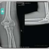 March 1, 2025 Management of Adult Lateral Condyle Fracture: Unveiling a Complex Elbow Injury: A Case Report
 April 10, 2024 Terrible Triad of the Elbow with Ipsilateral Complete Triceps Tearing, Distal Radius and Scaphoid Waist Fracture: A Case Report
 March 10, 2024 Management of Ipsilateral Terrible Triad Injury of Elbow and Concomitant Proximal Humerus Fracture: A Case Report and Literature Review