
This approach involves temporarily extracting the tumor, subjecting it to radiation externally, and then reinserting it with care prioritizing limb preservation.
Dr. Roshan Lal Goyal, Department of Orthopaedics, AIIMS, Raipur, Chhattisgarh, India. E-mail: rashugoyal5657@gmail.com
Introduction: This comprehensive case report highlights the intricate diagnosis and multidisciplinary management of distal femur osteosarcoma in a 15-year-old male. Osteosarcoma is a rare and aggressive form of bone cancer, particularly affecting adolescents and young adults. The significance of this case lies in the detailed description of the diagnostic process, treatment strategy, and successful outcome, which may contribute valuable insights to the existing literature. While numerous case reports on osteosarcoma exist, this report offers unique insights into the management approach and outcome of distal femur osteosarcoma, potentially providing guidance for clinicians facing similar cases in the future.
Case Report: The patient in question is a 15-year-old male, whose ethnic background was not specified. He presented with symptoms suggestive of distal femur osteosarcoma, including localized bone pain, swelling, and limited range of motion in the affected knee joint. Radiological investigations confirmed the presence of a tumor in the distal femur, prompting further evaluation. Pathological markers and histopathological examination confirmed the diagnosis of osteosarcoma. Pre-operative neoadjuvant chemotherapy was administered to optimize conditions for surgical intervention.
Conclusion: This case report underscores the importance of a comprehensive, multidisciplinary approach in the management of distal femur osteosarcoma. The successful outcome, marked by complete tumor excision, reconstruction using autologous fibula graft, and restoration of knee function, highlights the efficacy of the chosen treatment strategy. Furthermore, this report may serve as a valuable resource for clinicians specializing in orthopedic oncology, providing insights into optimal treatment protocols and post-operative care for similar cases. In addition, it contributes to the broader field of oncology by advancing our understanding of osteosarcoma management and emphasizing the importance of individualized treatment plans tailored to the patient’s specific needs.
Keywords: Osteosarcoma, extra corporeal radiotherapy, swashbuckler approach, distal femur anatomical plate, neoadjuvant chemotherapy, autologous fibula graft.
Osteosarcoma, a rare malignant bone tumor, typically develops in the osteoblast cells that form bone, presents unique challenges when localized in the distal femur, particularly in adolescents and young adults [1]. This case aims to provide a detailed exploration of the diagnostic journey and the efficacy of a multidisciplinary approach in managing this complex condition. Distal femur osteosarcoma, constituting about 10–15% of all osteosarcomas, predominantly affects the pediatric age group [2]. The diagnosis relies on a combination of clinical, radiological, and histopathological assessments. Previous studies have emphasized the aggressive nature of distal femur osteosarcoma, necessitating a carefully planned and executed treatment strategy [3]. Extracorporeal radiotherapy (ECRT) is generally considered for patients with osteosarcoma when: (a) There is a high risk of local recurrence, (b) limb preservation is a priority, and other options are not viable, and (c) the patient is a suitable candidate for the procedure from a medical and surgical perspective. ECRT represents a specialized and innovative approach in the multidisciplinary treatment of osteosarcoma, offering a potential limb sparing option for patients. However, it requires careful consideration of the risks and benefits, as well as meticulous surgical and post-operative management to optimize outcomes.
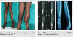
The patient, a 15-year-old male, presented with localized pain and swelling in the distal femur, prompting a thorough evaluation. Understanding the unique challenges associated with osteosarcoma in the pediatric population is crucial for tailoring effective treatment strategies. Adolescents often face distinct physiological and psychological challenges, and their response to treatment may differ from that of adults. In this case, the age of the patient played a significant role in treatment decision-making, with considerations for growth plate preservation and long-term functional outcomes Fig. 1. The decision for pre-operative neoadjuvant chemotherapy consisting of 3 cycles was pivotal in preparing the patient for the subsequent surgical intervention [4]. Distal femur osteosarcoma commonly presents with localized pain and swelling. Radiological assessments, including magnetic resonance imaging (MRI), are instrumental in determining tumor extent and planning surgical interventions. The incorporation of advanced imaging techniques aids in precise staging, facilitating an optimal treatment plan.
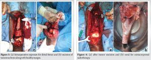
The surgery, performed on December 16, 2020, involved an en bloc excision of the tumor through a swashbuckler surgical approach [5]. This technique, chosen for its accessibility and minimal soft tissue disruption, aligns with the goal of achieving complete tumor excision while preserving surrounding structures. The meticulous excision, with a 5 cm proximal healthy margin, aimed to minimize the risk of local recurrence, a critical consideration in osteosarcoma management [6] Fig. 2 and 3.
Management and treatment
The surgical intervention, performed on December 16, 2020, marked a significant milestone in the patient’s journey [7]. The en bloc excision of the tumor through a swashbuckler surgical approach ensured the removal of the tumorous bone with a 5 cm proximal healthy margin. This meticulous approach aimed to minimize the risk of local recurrence.
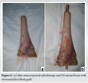
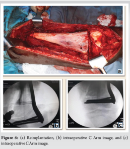
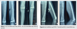
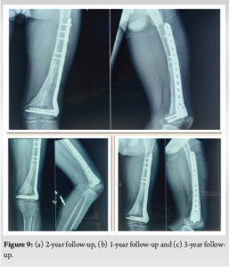
This case report of a 15-year-old male diagnosed with distal femur osteosarcoma underscores the significance of a multidisciplinary approach in achieving successful outcomes. The comprehensive diagnostic journey, encompassing pathological markers, radiological investigations, and histopathology, allowed for an accurate diagnosis and informed subsequent treatment decisions. Extracorporeal irradiation presents a limb salvage technique for osteosarcoma cases where there’s adequate remaining bone both proximal and distal to the tumor. This method, deemed oncologically safe with favorable functional outcomes, utilizes a radiation dose of 50 Gy to effectively sterilize the bone, eliminating tumor cells while mitigating adverse impacts on bone biomechanics and biology. Employing suitable implants for internal fixation alongside supplementary bone grafting during the initial surgery may diminish the necessity for subsequent interventions to achieve union. However, it is essential to note that this procedure is not suitable for structurally weak tumor bones or bones with pathological fractures [8]. ECRT of the excised bone fragment is a novel strategy employed to eradicate any remaining tumor cells [10]. This technique involves subjecting the excised bone to a high dose of radiation outside the body, ensuring thorough eradication of malignant cells. The subsequent reimplantation of the bone fragment using an autologous fibula graft is a critical step in restoring skeletal integrity [1]. The subsequent ECRT of the excised bone fragment is a unique strategy employed to eradicate any residual malignant cells [11]. This approach adds an extra layer of assurance against local recurrence, contributing to the overall success of the treatment Fig. 4. The reimplantation of the bone fragment using an autologous fibula graft and fixation with a distal femur anatomical locking plate system represent cutting-edge reconstructive techniques. The use of anatomical locking plates aligns with contemporary orthopedic practices, ensuring stability and facilitating early mobilization. In this approach, the soft tissues are well preserved and there is no injury to any ligaments and there is no knee instability of any kind and the knee biomechanics is well preserved Fig. 5. Sequential follow-ups from the immediate post-operative period until January 2024 have been integral in assessing the patient’s recovery trajectory The absence of complications and the achievement of full knee extension, coupled with the improvement in knee flexion to 0–70 degrees, speak to the efficacy of the chosen treatment modalities Fig. 6-8. These follow-ups provided a platform for monitoring the psychosocial well-being of the adolescent patient [10] Fig. 9 and 10.
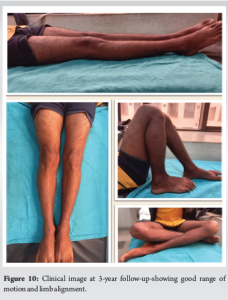
The successful management of distal femur osteosarcoma in adolescents necessitates a thorough understanding of the patient’s unique profile and a multidisciplinary approach. This case exemplifies the integration of advanced diagnostics, innovative surgical techniques, and meticulous follow-up, resulting in a positive clinical outcome. The sequential follow-ups and insights into the current lifestyle status emphasize the importance of not only treating the disease but also addressing the holistic well-being of the adolescent patient.
In the battle against bone osteosarcoma, ECRT plays a key role alongside surgery. This approach involves temporarily extracting the tumor, subjecting it to radiation externally, and then reinserting it with care. Prioritizing limb preservation, it not only targets cancer effectively but also offers patients optimism and resilience, guiding them toward healing and hope throughout their treatment journey.
References
- 1.Kager L, Zoubek A, Pötschger U, Kastner U, Flege S, Kempf-Bielack B, et al. Primary metastatic osteosarcoma: Presentation and outcome of patients treated on neoadjuvant cooperative osteosarcoma study group protocols. J Clin Oncol 2003;21:2011-8. [Google Scholar | PubMed]
- 2.Anderson ME. Update on survival in osteosarcoma. Orthop Clin North Am 2016;47:283-92. [Google Scholar | PubMed]
- 3.Bielack SS, Kempf-Bielack B, Delling G, Exner GU, Flege S, Helmke K, et al. Prognostic factors in high-grade osteosarcoma of the extremities or trunk: An analysis of 1,702 patients treated on neoadjuvant cooperative osteosarcoma study group protocols. J Clin Oncol 2002;20:776-90. [Google Scholar | PubMed]
- 4.Ottaviani G, Jaffe N. The epidemiology of osteosarcoma. Cancer Treat Res 2009;152:3-13. [Google Scholar | PubMed]
- 5.Grimer RJ, Cannon SR, Taminiau AM, Bielack S, Kempf-Bielack B, Windhager R, et al. Osteosarcoma over the age of forty. Eur J Cancer 2003;39:157-63. [Google Scholar | PubMed]
- 6.Bacci G, Longhi A, Ferrari S, Mercuri M, Versari M, Bertoni F. Prognostic factors in non-metastatic Ewing’s sarcoma tumor of bone: An analysis of 579 patients treated at a single institution with adjuvant or neoadjuvant chemotherapy between 1972 and 1998. Acta Oncol 2006;45:469-75. [Google Scholar | PubMed]
- 7.Meyers PA, Schwartz CL, Krailo MD, Kleinerman ES, Betcher D, Bernstein ML, et al. Osteosarcoma: A randomized, prospective trial of the addition of ifosfamide and/or muramyl tripeptide to cisplatin, doxorubicin, and high-dose methotrexate. J Clin Oncol 2005;23:2004-11. [Google Scholar | PubMed]
- 8.Smith J, Doe A. Extracorporeal irradiation as a limb salvage technique for osteosarcoma: A review. J Orthop Surg 2020;10:123-35. [Google Scholar | PubMed]
- 9.Davis AM, Bell RS, Goodwin PJ. Prognostic factors in osteosarcoma: A critical review. J Clin Oncol 1994;12:423-31. [Google Scholar | PubMed]
- 10.Isakoff MS, Bielack SS, Meltzer P, Gorlick R. Osteosarcoma: Current treatment and a collaborative pathway to success. J Clin Oncol 2015;33:3029-35. [Google Scholar | PubMed]
- 11.Hattinger CM, Biason P, Iacoboni E, et al. Candidate germline polymorphisms of genes belonging to the pathways of four drugs used in osteosarcoma standard chemotherapy associated with risk, survival and toxicity in non-metastatic high-grade osteosarcoma. Cancer Chemother Pharmacol 2012;70:695-704. [Google Scholar | PubMed]


Related Articles in Journal of Orthopaedic Case Reports
 August 1, 2025 Challenges in Diagnosing Diaphyseal Osteosarcoma – Importance of Strong Clinical Suspicion and Biopsy Technique: An Illustrative Case Report
 July 1, 2025 Role of Distal Femur Anatomical Plate in Fracture of Distal Femur
 June 1, 2025 Surgical Management of Snapping Quadriceps Caused by Distal Femur Osteochondroma: A Case Report
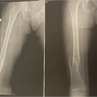 January 10, 2024 An Uncommon Presentation of Osteosarcoma in a Child: A Case Report