
Tophaceous deposits can cause massive destruction of bone mimicking malignancies, however, accurate diagnosis and complete excision and reconstruction with an intercalary graft can give satisfactory results.
Dr. P Janardhana Aithala, Department of Orthopaedics, Yeneoya Medical College, Deralakatte, Mangaluru, Karnataka, India, E-mail: janardanaaithala@yahoo.com
Introduction: Gout is characterized by the deposition of monosodium urate crystals and the formation of tophaceous deposits, which can lead to arthritis and bony erosions. While bony erosions in gouty arthritis are common, massive erosions of bone with the destruction of complete bone are rarely described.
Case Report: We report a case of gouty arthritis with bilateral first metatarsophalangeal (MTP) joint involvement with bone erosion involving adjacent bones. Both sides were treated by surgical treatment with the excision of entire tophaceous deposits, the first MTP joint and the adjacent involved bones followed by reconstruction using interposition grafts. The patient was able to walk comfortably at the end of 6 months, with good cosmetic correction and healing of the wound.
Conclusion: Surgical excision and reconstruction with intercalary graft for a massive tophaceous deposit around the first MTP joint can give satisfactory results.
Keywords: Gout, tophaceous, arthritis, metatarsophalangeal joint, surgical treatment.
Gout is characterized by the deposition of monosodium urate (MSU) crystals and the formation of tophaceous deposits in the first metatarsophalangeal (MTP) joints of feet, hands, wrists, elbows, and knees [1,2]. The disease initially begins with hyperuricemia leading to the deposition of MSU crystals in soft tissues and periarticular tissues, leading to acute inflammatory response and subsequently chronic disease [2,3]. Chronic tophaceous deposits can lead to arthritis and adjacent bony erosions [1,4]. The first MTP joint is commonly involved with erosions of articular surfaces and metaphyseal bone [1]. While bone erosions in gouty arthritis are common, massive erosion of bone with destruction of complete bone is rarely described. We report a case of gouty arthritis with bilateral MTP joint involvement of the great toe with bone erosion involving adjacent bones.
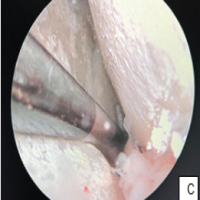
A 41-year-old male who was on irregular treatment for Gout presented to us with swelling around the base of the great toe and adjacent foot, which was progressively increasing in size leading to gross deformity and difficulty in walking (Fig. 1). The Patient does not give the history of alcohol intake. A radiograph of the right foot showed arthritis of the first MTP joint with subarticular erosions and sclerosis of adjacent bones consistent with gouty arthritis. On the left side, radiographs revealed an expansile lytic lesion involving the first metatarsal and entire proximal phalanx along with total destruction of the first MTP joint. Serum uric acid levels were elevated (10.3 mg/dL) consistent with gouty arthritis. Other blood parameters included elevated erythrocyte sedimentation rate (26 mm/h), elevated C-reactive protein (22 mg/L), Hb (13 g%), and total count (7600 cells/cumm). The previous treatment history was unclear, but the patient was taking allopurinol irregularly and native medicines. There were no neurovascular deficits. Although the history of gout and classical involvement of the right side pointed to the possibility of gout, there was a suggestion of alternate possibilities, such as chondrosarcoma, aggressive giant cell tumor, and tuberculosis for the left side lesion in view of the nature of destruction. Hence, magnetic resonance imaging (MRI) of the left foot was done, which was suggestive of tophaceous deposition (Fig. 2). An incision biopsy was done from the left side which confirmed gout. The patient also had tophaceous deposits in the ankle and both knee joints. Our next challenge was to excise the tophaceous deposits and reconstruct the massive defect. The patient underwent two-stage procedures (Fig. 3). The right side was operated first, as we contemplated a better prognosis on the right side while we were thinking of the possibility of amputation of the first ray on the left side. On the right side excision of the MTP joint along with the removal of all the tophi in surrounding tissues as well as an adjacent proximal phalanx and first metatarsal was done and the resultant bony defect was bridged with tricortical iliac crest autograft to maintain the length of 1st ray and achieve fusion of the first MTP joint. The limb was placed in a below-knee plaster slab for 3 weeks. Post-operative management included limb elevation and prophylactic antibiotics of 3 doses. Sutures were removed after 2 weeks, and there were no wound-related complications. The patient was mobilized non-weight bearing with a walker after wound healing and partial weight bearing after 5 weeks. Encouraged by good results on the right side, we decided to reconstruct the first ray on the left side, and a second surgery was done 2 months after the first surgery. The entire mass of lesion containing the first MTP joint, adjacent proximal phalanx, and near total first metatarsal along with soft tissue deposits were removed and extensor tendon of the great toe was sacrificed. The size of defect was measured and reconstructed with a fibular auto graft which was stabilized with K-wires between the stump of the first metatarsal and distal phalanx. The patient was treated with a below-knee plaster slab for the left side and non-weight bearing for 6 weeks followed by gradual weight bearing with a walker. There were no wound-healing problems on both sides. The patient was able to walk without support 3 months after the second surgery. The patient was also placed on medical treatment with Febuxostat 80 mg/day which was reduced to 40 m/day after 6 months. At 6 months follow-up, the patient had good relief of pain and was able to walk comfortably, while X-ray shows the incorporation of fibular graft and fusion across the distal phalanx and a stump of first metatarsal on the left side and incorporation of iliac crest graft on the right side (Fig. 4). The patient is allowed to resume work within his comfort, and adviced to continue febuxostat 40 mg once a day with regular monitoring of serum uric acid levels.


Bony erosions are common in gouty arthritis. Wu et al. [4] and others found bone erosions in 44% of gout patients using ultrasound, among these 78.4% of erosions were found in the first MTP joint. There is a debate, whether the bony erosions are due to an outside in mechanism from tophi deposits over the surface of the bone or an inside out mechanism due to intraosseous deposits. A study by Towiwat et al. [5] showed that MSU crystal deposition is present within the joint, on the bone surface, and within bone erosion, but it is not observed within the bone in the absence of a cortical break. These data support the concept that MSU crystals deposit outside bone and contribute to bone erosion through an “outside-in” mechanism [5], however, in large bony erosions intraosseous tophi were thought to be responsible for the bone erosions [6]. While the combination of a history of gout, clinical presentation, and radiological features are sufficient to make a diagnosis, sometimes advanced imaging modalities, such as ultrasound, computed tomography, and MRI are required, which helps to make early diagnosis and understand the pathology and extent of soft tissue involvement better [7]. Gout does not have a cure, but with proper and timely treatment, complications and end-stage arthropathy can be minimized. Medical treatment remains first line of treatment in early bone erosions, but in gross tophi deposits and bone erosions, surgery may have to be considered for debulking of large deposits, managing painful joint arthritis, joint instability, for skin breakdown, ulcerations, associated infections, functional disability to wear shoes, poorly controlled pain, and associated nerve compression and entrapment [6, 8-11]. Surgery also helps to restore the function faster [8]. Various surgical treatment options are described in the literature. Surgical options described are shaving of tophaceous deposits, excision of deposits, arthrodesis of joint, and rarely amputation [8,10,12]. Recently minimally invasive techniques, such as endoscopic resection of tophaceous deposits have been described [13]. Kim et al. [12] found that simple excision of tophi deposits will not help if more than 50% of articular cartilage has been damaged and hence, recommended arthrodesis. Many authors [9,10,11,14,15] advise early surgery like shaving off tophi to reduce urate load; however, there is a risk of sinus formation if surgery is done after sepsis has developed. Metaphyseal lesions need to be curetted, while amputation can be a last resort [15,16] to improve the function of the remaining part of the feet or hand. It is also essential to continue medical management after surgical treatment as it is impossible to remove all the deposits [10,14]. Our case is a rare case of massive tophaceous deposit including the involvement of entire adjacent bones in one of the limbs (left foot). Such involvement is rare; the radiographic images were suspicious of more serious pathologies, such as malignancies. MRI helped us to establish the diagnosis. On MRI, tophi exhibit low signal on T1-weighted images and medium-to-high signal on T2-weighted (T2W) images [7,17]. Reconstruction of such a massive defect was a challenge and to the best of our knowledge, we could not find a report where entire phalanx and adjacent metatarsal were reconstructed. Kim et al. [12] treated the first MTP joint arthritis patients surgically either with excision (group A, 9 patients,) or with excision and arthrodesis (group B, 7 patients) and found that the results were better in arthrodesis group. However, authors do not mention whether any of these patients had massive bone defects requiring interpositional graft. Wünschel et al. [18] described a rare case report of bilateral MTP joint involvement, treated with excision of tophaceous deposits and interposition arthroplasty, authors felt that arthrodesis is not a good option. Stapleton et al. [11] described a case report of the first MTP arthritis complicated with infection, which was treated with a staged procedure; first excision, debridement, antibiotic spacer, and external fixation, followed by removal of spacer and arthrodesis using iliac crest graft. Although authors have addressed significant bone defects, it was limited to adjacent areas of the proximal phalanx and first metatarsal, while our case on the left side showed massive bone defects involving the entire proximal phalanx and near total first metatarsal bone. We attempted reconstruction in view of the absence of infection. We decided to excise the entire phalanx and metatarsal to completely eradicate tophaceous deposits. While the defect on the right side was small and hence, we could easily manage with iliac crest bone graft as described by Stapleton et al. [11], defect on the left side was huge and iliac crest graft would have been insufficient. To restore stability, we decided to use cortical graft and hence fibular autograft was used. Our results at 6 months show that the patient has acceptable cosmesis on the left side with a good incorporation of fibular graft and the patient was able to walk comfortably. On the right side, there was some residual valgus deformity, but deformity is present largely at the IP joint which was not addressed, and as patient does not have any pain related to the deformity, no further interventions were suggested.
Patient perspective
At the past follow-up (3-year follow-up) patient was happy with the appearance of both feet, able to walk without much pain compared to pre-operative status.
We report a rare case of massive tophaceous deposits involving entire adjacent bones of the great toe MTP joint and the complete removal of tophaceous deposits along with involved bones with reconstruction of the defect with fibular graft or tricortical graft depending on the extent of bone removed can give good functional results.
- Tophaceous deposits can cause massive destruction of bone mimicking malignancies
- MRI has a characteristic presentation in gouty arthritis with low signal in T1 and medium to high signal in T2 weighted images, which helps in diagnosis
- Complete excision and reconstruction with an intercalary graft can give satisfactory results.
References
- 1.Bloch C, Hermann G, Yu TF. A radiologic reevaluation of gout: A study of 2,000 patients. AJR Am J Roentgenol 1980;134:781-7. [Google Scholar | PubMed]
- 2.Perez-Ruiz F, Dalbeth N, Bardin T. A review of uric acid, crystal deposition disease, and gout. Adv Ther 2015;32:31-41. [Google Scholar | PubMed]
- 3.Bardin T, Richette P. Definition of hyperuricemia and gouty conditions. Curr Opin Rheumatol 2014;26:186-91. [Google Scholar | PubMed]
- 4.Wu M, Liu FJ, Chen J, Chen L, Wei C, Hu ZM, et al. Prevalence and factors associated with bone erosion in patients with gout. Arthritis Care Res (Hoboken) 2019;71:1653-9. [Google Scholar | PubMed]
- 5.Towiwat P, Doyle AJ, Gamble GD, Tan P, Aati O, Horne A, et al. Urate crystal deposition and bone erosion in gout: “Inside-out” or “outside-in”? A dual-energy computed tomography study. Arthritis Res Ther 2016;18:208. [Google Scholar | PubMed]
- 6.Dalbeth N, Clark B, Gregory K, Gamble G, Sheehan T, Doyle A, et al. Mechanisms of bone erosion in gout: A quantitative analysis using plain radiography and computed tomography. Ann Rheum Dis 2009;68:1290-5. [Google Scholar | PubMed]
- 7.McQueen FM, Doyle A, Reeves Q, Gao A, Tsai A, Gamble GD, et al. Bone erosions in patients with chronic gouty arthropathy are associated with tophi but not bone oedema or synovitis: New insights from a 3 T MRI study. Rheumatology (Oxford) 2014;53:95-103. [Google Scholar | PubMed]
- 8.Kasper IR, Juriga MD, Giurini JM, Shmerling RH. Treatment of tophaceous gout: When medication is not enough. Semin Arthritis Rheum 2016;45:669-74. [Google Scholar | PubMed]
- 9.Larmon WA. Surgical management of tophaceous gout. Clin Orthop Relat Res 1970;71:56-69. [Google Scholar | PubMed]
- 10.Lee SS, Sun IF, Lu YM, Chang KP, Lai CS, Lin SD. Surgical treatment of the chronic tophaceous deformity in upper extremities-the shaving technique. J Plast Reconstr Aesthet Surg 2009;62:669-74. [Google Scholar | PubMed]
- 11.Stapleton JJ, Rodriguez RH, Jeffries LC, Zgonis T. Salvage of the first ray with concomitant septic and gouty arthritis by use of a bone block joint distraction arthrodesis and external fixation. Clin Podiatr Med Surg 2008;25:755-62. [Google Scholar | PubMed]
- 12.Kim YS, Park EH, Lee HJ, Koh YG. First metatarsophalangeal joint arthrodesis for the treatment of tophaceous gouty arthritis. Orthopedics 2014;37:e141-7. [Google Scholar | PubMed]
- 13.Lui TH. Endoscopic resection of the gouty tophi of the first metatarsophalangeal joint. Arch Orthop Trauma Surg 2008;128:521-3. [Google Scholar | PubMed]
- 14.Fang ZH, Waizy H. Current concepts in the treatment of gouty arthritis. Orthop Surg 2013;5:6-12. [Google Scholar | PubMed]
- 15.Sener EE, Güzel VB, Takka S. Surgical management of tophaceous gout in the hand. Arch Orthop Trauma Surg 2000;120:482-3. [Google Scholar | PubMed]
- 16.Yetkin H, Takka S, Kanatli U. Surgical treatment of chronic tophaceous gout arthritis in the feet: A case report. Foot Ankle Surg 1999;5:155-7. [Google Scholar | PubMed]
- 17.Yu JS, Chung C, Recht M, Dailiana T, Jurdi R. MR imaging of tophaceous gout. AJR Am J Roentgenol 1997;168:523-7. [Google Scholar | PubMed]
- 18.Wünschel M, Wülker N, Walter C. Surgical treatment of a young patient with bilaterally destroyed first metatarsophalangeal joints suffering from gout. J Am Podiatr Med Assoc 2012;102:334-7. [Google Scholar | PubMed]


Related Articles in Journal of Orthopaedic Case Reports
 March 1, 2025 Arthroscopic Treatment of Isolated Tophaceous Gout of the Knee: A Rare Case Report
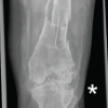 February 1, 2026 Case Report of Post-Traumatic Monoarticular Tuberculosis of the Knee in a Healthy Young Adult: Diagnostic and Therapeutic Challenges in a Non-Endemic Setting
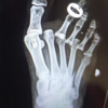 February 1, 2026 Closed Traumatic Dislocation of 2nd–4th Metatarsophalangeal Joints with Associated 5th Metatarsal Base Fracture: A Rare Case Report
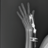 February 1, 2026 A Rare Case: Septic Arthritis of The Wrist