The treatment of a patient who presented with a Pseudoaneurysm as a complication that arose from extra-capsular hip fractures fixed with a cephalomedullary nail in an 89-year-old female.
Dr. Mohamed Elzayat, Department of Orthopaedic, Sligo University Hospital, Sligo, Ireland. E-mail: zayat_mohamed@hotmail.com
Introduction: Hip fractures are within the most injuries coming to the emergency department, and one of the highest operations done in the trauma section of the orthopedics department. Within this, hip fractures treatment can vary depending on where the fracture is situated within the femur, either intracapsular that requires a hip hemiarthroplasty or extracapsular with require in situ fixation, with each of these methods or any surgical procedure there are risks that can happen, in this case report, we are highlighting a rare complication of a hip fixation and how it was managed.
Case Report: An 89-year-old lady presented to our emergency department with a 6-week history of left thigh pain and swelling. She sustained a left hip fracture 8 weeks before this presentation for which she had an uneventful surgery. On examination her left thigh was swollen, there was no pitting edema and her pulses were intact. A Doppler ultrasound showed a large pseudoaneurysm arising from the profunda femoris artery. A computed tomography angiogram was subsequently performed to confirm the diagnosis. The patient had a successful embolization of the pseudoaneurysm. This was confirmed with ultrasound 24 h later, which showed no refilling of the pseudoaneurysm. The patient was discharged home in good condition.
Conclusion: Arterial vessel damage following hip fractures is very rare. It may be caused by the fracture itself or more commonly as a consequence of intertrochanteric fracture fixation. A high index of suspicion should be maintained as the symptoms can be non-specific and the diagnosis can be challenging.
Keywords: Pseudoaneurysm, fracture fixation, orthopedics, intervention radiology.
Hip fractures currently represent one of the most common causes of human morbidity and mortality in the elderly, with 1/3 of women and 1/12 of men experiencing a hip fracture at some point in their life [1]. Fractures of the hip are more common among older individuals, with approximately 86% of the fractures occurring in individuals ≥65 years of age [2]. Such injuries carry a high risk of functional decline, long-term disability, and high healthcare costs [3]. The treatment strategy for hip fractures includes hip hemiarthroplasty for intracapsular fractures and internal fixation for extracapsular fractures, such as intertrochanteric utilizing a cephalomedullary nail [4]. Pseudoaneurysms are rare but potentially major vascular complications in the surface treatment of hip fractures. These complications are either a result of a vascular injury during the initial trauma, or manipulation during surgery [3, 5]. The reported incidence of pseudoaneurysms following hip fracture is extremely low (about 0.21%), but it can result in devastating complications if left untreated [6, 7]. Profunda femoris artery injury is reported due to several causes including bone spikes, implant dislocation, surgical tools, etc., which would result in pseudoaneurysm formation [5, 8, 9]. We report a case of pseudoaneurysm of the profunda femoris artery after femoral nailing for an intertrochanteric femoral fracture in an 89-year-old male patient. We describe the diagnosis, management, and outcomes of this uncommon complication.
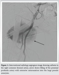
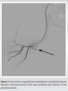
An 89-year-old woman had a 6-week history of left thigh pain and swelling. She had sustained a left intertrochanteric hip fracture 8 weeks prior and had an uneventful proximal femoral nailing fixation. Her post-operative recovery was uneventful and she was discharged free of in-hospital complications. However, the patient then experienced worsening pain, swelling, and decreased mobility. She saw her family doctor several times and ultimately landed in the hospital for further investigation. On examination, her left thigh was swollen, without pitting edema, and with preserved pulses. Severe symptoms raised suspicion of a deep vein thrombosis (DVT), but Doppler ultrasound excluded this diagnosis, demonstrating a 10 cm pseudoaneurysm stemming from the profunda femoris artery. A follow-up computed tomography (CT) angiogram confirmed the diagnosis. Interventional radiology angiogram image under fluoroscopy, demonstrating catheter in the right common femoral artery with large defect in the proximal profunda artery with extensive extravasation into the large pseudo aneurysm (Fig. 1), and after the embolization identifying the retained flow in the common femoral and superficial femoral arteries but occlusion of the proximal profunda femoris with no filling of the pseudo aneurysm noted (Fig. 2).
The pseudoaneurysm was successfully embolized, and ultrasound follow-up after 24 h revealed no refilling. She was then released in good condition.
Hip fractures represent a common traumatic injury among older people, being associated with one of the greatest long-term functional consequences. Most hip fractures are treated with surgical fixation; however, vascular complications (e.g., pseudoaneurysms) are rare but significant [1,6]. Pseudoaneurysm is rare after hip fracture, with an estimated incidence of 0.21%, occurring as a result of the surgical fixation itself rather than the initial injury, mortality 1 year post hip fracture is about 27.3% a population study has shown [7]. The development of pseudoaneurysms in hip fractures is frequently associated with the mechanical interruption of blood vessels by either displaced bone fragments or surgical tools. Due to its location near the fracture site, the profunda femoris artery (branch of the femoral artery) is at risk. Bone spicules, implant tips, or retraction of surrounding tissue can damage arteries during fracture fixation [5,9]. Moreover, atherosclerotic vessels manipulated during fracture repair may compromise their delicate endothelium and aggravate vascular injury and bleeding [2]. A pseudoaneurysm is perhaps one of the more difficult post-operative complications to diagnose, as its clinical picture often overlaps with other post-operative complications such as hematoma or DVT. A definitive diagnosis can be confirmed through imaging studies such as Doppler ultrasound and CT angiography [3,5]. After diagnosis, treatment could be conservative management, thrombin injection, and endovascular techniques, such as embolization, treatment of choice at present due to minimally invasive techniques, and good outcomes [9]. Our case underlines the need to keep a high index of suspicion for vascular complications after hip fracture fixation, especially in the setting of non-specific symptoms including swelling or pain. However, early identification and timely intervention perhaps with embolization can avoid dire complications, such as rupture and hemorrhage [5,10]. A review provided by this literature suggested that complex fractures and surgical approaches raise the risk of vascular injury. These complications are especially common with the use of cephalomedullary nailing or intramedullary fixation [2,4]. Pseudoaneurysms are a rare complication of hepato-billiary surgery but should be kept in mind among the differential diagnosis of post-operative complications, particularly in cases of new/unusual swelling or pain. The difference between aneurysm (true) and pseudoaneurysm (false) is that the true aneurysm contains all the layers of the vessel wall, while the false aneurysm (pseudoaneurysm) contains one or two of the layers (the three key layers of a blood vessel are tunica intima, tunica media, and tunica externa). The pseudoaneurysm could be caused by a trauma which is like the one seen in our case.
Introduction vascular complications after hip fracture fixation are infrequent but grave. This article presents a case that illustrates the potential for superficial femoral nailing to cause a profunda femoris artery pseudoaneurysm and for the condition to present without making for an uncomplicated post-operative course. Early diagnosis and intervention, including embolization, are necessary to avoid death. This case highlights the need to keep your index of suspicion high for vascular insult in patients with hip fractures, especially in the post-operative period.
Rare but serious vascular complications, including pseudoaneurysms, can occur after hip fracture fixation and are especially associated with femoral nailing procedures. Patients with unexplained pain, swelling, or mobility alterations after surgery require a high level of suspicion. Due to imaging perspectives (e.g., Doppler ultrasound, CT angiography), timely interventions (e.g., embolization) can eliminate life-threatening events, such as rupture and hemorrhage.
References
- 1.Zuckerman JD. Hip fractures. N Engl J Med 2011;364:1768-78. [Google Scholar]
- 2.Amarilla-Donoso FJ, López-Espuela F, Roncero-Martín R, Leal-Hernandez O, Puerto-Parejo LM, Aliaga-Vera I, Toribio-Felipe R, Lavado-García JM. Quality of life in elderly people after a hip fracture: a prospective study. Health Qual Life Outcomes. 2020 Mar 14;18(1):71. [Google Scholar]
- 3.Schnell S, Friedman SM, Mendelson DA, Bingham KW, Kates SL. The 1-year mortality of patients treated in a hip fracture program for elders. Geriatr Orthop Surg Rehabil 2010;1:6-14. [Google Scholar]
- 4.Koval KJ, Zuckerman JD. Hip fractures: Current treatment modalities. Orthopedic Clin North Am 2010;41:1-16. [Google Scholar]
- 5.Piolanti N, Giuntoli M, Nucci AM, Battistini P, Lisanti M, Andreani L. Profunda femoris artery pseudoaneurysm after intramedullary fixation for a pertrochanteric hip fracture. J Orthop Case Rep 2017;7:74-7. [Google Scholar]
- 6.Bowden S, Jaberi A, Roche-Nagle G. Large pseudoaneurysm arising from the deep femoral artery after hip fracture fixation. J Surg Case Rep 2020;2020:rjaa408. [Google Scholar]
- 7.Panula J, Pihlajamäki H, Mattila VM, Jaatinen P, Vahlberg T, Aarnio P, Kivelä SL. Mortality and cause of death in hip fracture patients aged 65 or older: a population-based study. BMC Musculoskelet Disord. 2011 May 20;12:105. [Google Scholar]
- 8.Labronici PJ, Santos Filho FC, Diamantino YL, Loureiro E, Ezequiel MC, Alves SD. Proximal femur fracture and vascular injury in adults-case report. Rev Bras Ortop (Sao Paulo) 2019;54:343-6. [Google Scholar]
- 9.Sarioglu O, Capar AE, Belet U. Interventional treatment options in pseudoaneurysms: Different techniques in different localizations. Pol J Radiol 2019;84:e319-27. [Google Scholar]
- 10.Jorissen RN, Lang C, Visvanathan R, Crotty M, Inacio MC (2020) The effect of frailty on outcomes of surgically treated hip fractures in older people. Bone 136:115327. [Google Scholar]