Intra-articular lipoma is a rare entity that requires a high index of suspicion for accurate diagnosis. MRI is an invaluable diagnostic tool for reliable diagnosis of intra-articular masses.
Dr. Dhrushith Etakkepravan Puthanveetil, Department of Orthopaedics, Sree Uthradom Thirunal Academy of Medical Sciences, Thiruvananthapuram - 695028, Kerala, India. E-mail: epdhrushith@gmail.com
Introduction: Most soft-tissue tumors in the foot and ankle are benign. While lipomas are the most prevalent type of soft-tissue tumor, intra-articular lipomas are exceptionally rare. Most documented intra-articular lipomas involve the knee joint, and there have been only a few case reports of an intra-articular lipoma in the ankle.
Case Report: We present the case of a 77-year-old woman with a recurrent, progressively enlarging ankle mass that had persisted for 19 years following surgery 15 years prior. At present, the swelling is limiting her range of motion. On examination, a firm, non-tender mass was observed on the medial side of the left ankle joint without any signs of inflammation. The mass was non-compressible, immobile, and did not transilluminate. The clinical diagnosis suggested a soft-tissue ganglion. A radiograph showed soft-tissue opacity over the anteromedial aspect of the tibiotalar joint. Magnetic resonance imaging (MRI) revealed a well-defined, multilobulated, encapsulated lesion at the tibiotalar joint’s medial side, with intra- and extra-articular components. The patient underwent surgical excision of the tumor, and histopathological examination confirmed the presence of mature adipose tissue consistent with an intra-articular lipoma with fibrous septa. At the follow-up visit, the patient reported a complete resolution of symptoms and had no further complaints.
Conclusion: An intra-articular lipoma of the ankle is an extremely rare tumor. Clinical examination is essential in diagnosing the lesion. MRI is primordial for both differential diagnosis and preoperative planning. Depending on the tumor size, an excision can be performed through open arthrotomy or arthroscopy.
Keywords: Ankle joint, intra-articular tumors, lipoma, magnetic resonance imaging.
Lipomas are adipose tumors often located in the subcutaneous tissues. While they are the most prevalent type of soft-tissue tumor, intra-articular lipomas are exceptionally rare. Most documented intra-articular lipomas involve the knee joint, and there have been only a few case reports of an intra-articular lipoma in the ankle [1]. Lipomas have been identified in all age groups but usually appear between 40 and 60 [2]. Despite an equal sex distribution, males tend to have multiple lipomata, whereas women have solitary lipomata [3]. Lipoma size can range from a few millimeters to ≥5 cm, although they tend to be smaller in the foot [3]. These slow-growing, nearly always benign tumors are generally present as non-painful, round, mobile masses with a characteristic soft, doughy feel. Most lipomas are best left alone, but rapidly growing or painful lipomas can be treated with various procedures ranging from steroid injections to tumor excision. Lipomas must be distinguished from liposarcoma, which can have a similar appearance.
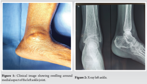
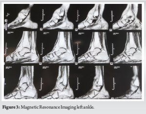
The reported case is for a 77-year-old Indian lady who presented with recurrent progressively enlarging ankle mass of 19 years duration, for which she underwent surgery 15 years ago. Even though the Swelling is not associated with pain, it is progressively increasing in size, limiting her range of motion in the left ankle. On examination, a firm, non-tender mass is present on the medial side of the left ankle joint without any signs of inflammation like redness or the local rise of temperature. The mass was non-compressible, immobile, and did not transilluminate. A scar was present over the swelling, which healed by primary intention (Fig. 1). The clinical diagnosis suggested a soft-tissue ganglion. A radiograph showed soft-tissue opacity over the anteromedial aspect of the tibiotalar joint (Fig. 2). Magnetic resonance imaging (MRI) revealed a well-defined, multilobulated, encapsulated lesion at the tibiotalar joint’s medial side, with intra- and extra-articular components and an analogous signal intensity to fat (Fig. 3).

The patient underwent surgical excision of the tumor, and histopathological examination confirmed the presence of mature adipose tissue consistent with an intra-articular lipoma with fibrous septa. At the follow-up visit, the patient reported a complete resolution of symptoms and had no further complaints.
Operation
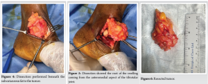
We performed an excision of the tumor under spinal anesthesia. An anteromedial approach to the ankle was used, and the skin incision followed the scar. Dissection was then performed beneath the subcutaneous fat to the tumor. Any tissue cutting was performed under direct visualization using a no.15 scalpels or scissors around the tumor (Fig. 4). Once a portion of the tumor had been dissected from the surrounding tissue, hemostats or clamps were attached to it to provide traction to remove the remainder of the growth. We encountered a soft ovoid mass measuring about 6 × 4.5 cm, with a fibrous capsule that was adhered to deep planes. Dissection showed the root of the swelling coming from the anteromedial aspect of the tibiotalar joint (Fig. 5). Once freed, the tumor was delivered whole, and the tibiotalar joint was exposed. Specimens were submitted for histologic analysis (Fig. 6).
Following the removal of the tumor, adequate hemostasis was achieved. The dead space was closed beneath the skin using buried, interrupted 3–0 Vicryl sutures. The skin was then closed with interrupted 3–0 nylon sutures. A pressure dressing was placed to reduce the incidence of hematoma formation. Outcome and follow-up: The case coursed with a favorable post-operative evolution. Although the surgical site incision had serous discharge, it subsided with antibiotics. The patient suffered from post-operative rigidity, which was resolved with physiotherapy sessions. She was discharged without symptoms and with a complete range of motion. The patient was given routine wound care instructions. The sutures were removed after 14 days.
Histological examination
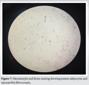
Microscopic examination with hematoxylin and eosin staining of the specimen revealed images of mature adipocytes without an atypical nucleus and separated by fibrous septa (Fig. 7).
Lipomas are commonplace soft-tissue tumors found anywhere in the body [4]. However, Intra-articular lipomas are an infrequent entity [2]. Most documented intra-articular lipomas involve the knee joint, and there have been only a few case reports of a true intra-articular lipoma in the ankle [1]. They most commonly occur in the fifth decade of life. Despite an equal sex distribution, males tend to have multiple lipomata, whereas women have solitary lipomata. Lipoma size can range from a few millimeters to ≥5 cm, although they tend to be smaller in the foot. Patients usually present with a non-tender mass which is soft to palpation. Lipomata can often be present for many years without causing any noticeable effect. Diagnosing an intra-articular lipoma can be challenging, mainly when it is small and not visible on standard radiographs. Ultrasound demonstrates a hyperechoic mass, with the lesion usually being encapsulated. If a lesion is detected, it typically appears as a well-defined area of radiolucency. MRI is the preferred method for further evaluation, as it effectively identifies intra-articular masses and soft-tissue lesions [5]. On MRI, lipomas exhibit high signal intensity on T1-weighted and T2-weighted sequences, similar to subcutaneous fatty tissue. However, they can also present non-specific characteristics on MRI, such as a signal intensity resembling fluid, which may result from mucoid degeneration [6]. Incomplete fat-suppressive images can indicate either an atypical lipomatous tumor or liposarcoma. A differential diagnosis should rule out ganglia, intra-articular liposarcoma, schwannoma, and tenosynovial giant cell tumor (TGCT) [3, 7]. Ganglia are the most prevalent soft-tissue tumor in the foot and ankle. A ganglion is a cyst filled with mucinous fluid. Although benign, ganglia can exert localized pressure, leading to pain or neurological symptoms. The most common presenting complaint is related to these mass effects. Ganglia may also be associated with tarsal tunnel syndrome, a compressive neuropathy within the tarsal tunnel. Other potential causes of tarsal tunnel compression include lipomas, vascular engorgement, and synovial or bony abnormalities. Macroscopically, ganglia appear firm, often oval-shaped, with a smooth surface, and are dense to the touch; they contain varying amounts of mucinous substance [8]. Low-grade liposarcoma typically affects middle-aged individuals and usually presents as a painless, slow-growing, locally aggressive tumor that rarely metastasizes. Intra-articular liposarcoma is uncommon. On MRI, it is seen as a large lesion characterised by thick septa, along with non-lipomatous soft-tissue containing a low amount of fatty component [9]. A schwannoma (neurilemmoma) is a benign nerve sheath tumor that originates from the Schwann cells responsible for myelin production. While the exact cause is unknown, schwannomas are slow-growing, benign tumors often associated with neurofibromatosis Type II. They can present as a painful, slowly growing mass or remain asymptomatic for years. Localised compression from the tumor can cause paresthesia along the affected peripheral nerve and may yield a positive Tinel’s sign [3]. TGCT are rare, benign proliferations of cells originating from the synovium, tendon sheath, or bursae. TGCT can present in either solitary (nodular) or diffuse forms. The diffuse type has a higher recurrence rate and is more locally aggressive. Previously, the diffuse form was known as pigmented villonodular synovitis, while the nodular form was called giant cell tumor of tendon sheath [10]. The nodular form is less destructive compared to the diffuse type. Clinically, it presents as a small nodule or firm tissue that may be connected by a stalk (pedunculated lesions). Localised tumors are more common in smaller joints, such as the hands and feet, and can lead to locking or a “catching” sensation. Diffuse TGCT consists of a proliferation of synovial villi and nodules and can be monoarticular or polyarticular. Although the underlying condition is benign, it is aggressive and can result in joint erosion. The tumor appears brownish and pigmented during surgery due to hemosiderin deposition within the tissues [11]. The synovium is hypertrophied and typically affects the entire surrounding synovium. Histopathologically, intra-articular lipoma consists of mature adipocytes covered with a synovial membrane and may contain a vascular fibrous septum. That is why it is a true neoplasm of uncertain etiology. The natural history of this condition has not been thoroughly examined, but it is recognized to grow slowly and typically remains asymptomatic until symptoms arise from a space-occupying lesion [9]. Lipomas can be managed non-operatively if they are asymptomatic. However, marginal surgical excision is recommended if symptomatic, growing, or showing non-lipomatous components on imaging. The gold-standard treatment for intra-articular lipoma has not yet been established. Both arthroscopic excision and open arthrotomy have been performed, with previous studies indicating no recurrences following arthroscopic excision, suggesting it is a valid approach when feasible. In our case, arthroscopy was not considered an option due to the large size of the patient’s lesion, so we opted for a limited arthrotomy as a more practical solution.
An intra-articular lipoma of the ankle is an extremely rare tumor. Clinical examination is essential in diagnosing the lesion. MRI is primordial for both differential diagnosis and preoperative planning. Depending on the tumor size, an excision can be performed through open arthrotomy or arthroscopy.
Intra-articular lipoma is a rare entity that requires a high index of suspicion for accurate diagnosis. MRI is an invaluable diagnostic tool for reliable diagnosis of intra-articular masses.
References
- 1.Alsahlawi HS, Hassan A, Hasan SM, Alshaikh SA, Al-Aradi HA. A rare case of an intra-articular true lipoma of the ankle. Skeletal Radiol 2023;52:797-801. [Google Scholar]
- 2.Kheok S, Ong K. Benign periarticular, bone and joint lipomatous lesions. Singapore Med J 2017;58:521-7. [Google Scholar]
- 3.Thomson L, Putt O, Rennie WJ, Ashford RU, Mangwani J. Benign soft tissue tumours of the foot and ankle: A pictorial review. J Clin Orthop Trauma 2023;37:102105. [Google Scholar]
- 4.Marui T, Yamamoto T, Kimura T, Akisue T, Nagira K, Nakatani T, et al. A true intra-articular lipoma of the knee in a girl. Arthroscopy 2002;18:E24. [Google Scholar]
- 5.Yilmaz E, Karakurt L, Akpolat N, Özdemir H, Belhan O, İncesu M. Intra-articular lipoma of the knee joint in a girl. Arthroscopy 2005;21:98-102. [Google Scholar]
- 6.Bankaoglu M, Ugurlar OY, Ugurlar M, Sonmez MM, Eren OT. Intra-articular lipoma of the knee joint located in the posterior compartment: A rare location. North Clin Istanb 2016;4:89-92. [Google Scholar]
- 7.Macdonald DJ, Holt G, Vass K, Marsh A, Kumar C. The differential diagnosis of foot lumps: 101 cases treated surgically in North Glasgow over 4 years. Ann R Coll Surg Engl 2007;89:272-5. [Google Scholar]
- 8.Rozbruch SR, Chang V, Bohne WH, Deland JT. Ganglion cysts of the lower extremity: An analysis of 54 cases and review of the literature. Orthopedics 1998;21:141-8. [Google Scholar]
- 9.Dalla Rosa J, Nogales Zafra JJ. Large intra-articular true lipoma of the knee. BMC Musculoskelet Disord 2019;20:110. [Google Scholar]
- 10.Ravi V, Wang WL, Lewis VO. Treatment of tenosynovial giant cell tumor and pigmented villonodular synovitis. Curr Opin Oncol 2011;23:361-6. [Google Scholar]
- 11.Mastboom MJ, Palmerini E, Verspoor FG, Rueten-Budde AJ, Stacchiotti S, Staals EL, et al. Surgical outcomes of patients with diffuse-type tenosynovial giant-cell tumours: An international, retrospective, cohort study. Lancet Oncol 2019;20:877-86. [Google Scholar]


