
Early recognition and intervention are crucial in patients with scleroderma and Raynaud’s syndrome to prevent the progression of infections such as osteomyelitis, particularly in the presence of digital ulcers, as delayed treatment can lead to severe complications, including necrosis and permanent disability. This article emphasizes the critical importance of early diagnosis and intervention in patients with autoimmune diseases such as scleroderma.
Dr. Jared B Hinton, Department of Medicine, Northeast Ohio Medical University, Rootstown, Ohio, USA. E-mail: jhinton@neomed.edu
Introduction: Osteomyelitis is a serious bone infection commonly caused by bacterial pathogens, with Staphylococcus aureus being the most prevalent. The condition poses significant challenges in patients with underlying autoimmune disorders such as scleroderma and Raynaud’s syndrome, where vascular dysfunction and immunosuppression heighten infection risks. This case report illustrates the complex interplay between these conditions and underscores the importance of early diagnosis and comprehensive management to prevent severe complications.
Case Report: We present the case of a 79-year-old female with a history of Raynaud’s syndrome and suspected scleroderma who developed osteomyelitis of the left middle finger following a paronychial infection. The patient experienced persistent pain and swelling despite multiple debridement procedures. Physical examination revealed necrosis and gangrene of the affected finger, and magnetic resonance imaging confirmed osteomyelitis. Cultures identified Corynebacterium accolens, an uncommon pathogen in this context. The patient underwent surgical debridement, followed by antibiotic therapy and vasodilators. Her condition improved, with no signs of infection at follow-up.
Conclusion: This case highlights the critical need for early and thorough assessment of infections in patients with autoimmune conditions such as scleroderma and Raynaud’s syndrome. The vascular dysfunction inherent in these diseases can exacerbate infections, leading to severe outcomes such as osteomyelitis. A multidisciplinary approach involving early surgical intervention and tailored medical management is essential to optimize patient outcomes.
Keywords: Osteomyelitis, Raynaud’s syndrome, scleroderma, autoimmune disease, vascular dysfunction.
Osteomyelitis presents a considerable challenge in both infectious diseases and orthopedics, as it is a bone infection predominantly caused by bacterial pathogens, with Staphylococcus aureus being the most frequent culprit [1]. This condition can develop through the hematogenous spread, direct inoculation following trauma or surgery, or through contiguous spread from nearby soft-tissue infections [2]. Clinically, osteomyelitis can present acutely, with severe localized pain, fever, and systemic signs of infection, or chronically, with persistent but less intense symptoms that may lead to complications such as necrotic bone tissue [2,3]. Diagnosis involves a combination of imaging techniques, such as magnetic resonance imaging (MRI) or computed tomography scans and microbiological cultures, while treatment typically necessitates a multifaceted approach, including prolonged antibiotic therapy and surgical debridement [3]. Raynaud’s syndrome and scleroderma share significant pathophysiological interconnections with osteomyelitis. Raynaud’s syndrome, a vascular disorder marked by episodic vasospasm of small arteries and arterioles, primarily affects the extremities and is often triggered by cold exposure or emotional stress [4]. It can occur independently as primary Raynaud’s disease or in association with systemic diseases such as scleroderma as secondary Raynaud’s phenomenon [5,6]. Scleroderma, or systemic sclerosis, is a complex autoimmune disease characterized by widespread vascular abnormalities, chronic inflammation, and progressive fibrosis of the skin and internal organs [7,8]. The vascular dysfunction and systemic inflammation seen in scleroderma and secondary Raynaud’s phenomenon contribute to vasculopathy, delayed wound healing, and ischemic complications, increasing susceptibility to infections such as osteomyelitis [9-13]. Digital ulcers in scleroderma patients, exacerbated by Raynaud’s phenomenon, serve as potential entry points for bacteria, heightening the risk of infection. The chronic vasculopathy in scleroderma and secondary Raynaud’s phenomenon not only delays wound healing but also increases ischemic complications, predisposing patients to osteomyelitis [13]. Digital ulcers, a hallmark of severe Raynaud’s in scleroderma, provide direct bacterial entry points, further amplifying infection risks. Additionally, immunosuppressive therapies used in scleroderma weaken host defenses, compounding susceptibility to infections [14,15]. These interconnected mechanisms highlight the multifaceted challenges in managing these conditions, requiring a comprehensive approach that addresses vascular dysfunction, immune dysregulation, and infection control. This case report highlights a unique presentation of osteomyelitis in the context of systemic sclerosis and Raynaud’s phenomenon, emphasizing the intricate interplay between autoimmune and infectious processes.
A 79-year-old female presented to the clinic with persistent, severe pain localized to her left middle finger after being referred from the emergency room following months of treatment by her family physician and multiple emergency room visits. She had been diagnosed with infected paronychia and felon. Initially prescribed clindamycin, the infection progressed. The patient underwent surgical lancing in the emergency room on her first visit and was switched to cefuroxime. Initial cultures were negative. The patient had a second surgical procedure with lancing and irrigation due to reaccumulating purulence and pain. The finger remained swollen and purple (Fig. 1), with the patient rating her pain as 10 out of 10, primarily upon waking. She described the pain as sharp, burning, dull, and aching, exacerbated by any contact with the nail. Despite debridement on four separate occasions by two providers, the symptoms persisted. Her medications included ibuprofen, acetylsalicylic acid, and bisoprolol-hydrochlorothiazide. She reported cold intolerance in her hands, consistent with Raynaud’s for the last 40 years, but denied calcinosis or telangiectasias.

Physical examination revealed that the patient was alert, though her daughter noted increasing forgetfulness, requiring nursing home assistance. Her hands were cool to the touch and displayed a purplish color at room temperature. The left middle finger exhibited necrosis with purulence and gangrene on the radial aspect of the distal phalanx, consistent with digital ulceration. Similar ulcerations were noted on the ring finger, indicating Raynaud’s phenomenon. MRI findings demonstrated subcutaneous edema around the middle and proximal phalanges of the fingers and evidence of flexor tenosynovitis in the middle finger. Previous cultures identified Corynebacterium accolens in the paronychial infection. The patient was scheduled for debridement surgery and diagnostic tests due to a high suspicion of undiagnosed systemic sclerosis. Tests included rheumatoid factor, ANA, HLA-B27 antibody testing, and others for pre-operative planning. The patient had a positive anticentromere antibody (>8 AI) and a positive ANA screen, along with Vitamin D deficiency. Radiographs revealed osteolysis of the distal phalanx, consistent with osteomyelitis, and MRI confirmed bone marrow edema and flexor tenosynovitis. During surgery, the patient underwent exploration of the necrotic digital ulcer and debridement of the radial aspect of the distal phalanx. C-arm fluoroscopy confirmed osteolysis of the phalanx (Fig. 1). Irrigation and debridement of skin, subcutaneous tissue, and bone were performed, followed by partial ostectomy (Fig. 1). Microscopic analysis revealed acute osteomyelitis with fibrosis, clusters of neutrophils, and necrotic debris. No abnormal cells were detected. Further culturing confirmed C. accolens, with Gram stain showing Gram-positive rods. Following the procedure, the patient was treated with doxycycline, Vitamin D3, and vasodilators. A 2-week follow-up showed intolerance to topical nitroglycerin, which was switched to topical nifedipine and pentoxifylline. By the 4-week follow-up, the patient was tolerating treatment, with the wound healing well. Antibiotics were discontinued at 3 months, and 2 months post-treatment; the patient showed no signs of infection (Fig. 2).

This case highlights the complex interplay between Raynaud’s syndrome, scleroderma, and osteomyelitis, emphasizing the need for a comprehensive approach to managing these interconnected conditions. The vascular dysfunction in Raynaud’s likely played a pivotal role in the progression of the paronychial infection into osteomyelitis. The presence of necrosis and persistent pain, despite multiple debridement procedures, illustrates the significant challenges in managing infections complicated by underlying vascular and autoimmune conditions. The identification of C. accolens in this context suggests a potential link between systemic inflammation, autoimmune processes, and severe infections such as osteomyelitis, particularly in patients with scleroderma [15]. The persistence of symptoms despite repeated debridement underscores the importance of recognizing osteomyelitis in patients with Raynaud’s syndrome and scleroderma. This case demonstrates the critical necessity for thorough and potentially repeated microbiological investigations, as initial cultures may not reveal uncommon organisms like C. accolens. A multidisciplinary approach is, therefore, essential for optimizing patient outcomes. This includes rigorous infection monitoring, particularly in patients with scleroderma, who are at higher risk of complications due to vascular and immune dysfunction [4]. Effective management also requires balancing immunosuppressive therapies, which control autoimmune inflammation but increase the risk of infections.
This report underscores the importance of recognizing the interconnections between osteomyelitis, Raynaud’s syndrome, and scleroderma. Patients with autoimmune diseases such as scleroderma are particularly susceptible to infections, especially in the presence of digital ulcers exacerbated by Raynaud’s vascular dysfunction. Immunosuppressive treatments, while necessary for controlling inflammation, heighten infection risks, necessitating careful monitoring. Managing these patients requires addressing vascular, immune, and infectious dimensions through a tailored, multidisciplinary strategy to prevent complications and optimize outcomes. Regular screening and prompt treatment of infections are critical, alongside therapies aimed at improving vascular health and immune function. Further research is needed to explore the relationship between autoimmune diseases and osteomyelitis, as well as to develop more effective treatment strategies for these complex cases.
This article emphasizes the critical importance of early diagnosis and intervention in patients with autoimmune diseases such as scleroderma, who are at increased risk of severe infections such as osteomyelitis. The interconnections between Raynaud’s syndrome, scleroderma, and osteomyelitis underscore the need for heightened clinical awareness and a multidisciplinary approach to prevent and manage complications effectively.
References
- 1.Lew DP, Waldvogel FA. Osteomyelitis. Lancet 2004;364:369-79. [Google Scholar | PubMed]
- 2.Berbari EF, Kanj SS, Kowalski TJ, Darouiche RO, Widmer AF, Schmitt SK, et al. 2015 infectious diseases society of America (IDSA) clinical practice guidelines for the diagnosis and treatment of native vertebral osteomyelitis in adults. Clin Infect Dis 2015;61:e26-46. [Google Scholar | PubMed]
- 3.Bury DC, Rogers TS, Dickman MM. Osteomyelitis: Diagnosis and treatment. Am Fam Physician 2021;104:395-402. [Google Scholar | PubMed]
- 4.Wigley FM. Clinical practice. Raynaud’s phenomenon. N Engl J Med 2002;347:1001-8. [Google Scholar | PubMed]
- 5.Herrick AL. The pathogenesis, diagnosis and treatment of Raynaud phenomenon. Nat Rev Rheumatol 2012;8:469-79. [Google Scholar | PubMed]
- 6.Hughes M, Herrick AL. Raynaud’s phenomenon. Best Pract Res Clin Rheumatol 2016;30:112-32. [Google Scholar | PubMed]
- 7.Varga J, Abraham D, Trojanowska M, editors. Scleroderma: From Pathogenesis to Comprehensive Management. Berlin: Springer International Publishing; 2017. [Google Scholar | PubMed]
- 8.Denton CP, Khanna D. Systemic sclerosis. Lancet 2017;390:1685-99. [Google Scholar | PubMed]
- 9.Khanna D. Diagnosis and treatment of systemic and localized scleroderma. Expert Rev Dermatol 2011;6:287-302. [Google Scholar | PubMed]
- 10.Van Den Hoogen F, Khanna D, Fransen J, Johnson SR, Baron M, Tyndall A, et al. 2013 classification criteria for systemic sclerosis: An American college of rheumatology/European league against rheumatism collaborative initiative. Arthritis Rheum 2013;65:2737-47. [Google Scholar | PubMed]
- 11.Gabrielli A, Avvedimento EV, Krieg T. Scleroderma. N Engl J Med 2009;360:1989-2003. [Google Scholar | PubMed]
- 12.Sapadin AN, Fleischmajer R. Treatment of scleroderma. Arch Dermatol 2002;138:99-105. [Google Scholar | PubMed]
- 13.Lambova S, Batalov A, Sapundzhiev L, Müller-Ladner U. Digital ulcers in systemic sclerosis - frequency, subtype distribution and clinical outcome. Curr Rheumatol Rev 2013;9:268-73. [Google Scholar | PubMed]
- 14.Pauling JD, Hughes M, Pope JE. Raynaud’s phenomenon-an update on diagnosis, classification and management. Clin Rheumatol 2019;38:3317-30. [Google Scholar | PubMed]
- 15.Yayla ME, Yurteri EU, Torgutalp M, Eroğlu DŞ, Sezer S, Dinçer AB, et al. Causes of severe infections in patients with systemic sclerosis and associated factors. Turk J Med Sci 2022;52:1881-8. [Google Scholar | PubMed]


Related Articles in Journal of Orthopaedic Case Reports
 November 1, 2025 Complications and Functional Outcomes in Open Tibia-Fibula Fractures: A Retrospective Analysis from a Tertiary Care Centre
 October 1, 2025 A Missed Case of Langerhans Cell Histiocytosis of the Proximal Femur after Total Hip Arthroplasty in an Adult: A Case Report
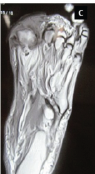 September 1, 2025 Role of Biomarkers in Diabetic Foot Osteomyelitis
 August 1, 2025 Disseminated Skeletal Cryptococcosis: A Case Report