
Although an incidental diagnosis, a multipartite patella can cause symptoms after a trivial injury. It should always be differentiated from its traumatic counterparts and, in most patients, can be treated conservatively. However, surgery should be considered in resistant patients
Dr. M L V Sai Krishna, Assistant Professor, Department of Orthopaedics, Great Eastern Medical School and Hospital, Srikakulam, Andhra Pradesh, India. E-mail: krishna.mlv.sai@gmail.com
Introduction: Multipartite patella is an incidental diagnosis, rarely symptomatic, and described scantily in the literature. Symptoms are secondary to direct injury or repetitive micro-trauma, resulting in the separation of fibro-cartilaginous joints across the multiple patellar components. Treatment is usually conservative, and occasionally, in resistant cases, surgery is advised.
Case Report: We present a 50-year-old with a tripartite patella who presented after a history of falls and incidentally discovered a bipartite patella of the other knee. The symptoms of the tripartite patella were managed conservatively.
Conclusion: Symptomatic multipartite patella should be distinguished from traumatic patella fracture. In old patients, a high index of suspicion is required to differentiate between a traumatic disruption of the multipartite patella with quadriceps avulsion and an avulsion fracture of the patella. Suppose there is a high index of suspicion. In that case, magnetic resonance imaging should be preferred to explain the signs and symptoms by noting bone marrow edema, partial or complete rupture of quadriceps, and quadriceps fat pad edema. We suggest that surgical decisions to either fix the fragment or excise be taken intraoperatively based on size, site, amount of articular surface, and associated tendon avulsion.
Keywords: Knee, Tripartite patella, Bipartite patella
The patella is an integral component of the knee extensor mechanism and functions as a fulcrum for effective quadriceps function. Multipartite (Bi/Tri) patella is an embryological anomaly emanating from an aberration in the fusion of ossification centers [1]. The ossification center of the patella appears at the age of 3–5 years, and the accessory ossific nucleus appears by 8–12 years. These ossification centers fail to unite, and eventually, synchondrosis develops [2]. Multipartite patella is an incidental diagnosis and rarely symptomatic. The reported incidence of the bipartite variant is around 0.6–2%, of which 50% are bilateral [3]. Only 2% of these patients with multipartite patella may present with primary anterior knee pain [4]. Symptoms are secondary to direct injury or repetitive micro-trauma resulting in the separation of fibro-cartilaginous joints across the multiple patellar components. Friction between these patellar fragments due to abnormal movement causes bone and soft tissue edema. The anterior knee pain is typically localized to abnormal free fragments accentuated with knee extension [4]. Treatment of symptomatic multipartite patella should begin with non-operative measures; however, when it fails, the further line of management is surgery to address primary anterior knee pain, traumatic disruption of synchondrosis, or associated tendon avulsion. There are only a handful of case reports on tripartite patella in the literature.
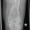
Our patient was a 50-year-old male who presented with a history of a fall from stairs (5 feet) and reported pain, swelling, and abrasion over the right knee. On clinical examination, there was no ligament instability. Although there was a 20° extensor lag, we were not able to comment on the disruption of the extensor mechanism as the patient was in pain. We suspected a right patella fracture with possible disruption of the extensor mechanism. Standard radiographs (Fig. 1 and 2) showed rounded and sclerosed cortical edges of patellar fragments. Computed tomography (Fig. 3) scan confirmed the presence of tripartite patella involving the superolateral and lateral part (type 3 – according to Oohashi classification [5]) of the right patella, with an irregular appearance of the synchondrosis. In addition, we went on to review the left knee (Fig. 4) which showed type III bipartite patella (according to Saupe classification [6]) patients had been asymptomatic before the traumatic event.


Non-operative management, including rest, non-steroidal anti-inflammatory drugs, and immobilization in a cylindrical cast for 2 weeks, was advised. The cast was discontinued after 2 weeks, and physical therapy was started, including hamstring and quadriceps strengthening exercises. Extracorporeal shock wave therapy was also prescribed. The patient was compliant with physical therapy despite having mild knee pain and, at 6 weeks follow-up, regained full strength of the extensor mechanism without any residual extensor lag. Follow-up radiographs were unremarkable. The patient was symptom-free by the end of 3 months.
A tripartite patella is a peculiar congenital anatomical variant emanating from a deviation of normal patellar ossification. Mostly, this is an incidental diagnosis characterized by well-corticated, rounded edges of the ossified segments, contrary to a patella fracture in a radiograph. The synchondrosis between patellar fragments is composed of fibro-cartilaginous tissue. Multipartite patellae are mostly asymptomatic. However, strenuous activities or repetitive micro injuries might cause separation of synchondroses, thus causing anterior knee pain [4]. Exaggerated motion between the disrupted fragments leads to a combination of impaction and friction, which eventually results in bone and soft-tissue edema and pain [7]. The other lesser-known reasons for pain were patellofemoral maltracking and dysplastic trochlea [8]. In our patient, the tripartite patella was diagnosed after trauma, and the other side bipartite patella was an incidental diagnosis. Maras Ozdemir et al. [9] successfully correlated anterior knee pain in multipartite patella with edema in the fat pad of the quadriceps. The symptomatic multipartite patella is often managed conservatively by activity modification, immobilization, non-steroidal anti-inflammatory drugs, physical therapy, and local corticosteroid injections [10,11]. In our patient, the symptoms of multipartite patella were managed conservatively. Surgical procedures, reserved for resistant cases, include open or arthroscopic excision of the accessory patella, releasing the vastus lateralis insertion, lateral retinacular release, or surgical fixation with or without bone graft and should be considered after a trial of conservative treatment [12-14]. Felli et al. [12] reported a case of arthroscopic excision of the superolateral fragment. In addition, they released the lateral retinaculum to decrease traction force on the patella. McKee reviewed all the possible treatment options being used for the multipartite patella and suggested that accessory fragment excision, release of vastus lateralis offer the greatest alleviation of symptoms than the screw fixation [15]. They strongly recommend arthroscopy as it offers careful evaluation and management of associated lesions and allows dynamic analysis of the lateral retinaculum before release [16]. Open reduction and internal fixation should be considered following traumatic disruption if the fragment is large enough with a considerable amount of articular cartilage. In our patient, the bipartite patella was an incidental diagnosis, and the tripartite patella was first diagnosed post-trauma, and the complaints were resolved with conservative treatment.
Symptomatic multipartite patella should be distinguished from traumatic patella fracture. In old patients, a high index of suspicion is required to differentiate between a traumatic disruption of the multipartite patella with quadriceps avulsion and an avulsion fracture of the patella. Suppose there is a high index of suspicion. In that case, magnetic resonance imaging should be preferred to explain the signs and symptoms by noting bone marrow edema, partial or complete rupture of quadriceps, and quadriceps fat pad edema. We suggest that surgical decisions to either fix the fragment or excise be taken intra-operatively based on size, site, amount of articular surface, and associated tendon avulsion.
Traumatic disruption of patellar synchondrosis, repetitive strain injury, or associated malalignment could be the possible explanation of symptoms in the multipartite patella, and most of the time, the diagnosis is incident and can be treated conservatively.
References
- 1.Ma J, Shi F, Huang C, Gu S. Forensic identification of bipartite patella misdiagnosed as patella fracture. J Forensic Sci 2017;62:1089-91. [Google Scholar | PubMed]
- 2.Ogden JA. Radiology of postnatal skeletal development. X. Patella and tibial tuberosity. Skeletal Radiol 1984;11:246-57. [Google Scholar | PubMed]
- 3.Green WT Jr. Painful bipartite patellae. A report of three cases. Clin Orthop Relat Res 1975;110:197-200. [Google Scholar | PubMed]
- 4.Skiada V, Perdikakis E, Plotas A, Lahanis S. MR imaging of anterior knee pain: A pictorial essay. Knee Surg Sports Traumatol Arthrosc 2013;21:294-304. [Google Scholar | PubMed]
- 5.Oohashi Y, Koshino T, Oohashi Y. Clinical features and classification of bipartite or tripartite patella. Knee Surg Sports Traumatol Arthrosc 2010;18:1465-9. [Google Scholar | PubMed]
- 6.Saupe E. Beitrag zur Patella bipartita. Fortsch Röntgenstr 1921;28:37-41. [Google Scholar | PubMed]
- 7.Kavanagh EC, Zoga A, Omar I, Ford S, Schweitzer M, Eustace S. MRI findings in bipartite patella. Skeletal Radiol 2007;36:209-14. [Google Scholar | PubMed]
- 8.Atay M. The effect of patellofemoral maltracking and patella type on symptomatic bipartite patella. Cureus 2023;15:e34076. [Google Scholar | PubMed]
- 9.Maras Ozdemir Z, Gormeli CA, Sagir Kahraman A, Demirtas G, Gormeli G. Unusual symptomatic multipartite patella associated with quadriceps fat pad edema. J Belg Soc Radiol 2016;100:49. [Google Scholar | PubMed]
- 10.Faizan M, Jilani LZ, Bin Sabir A, Abbas M. Unusual cause of anterior knee pain. Saudi Med J 2016;37:910-2. [Google Scholar | PubMed]
- 11.Marya KM, Yadav V, Devagan A, Kundu ZS. Painful bilateral bipartite patellae--case report. Indian J Med Sci 2003;57:66-7. [Google Scholar | PubMed]
- 12.Felli L, Fiore M, Biglieni L. Arthroscopic treatment of symptomatic bipartite patella. Knee Surg Sports Traumatol Arthrosc 2011;19:398-9. [Google Scholar | PubMed]
- 13.Gorva AD, Siddique I, Mohan R. An unusual case of bipartite patella fracture with quadriceps rupture. Eur J Trauma 2006;32:411-3. [Google Scholar | PubMed]
- 14.Hines KE, Liu DS, Steele AE, Gabriel D, Prabhat A, Yen YM, et al. Treatment of symptomatic bipartite patella in patients <21 years of age: A systematic review and treatment algorithm. J Child Orthop 2024;19:75-82. [Google Scholar | PubMed]
- 15.McKee CE. Multipartite patella: A review of diagnostic techniques and management of the symptomatic patient. Clin Anat 2024;37:710-8. [Google Scholar | PubMed]
- 16.Loewen A, Ge SM, Marwan Y, Burman M, Martineau PA. Arthroscopic management for bipartite patella: A systematic review. Orthop J Sports Med 2021;9(8):23259671211022248. [Google Scholar | PubMed]


Related Articles in Journal of Orthopaedic Case Reports
 February 1, 2026 Case Report of Post-Traumatic Monoarticular Tuberculosis of the Knee in a Healthy Young Adult: Diagnostic and Therapeutic Challenges in a Non-Endemic Setting
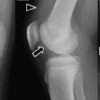 February 1, 2026 A Case of Fixation Using Poly-L-Lactic Acid Pins for Chronic Juvenile Massive Osteochondritis Dissecans of the Knee
 February 1, 2026 Conventional Total Knee Arthroplasty in Severe Anterolateral Femoral Bowing: Lateralized Femoral Entry Point to Approach Navigation Level Alignment – A Case Report
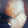 February 1, 2026 Gouty Arthritis Versus Chondrocalcinosis in a Stiff Knee, A Diagnostic Dilemma – A Case Report