
The learning point of the article is understanding the surgical technique, choice of surgical implants, and rehabilitation protocol.
Dr. K P Chiranjeevi, Department of Orthopaedics, Apollo Hospital, Plot #13, Parsik Hill Road, Off Urban Road, Opp. Nerul Wonder Park, Sector 23, CBD Belapur, Navi Mumbai - 400614, Maharashtra, India. E-mail: chiranjeevipirabhu@gmail.com
Introduction: Ipsilateral tibia and fibula shaft fractures with trimalleolar fracture are quite rare in clinical practice.
Case Report: This is a case report of a 49-year-old female presented on March 6th, 2024, and was diagnosed to have an ipsilateral left comminuted distal tibia shaft and fibula shaft fracture with an anterior lacerated wound 2 cm over the fracture site with trimalleolar fracture after falling twice while walking. The patient was treated with wound debridement, intramedullary interlocking nailing for the left tibia shaft, and open reduction internal fixation with coracoclavicular screw for posterior malleolus, K-wires + FiberWire tension band wiring for medial malleolus, and K-wires for lateral malleolus on March 07th, 2024. K-wires from the lateral malleolus were removed and tibia nail dynamization was done on April 10th, 2024. All fractures united in 4 months and the patient was followed up for a period of 1 year post-operatively.
Conclusion: Various treatment options were possible, of which we chose implants and a sequence of fixation based on the fracture pattern being comminuted and an open fracture.
Keywords: Trimalleolar fracture, tibia-fibula shaft fracture, ankle injury, internal fixation.
Tibial-fibula shaft fractures are commonly encountered long bone fracture of the lower limbs. It occurs more commonly in men than women, AO-type 42-A1 was the most common fracture type as per Larsen et al. [1]. Trimalleolar ankle fractures have a rising incidence in the past decade, with up to 40/100,000 people per year Pflüger et al. [2]. The combination of these fractures very rarely reported in the literature. The mechanism of injury is a rotational force that results in a spiral fracture of the tibia. Lauge-Hansen cadaveric study shows that posterior malleolus fractures were also the result of a rotational force. The mechanism of injury of a trimalleolar fracture is supination and external rotation. In this complex situation, proper planning as per the fracture pattern is important for a favorable outcome.
Pre-op evaluation
A 49-year-old female came with an alleged history of twisting her ankle and fall, after that she got up and tried to walk and fell down again on March 06, 2024, following which she was brought to the hospital with complaints of pain, swelling, and deformity in left leg and ankle. She also had a 2 cm lacerated wound over the anterior aspect of the leg at the fracture site (GA type 2) with no neurovascular deficit. Stay sutures were applied after adequate wash under asceptic precaution. Radiographs were done (Fig. 1) and she was diagnosed to have left tibia shaft fracture, upper two third-one third junction with butterfly fragment and comminution (AO 42 B1) and fibula -shaft-comminuted fracture with extension till lateral malleolus. Trimalleolar fracture (AO 44 A 3) (lateral malleolus posterior-undisplaced fracture, posterior malleolus undisplaced fracture, and medial malleolus undisplaced fracture). Further evaluated with a computed tomography (CT) scan (Fig. 2) and ankle mortise was found to be stable. Fracture was temporarily stabilized with an above-knee pop-back slab and limb elevation given. Patient was planned for intramedullary nailing for tibia and ankle fixation the next day.

Figure 1: Pre-operative radiograph.

Figure 2: Pre-operative computed tomography scan.
Surgical technique
A third-generation cephalosporin was administered for prophylaxis before the induction of spinal anesthesia. The patient was positioned in a supine position the stay sutures were removed. The wound was not contaminated and was clean, so debridement was done and a wash given with adequate saline and betadine. Wound closed with Ethilon 3-0 interrupted sutures. The patient was positioned in a floppy lateral position for an undisplaced posterior malleolus fracture, which was fixed first. Posterior stab incision made the lateral border of the Tendo Achilles. An AO reduction clamp was applied to hold the fracture fragment anteroposteriorly and was checked in . Fracture was temporarily held with the help of a 1.6 mm K-wire and a Guide wire 1.5 mm passed posterior to anterior under an image intensifier. Guide wire was further advanced anteriorly till it pierced the skin, soft tissue dissected anteriorly, and the anterior surface of the tibia visualized and fixed anteroposteriorly with a 4.5 × 40 mm partially threaded cannulated cancellous screw. The patient was positioned in a supine position a longitudinal skin incision made infrapatellar. Extending it 3 cm proximal from the level of the tibial plateau. Entry taken with the help of a bone awl. Ball-tipped guide wire passed while the assistant holds the fracture in the reduced position. Sequential reaming done up to 10.5 mm. Fixation done with 9 × 360 mm expert tibial nail (Titanium) with 2 screws proximally and 3 locking screws distally. Fixation and alignment are satisfactory under C-ARM. Fibula shaft length was achieved. Nail entry wound was washed, closed in layers with vicryl sutures and skin with stapples. The distal fibula shaft was not fixed with a plate as severely comminuted and such long anatomic locking plates to cover the distal fibula to mid shaft were not available and the fracture was minimally displaced.
Lateral malleolus fracture was infra-syndesmotic and posterior oblique with stable syndesmosis. Hence, fixed with 2 parallel K-wires 1.6 mm percutaneously under C-ARM guidance. Intraoperatively mortise was again checked and was found to be stable. Curve incision made over medial malleolus, dissection done and the inverted periosteum at the fracture site was excised. Fracture was reduced with the help of AO type clamp and fixed with 2 K-wires size 1.6 mm, parallel and perpendicular to the fracture line. FiberWire number-2 applied in a figure of “8” fashion, such as a tension band wire, proximally passed through bone, and distally engaging K-wires at the entry point. Fixation and alignment were satisfactory under the image intensifier. Wash given wound was closed in layers with vicryl 3-0 interrupted sutures and skin with stapples Below-knee back slab applied. Post-operative radiograph satisfactory (Fig. 3).

Figure 3: Immediate post-operative radiograph.
Post-operative period
Skin stapples and sutures were removed 2 weeks after surgery. Patient advised to continue below-knee back slab and non-weight-bearing for 1 month. One month from the index surgery, the patient was admitted, and the 2 K-wires from the lateral malleolus was removed, and at the same time, dynamization of the tibial nail was done by removing the static screw to stimulate callus formation, as it was a compound injury, as a day care procedure.
Post-operative radiograph satisfactory (Fig. 4)

Figure 4: One-month radiograph after implant removal.
The patient was regularly followed up in the outpatient department (OPD).
Below-knee back slab applied and patient advised non-weight-bearing for 2 more weeks.
At 6 weeks, she was started on partial weight-bearing with a walker support and air cast boot walker.
At 8 weeks, the patient was advised to full-weight-bearing with an air cast boot walker.
At 16 weeks, X-ray was done, fracture united, and the patient was asked to mobilize normally without support and to resume normal activities.
At 6 months follow-up, the patient had a good range of motion (Fig. 5).

Figure 5: Clinical picture showing ankle range of motion at 6 months.
At 1-year follow-up, the patient had complete radiological union (Fig. 6) and had a full range of motion in ankle.

Figure 6: One-year follow-up radiograph.
Schottel et al. found that ipsilateral ankle fractures are commonly associated with tibial shaft fractures, specifically distal one-third spiral type injuries. Recognition of an associated ankle injury is important as it can alter operative and post-operative management [3]. Posterior malleolus fractures occurring concomitantly with ipsilateral distal third tibia shaft fractures are often missed by a plain radiograph. A pre-operative CT scan for tibial shaft fractures can drastically increase the chances of diagnosing an intra-articular fracture that may not be evident on a radiograph. Knowing these associated intra-articular fractures can prevent their displacement during intramedullary nailing of tibia shaft fractures [4]. Chen et al. found that an Intramedullary nailing for tibia shaft fracture and posterior malleolus screw fixation were effective and a straightforward treatment option [5]. The posterior malleolus needs surgical stabilization first so as to prevent it from getting further displaced while nailing the tibia. Kempegowda et al. also suggest the same sequence of fixation of the posterior malleolar fragment before nailing of the tibia in the associated fracture pattern to avoid intraoperative displacement and poor reduction [6]. Hence, in our study, we did fixation of the posterior malleolus first, followed by tibia nailing. Tibia nailing seems to be the gold standard for treatment of Gustilo Anderson Type 2 fractures and early debridement and fixation is recommended by several studies [7]. Compared to external fixator, intramedullary nailing had a significantly lower risk of post-operative superficial infection and malunion in patients with open tibial fractures [8]. Reamed intramedullary nails can have early deleterious effects on endosteal and cortical blood flow, canal reaming appears to have several positive effects on the fracture site, such as increasing extraosseous circulation, which is important for bone healing [9]. SS wire is biomechanically stronger than FiberWire when used for tension band wiring (TBW). However, FiberWire causes fewer hardware complications, such as prominence and pain and reduces the need for implant removal [10]. In our case, the lateral malleolus fracture was infra-syndesmotic and posterior oblique type with stable syndesmosis. The lateral malleolus was not fixed with a plate, as it is severely comminuted proximal supra malleolar fibula and shaft and also such long anatomic locking plates to cover the distal fibula to mid shaft were not available. Hence, fixed with 2 parallel K-wires 1.6 mm percutaneously under C-ARM guidance. Intraoperatively mortise was again checked and was found to be stable. The most common complications of open tibia fracture include infection and non-union. The higher the Gustilo grade of an open tibial fracture, the higher the risk of post-operative complications. Antimicrobial prophylaxis should be used to minimize the rate of infection [11].
Dynamization of the tibial nail allows micromotion to occur between fracture fragments, thereby stimulating bone formation and the development of callus. There is no consensus regarding the ideal time for dynamization [12]. After the removal of static screw interlocking screws in the dynamic hole moves about in the axial direction, causing a dynamic compression at the fracture site during weight bearing. Thereby reducing the chances of complications, such as Delayed union and non-union. Thus, dynamization was done at 1 month along with the removal of percutaneous lateral malleolus K-wires. Mobilization depends on various patient factors, such as age, bone mineral density, and callus formation. Patient is then followed up on a regular basis in OPD and sequential radiographs are done. At 6 weeks patient was started on active ankle range of motion and partial weight bearing with an additional air cast boot walker, gradually progressing to full weight bearing at 8 weeks. Patient was on deep vein thrombosis prophylaxis for 6 weeks post-primary surgery. At 4 months patient was encouraged to start her regular activities.
Fracture tibia-fibula shaft with trimalleolar fracture is a rare injury. Evaluating all tibia shaft fractures for ankle injury with radiograph and CT scan is important. A rare injury, as the patient had a history of falls twice. Adequate planning pre-operatively of the positioning of the patient, sequence of fixation, and choice of implant are vital in achieving good functional and clinical outcome.
In our study, we discuss the pre-operative evaluation, with radiograph and CT scan, sequence of fixation-debridement of open wound posterior malleolus fixation, followed by tibia nailing, followed by K-wires for fibula and FiberWire for medial malleolus, choice of implants-like the innovative use of FiberWire over SS wires for TBW, and follow-up protocol-nail dynamization to promote fracture healing.
References
- 1. Larsen P, Elsoe R, Hansen SH Graven-Nielsen T, Laessoe U, Rasmussen S. Incidence and epidemiology of tibial shaft fractures. Injury 2015;46:746-50. [Google Scholar] [PubMed]
- 2. Pflüger P, Braun KF, Mair O, Kirchhoff C, Biberthaler P, Crönlein M. Current management of trimalleolar ankle fractures. EFORT Open Rev 2021;6:692-703. [Google Scholar] [PubMed]
- 3. Schottel PC, Berkes MB, Little MT, Lazaro LE, Nguyen JT, Helfet DL, et al. Predictive radiographic markers for concomitant ipsilateral ankle injuries in tibial shaft fractures. J Orthop Trauma 2014;28:103-7. [Google Scholar] [PubMed]
- 4. Purnell GJ, Glass ER, Altman DT, Sciulli RL, Muffly MT, Altman GT. Results of a computed tomography protocol evaluating distal third tibial shaft fractures to assess noncontiguous malleolar fractures. J Trauma 2011;71:163-8. [Google Scholar] [PubMed]
- 5. Chen Q, Song L, Fang J, Qin X, Lv T, Li X. Effectiveness of diagnosis and treatment of spiral fracture of the distal third of the tibia combined with posterior malleolus fracture a series of ten cases. J Am Podiatr Med Assoc 2018;108:106-14. [Google Scholar] [PubMed]
- 6. Kempegowda H, Maniar HH, Richard R, Tawari A, Jove G, Suk M, et al. Posterior malleolar fractures associated with tibial shaft fractures and sequence of fixation. J Orthop Trauma 2016;30:568-71. [Google Scholar] [PubMed]
- 7. Pradeepkumar T, ChandrasekaranM, Dhiyanesh K, Krishnagopal R. Clinical and radiological outcome of intramedullary nailing in grade I and II (Gustilo-Anderson) compound diaphyseal fractures of tibia. Int J Orthop Sci 2019;5:263-9. [Google Scholar] [PubMed]
- 8. Liu J, Xie L, Liu L, Gao G, Zhou P, Chu D, et al. Comparing external fixators and intramedullary nailing for treating open tibia fractures: A meta-analysis of randomized controlled trials. J Orthop Surg Res 2023;18:13. [Google Scholar] [PubMed]
- 9. Bong MR, Kummer FJ, Koval KJ, Egol KA. Intramedullary nailing of the lower extremity: Biomechanics and biology. J Am Acad Orthop Surg 2007;15:97-106. [Google Scholar] [PubMed]
- 10. Noothan PT, Somashekara SA, Sunkappa SR, Karthik B, Rameshkrishnan K. A randomized comparative study of functional and radiological outcome of tension band wiring for patella fractures using ss wire versus fiberwire. Indian J Orthop 2023;57:876-83. [Google Scholar] [PubMed]
- 11. Lua J, Tan VH, Sivasubramanian H, Kwek E. Complications of open tibial fracture management: Risk factors and treatment. Malays Orthop J 2017;11:18-22. [Google Scholar] [PubMed]
- 12. Pesciallo CA, Garabano G, Alamino LP, Dainotto TL, Gaggiotti S, Del Sel H. Effectiveness of nail dynamization in delayed union of tibial shaft fractures: Relationship between fracture morphology, callus diameter, and union rates. Indian J Orthop 2021;56:386-91. [Google Scholar] [PubMed]


Related Articles in Journal of Orthopaedic Case Reports
 February 1, 2026 Bipolar Clavicle Fracture in Elderly: A Rare Case Report
 February 1, 2026 Medial Epicondyle Fractures Treated with Diverse Fixation Techniques: A Case Series
 December 1, 2025 Management of Pathological Subtrochanteric Fractures in Two Patients with Osteopetrosis
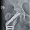 December 1, 2025 From Valgus-impacted to Displaced: Clinical and Technical Lessons in Femoral Neck Fracture Fixation with the Femoral Neck System