
This case highlights the importance of early imaging, a high index of suspicion, and a multidisciplinary approach to complex surgical complications. When conventional revision methods are not possible, intra-abdominal retrieval, with vascular surgery on standby, is essential to avoid life-threatening outcomes. The TFNA system recommends a fully engaged set screw to be turned back by ¼ to provide a reduction of excessive force at the bone-implant interface and promote fracture healing by controlled impaction.
Dr. Alexander Price, Department of Trauma & Orthopaedics, University Hospital Galway, Galway, Ireland. E-mail: alexanderprice23@rcsi.com
Introduction: Intrapelvic migration of cephalomedullary screws is an exceptionally rare but potentially life-threatening complication of internal fixation of intertrochanteric femur fractures. When displaced implants lie near major vascular structures, the clinical concerns rise significantly. This case highlights the importance of early recognition, detailed imaging, and multidisciplinary surgical coordination in managing this high-risk scenario.
Case Report: An 85-year-old male underwent short cephalomedullary nail fixation for a left intertrochanteric femur fracture. After a subsequent fall in a respite facility, investigations revealed superomedial migration of the lag screw into the pelvic cavity. Computed tomography angiography showed the screw tip lying within 2 cm of the iliac bifurcation, without evidence of hemorrhage or visceral injury. A multidisciplinary team, including orthopedic, vascular, and general surgeons, recommended operative retrieval. An initial exploratory laparotomy was aborted due to intraoperative bleeding risk. Definitive removal was achieved safely via a second laparotomy 2 weeks later, with an uneventful post-operative course.
Discussion: This case highlights a rare but dangerous complication. Unlike more routine cases of implant failure managed through direct hip revision alone, the intrapelvic trajectory and vascular proximity in this case required intra-abdominal retrieval. High-resolution imaging was critical in defining anatomy and risk, and the collaborative surgical approach ensured a successful outcome.
Conclusion: Superomedial migration of cephalomedullary screws can pose an imminent danger due to their proximity to critical pelvic structures. When standard revision techniques are not feasible, multidisciplinary surgical planning, guided by advanced imaging, becomes essential to avoid catastrophic outcomes.
Keywords: Intertrochanteric fracture, intrapelvic screw migration, cephalomedullary nail, laparotomy, orthopedic implant complication, case report.
Intertrochanteric femoral fractures are a common injury in the elderly population, typically resulting from low-energy falls in the context of osteoporotic bone [1]. With an aging global population, the incidence of these fractures continues to rise, placing increasing demands on orthopedic services [2]. Cephalomedullary nailing has become a preferred method of fixation due to its biomechanical stability and ability to facilitate early weight-bearing [3]. While outcomes are generally favorable, complications can and do arise. These include well-recognized failure patterns such as femoral head cutout, distal femoral fractures, and implant failure [4]. Less frequently encountered, but potentially far more serious, is the migration of the lag screw [5]. While lateral migration into surrounding soft tissues is more commonly documented, medial migration into the pelvic cavity remains a rare event, particularly when it occurs spontaneously or from relatively low-impact trauma [6]. Such migration carries significant risk due to the close proximity of major vascular and visceral structures. This has been documented in the literature, with a lag screw being retrieved via a Pfannestiel incision, where the screw was located deep in the pelvis, between the internal and external iliac vessels, and posterior to the ureter [7]. This report presents an unusual case of intrapelvic superomedial migration of a cephalomedullary lag screw following fixation of an intertrochanteric femoral fracture, requiring intra-abdominal retrieval, at a regional referral center for orthopedic surgery. After a low-energy fall, the patient was found to have superomedial displacement of the lag screw. Standard retrieval techniques using direct access to the hip were not feasible, as the position of the screw within the femoral head would not allow for this. The situation necessitated a staged surgical retrieval, with an initial laparotomy retrieval aborted due to the proximity of the screw to the iliac bifurcation. With further superomedial migration on serial imaging, a multidisciplinary team considered retrieval necessary, for the concern of further migration and vascular injury. This case highlights the critical importance of collaborative surgical planning, advanced imaging, and the safe management of complex implant-related complications.
An 85-year-old male was diagnosed with a closed left intertrochanteric femur fracture after a syncopal episode and fall onto his left side (Fig. 1).

His medical history included atrial fibrillation managed with a direct oral anticoagulant, hypertension, and a prior right total hip arthroplasty (THR) in 2009 with a revision in 2021. His American Society of Anesthesiology score was III. He required a walking frame at baseline.
The patient underwent internal fixation using a short trochanteric femoral nail – advanced (TFNA) proximal femoral nailing system (DePuy Synthes, Pennsylvania) the following day, performed by the consultant surgeon. The procedure was uneventful, and intraoperative radiographs confirmed appropriate positioning of the implant (Fig. 2).

Figure 2: Intraoperative X-ray – showing adequate implant position.
No information was available as to whether the lag screw was locked, using the “quarter-turn back” technique. This refers to a technique used with the TFNA system, specifically related to the set screw. This technique would be suggested, especially when fracture impaction is likely due to high levels of comminution, as in this case. The absence of intraoperative documentation regarding the degree of set screw engagement represents a potential limitation in understanding the mechanical factors contributing to migration in this case. Future operative records should ideally capture such technical details, as incomplete or excessive set screw engagement may critically influence construct stability. Two weeks postoperatively, after a witnessed fall in the ward, radiographs continued to show satisfactory implant alignment, without any concern of migration (Fig. 3).

Figure 3: Post-operative X-ray (2 weeks post-operative) – Showing continued satisfactory alignment.
In the absence of a fall, routine post-operative imaging would include serial radiographs performed at 2 weeks and then 6 weeks after the internal fixation. A week later, 3 weeks after his surgery, he was discharged to a respite facility for supervised rehabilitation, before planned return to a supported living facility.
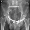
However, after a week at the rehabilitation facility, the patient sustained a second fall while attempting to mobilize. With an increase in left hip pain, repeat radiographs revealed significant superomedial migration of the cephalomedullary lag screw into the pelvis (Fig. 4).

Figure 4: Superomedial migration of lag screw.
A computed tomography (CT) scan and CT angiography demonstrated that the screw had traversed the acetabular dome and entered the pelvic cavity, with its tip located approximately 2 cm from the iliac bifurcation (Fig. 5).

Figure 5: Computed tomography angiogram demonstrating the proximity of the lag screw to the iliac bifurcation.
Importantly, there was no evidence of active hemorrhage, vascular compromise, or injury to solid pelvic organs.
After thorough investigations and given the proximity to major vascular structures, the patient was taken to the operating theater for exploratory laparotomy. Intraoperative findings revealed significant difficulty accessing the screw safely, and due to the high risk of catastrophic bleeding, the procedure was aborted without implant retrieval. The patient was monitored postoperatively and remained hemodynamically stable. Subsequent advanced imaging was performed 10 days later to assess the position of the screw, and further superomedial migration was noticed. The screw had migrated an approximate distance of 5 mm toward the iliac bifurcation (Fig. 6).

Figure 6: Computed tomography angiogram with further superomedial migration of lag screw.
After a multidisciplinary team meeting, inclusive of the orthopedic surgeons, the general surgeons, and the vascular surgeons, retrieval was recommended. A second exploratory laparotomy was performed by the senior general surgeon, 2 weeks after the initial laparotomy, through the previous wound, with the vascular surgery team on standby, and the cephalomedullary screw was successfully removed without intraoperative complications. Post-operative imaging revealed an unhealed fracture, and further surgical planning was commenced (Fig. 7). This involved consideration of a revision with THR (Fig. 8).

Figure 7: X-ray post-retrieval of screw showing ununited fracture.

Figure 8: X-ray with pre-operative planning for left hip arthroplasty.
After discussion with the patient and the family, further surgical intervention was decided against. They wanted to proceed with non-operative measures for fracture healing. The patient underwent intensive inpatient physiotherapy and occupational therapy to return to pre-injury functional levels. On subsequent outpatient clinic follow-up reviews, the patient continued to have limited mobility, but further surgical intervention was once again decided against, as it was believed that the risks would far outweigh the benefits. The patient and the family opted for the non-operative treatment option, understanding the risks involved. Serial imaging, now 4 months after his initial injury, revealed that the original fracture had progressed to union (Fig. 9). The patient continued with an intensive rehabilitation program and was able to mobilize with a rollator frame, with a shoe insert to accommodate the leg length discrepancy he had developed.

Figure 9: X-ray showing healed fracture after screw retrieval.
Intrapelvic migration of a cephalomedullary screw is an exceptionally rare yet serious complication following internal fixation of intertrochanteric femur fractures [5]. While lateral migration or cutout of lag screws is relatively well-documented, medial or superomedial migration into the pelvic cavity poses a significantly higher risk due to potential damage to adjacent vascular and visceral structures [4]. In this case, the lag screws trajectory placed it within close proximity of the iliac bifurcation, highlighting the potential for catastrophic vascular injury. The patient’s advanced age and medical comorbidities, including current anticoagulation for atrial fibrillation, hypertension, and prior contralateral THR, may have potentially influenced both surgical decision-making and rehabilitation. These factors underscore the importance of individualized risk assessment in elderly patients with multiple systemic and orthopedic comorbidities. The mechanisms contributing to screw migration are multifactorial. Poor bone quality, with osteoporosis, inadequate surgical fixation, mechanical loading from premature mobilization, or even repeated trauma, as occurred in this case, can contribute to failure [8]. This case demonstrates how even initially well-positioned implants may fail when exposed to new biomechanical stressors, such as repeated trauma. The TFNA system’s official surgical technique guide instructs the surgeon to fully tighten the lag screw to lock it in place, but then back the set screw off by ¼ turn once engaged [9]. This provides a reduction of excessive force at the bone-implant interface, allowing a sliding construct that promotes fracture healing by controlled impaction. The TFNA set screw has a fine thread pitch; therefore, backing it off more than the ¼ turn back may fully disengage it from the lag screw, which would convert the construct from a controlled sliding mechanism to a completely unlocked one. In weight-bearing, an unlocked lag screw can migrate medially into the pelvis, especially if the fracture site is unstable and the screw is not well centered. Preventive strategies to mitigate intrapelvic migration include meticulous implantation of the lag screw centering within the femoral head, appropriate fracture reduction to minimize varus malalignment, and adherence to manufacturer guidelines regarding set screw engagement. Post-operative early weight-bearing protocols should also be tailored to bone quality and fracture stability. Previous literature, such as the reports by Ortega-Yago et al., demonstrates the different retrieval techniques for intrapelvic migration of the lag screw, including open surgery, laparotomy, laparoscopy, or endovascular approaches [10]. They performed a recent literature review that identified only a single other patient who underwent screw retrieval via a laparotomy [10]. Our patient exhibited progressive superomedial migration and significant anatomical vascular threat, necessitating surgical retrieval via laparotomy. This would be exceedingly rare and has not been well documented in the existing literature. Advanced imaging, particularly CT angiography, played a pivotal role in pre-operative planning. Firstly, by identifying progressive displacement, and secondly, by providing better delineation of the screw positions in relation to vital vascular structures. The first attempt at retrieval was aborted due to intraoperative bleeding risk, where the risk of bleeding outweighed the need of screw retrieval, an example of appropriate intraoperative judgment, prioritizing patient safety over procedural completion. The role of multidisciplinary collaboration cannot be overstated. Engagement of vascular, general, and orthopedic surgical teams enabled an individualized operative approach and ensured intraoperative contingencies were addressed. This collaborative approach ultimately led to successful implant retrieval without complication. Currently, there is no universal imaging protocol recommended following cephalomedullary nailing. This case supports the potential value of early repeat imaging after post-operative falls or new pain, particularly within the first 6 weeks, to identify subtle implant migration before catastrophic progression. This case highlights the importance of close radiological surveillance following any post-operative trauma, especially if occurring on the recently operated joint or fracture. Subtle or early migration may be missed without high clinical suspicion, leading to delayed recognition and increased risk. Quantitative post-operative functional or quality-of-life assessments were not performed in this patient, primarily due to their advanced age and baseline mobility limitations. Future studies incorporating validated tools such as the Harris Hip Score could better evaluate long-term recovery following complex implant-related complications. Long-term follow-up beyond radiographic union was not feasible, as the patient and family elected against further surgical intervention, and long-term clinical follow-up was limited to local physiotherapy reviews. Given the rarity of intrapelvic screw migration, comparative or epidemiological data remain limited. Systematic reviews and registry data would be useful to identify procedural risk patterns and guide evidence-based preventive strategies. In summary, this report contributes to the sparse literature on intrapelvic screw migration and emphasizes the need for a high index of suspicion, early imaging, and multidisciplinary intervention when standard revision techniques are not viable. The safe management of such rare complications relies heavily on pre-emptive planning and adaptable surgical strategies.
Potential limitations
This case report is inherently limited by its single-patient review and lack of long-term follow-up beyond fracture union. Quantitative outcome data and detailed intraoperative variables were unavailable. Nevertheless, the report provides valuable insights into the multidisciplinary management of this rare and high-risk complication. Larger case series or registry-based analyses would be valuable to further define the incidence, risk factors, and management outcomes.
Superomedial migration of cephalomedullary lag screws, while rare, presents a high-risk complication due to the potential for vascular and visceral injury. This case highlights the importance of early imaging, a high index of suspicion, and a multidisciplinary approach to complex surgical complications. When conventional revision methods are not possible, intra-abdominal retrieval, with vascular surgery on standby, is essential to avoid life-threatening outcomes. The TFNA system recommends a fully engaged set screw to be turned back by ¼ to provide a reduction of excessive force at the bone-implant interface and promote fracture healing by controlled impaction.
Intrapelvic migration of cephalomedullary screws, though exceedingly rare, poses a significant risk of vascular injury due to their close proximity to major pelvic structures. This case highlights the critical importance of early detection through advanced imaging, a high index of suspicion after post-operative trauma, and the necessity of multidisciplinary collaboration. When standard retrieval methods are not viable, planned intra-abdominal screw removal can prevent life-threatening complications and ensure safe patient outcomes.
References
- 1. Attum B, Pilson H. Intertrochanteric Femur Fracture. In: StatPearls. Treasure Island, FL: StatPearls Publishing; 2025. Available from: https://www.ncbi.nlm.nih.gov/books/nbk493161 [Accessed 17 June 2025] [Google Scholar] [PubMed]
- 2. Adeyemi A, Delhougne G. Incidence and economic burden of intertrochanteric fracture: A medicare claims database analysis. JBJS Open Access 2019;4:e0045. [Google Scholar] [PubMed]
- 3. Niu E, Yang A, Harris AH, Bishop J. Which fixation device is preferred for surgical treatment of intertrochanteric hip fractures in the United States? A survey of orthopaedic surgeons. Clin Orthop Relat Res 2015;473:3647-55. [Google Scholar] [PubMed]
- 4. Petfield JL, Visscher LE, Gueorguiev B, Stoffel K, Pape HC. Tips and tricks to avoid implant failure in proximal femur fractures treated with cephalomedullary nails: A review of the literature. OTA Int 2022;5 2 Suppl:e191. [Google Scholar] [PubMed]
- 5. Scholl B, Tran S, Patel S, Vanlaningham CJ, Veenstra J. Cephalomedullary nail lag screw migration following open reduction and internal fixation of an intertrochanteric femur fracture in a patient with body mass index of 63. Cureus 2024;16:e69916. [Google Scholar] [PubMed]
- 6. Güven Ş, Naldöven ÖF, Alkan H, Erdoğan Y, Çepni Ş, Veizi E, et al. Laterally protruded cephalomedullary nail lag screws are a source of consistent thigh pain after pertrochanteric fracture. J Orthop Trauma 2024;38:320-6. [Google Scholar] [PubMed]
- 7. Robinson SJ, Fountain JR, Torella F, Pennie BH. Intrapelvic migration of a lag screw from a cephalomedullary femoral nail: A case report. Injury 2011;42:1384-6. [Google Scholar] [PubMed]
- 8. Sermon A, Hofmann-Fliri L, Zderic I, Agarwal Y, Scherrer S, Weber A, et al. Impact of bone cement augmentation on the fixation strength of TFNA blades and screws. Medicina (Kaunas) 2021;57:899. [Google Scholar] [PubMed]
- 9. DePuy Synthes. TFN-ADVANCED™ Proximal Femoral Nailing System: Surgical Technique Guide. West Chester, PA: DePuy Synthes; 2015. Available from: https://www.jnjmedtech.com/sites/default/files/2023-03/tfna_surgicaltechnique_en.pdf [Accessed 2 August 2025] [Google Scholar] [PubMed]
- 10. Ortega-Yago A, Balfagon-Ferrer A, Sanchez-Jimenez A, Barrés-Carsí M. Intrapelvic migration of cephalic screw: Report of two cases and review of the literature. London J Med Health Res 2025;25:9-16. [Google Scholar] [PubMed]


Related Articles in Journal of Orthopaedic Case Reports
 February 1, 2026 Intraoperative Diagnosis and Management of Testicular Dislocation During Pelvic Fracture Fixation: A Report of Two Cases and Literature Review
 February 1, 2026 Symptomatic Ventromedial Scapular Osteochondroma Presenting with Restriction of Shoulder Movements: A Case Report
 February 1, 2026 A Novel Technique of Rerouting Semitendinosus Graft for Medial Collateral Ligament and Medial Patellofemoral Ligament Reconstruction – In a Polytrauma Patient with Multiligament Injury: Kakran et al. Technique
 January 1, 2026 Primary Synovial Chondromatosis of the Elbow Joint Presenting with Ulnar Nerve Compression and Restricted Range of Motion: A Case Report