
Serum biochemical markers, such as calcium, phosphorus, alkaline phosphatase, and Vitamin D can serve as valuable adjuncts for monitoring radiographic fracture healing progression in tibial fractures
Dr. Vipin Kumar Mishra, Department of Orthopaedics, Government Medical College, Satna, Madhya Pradesh, India. E-mail: vipin9926@gmail.com
Background: Fracture healing is a complex biological process influenced by systemic biochemical factors that regulate bone formation and remodeling. Evaluating the association between biochemical markers and radiographic evidence of union may enhance understanding of fracture repair and help predict healing outcomes.
Materials and Methods: This prospective observational study included 63 adult patients with tibial shaft fractures managed either surgically or conservatively. Serial assessments of serum calcium, phosphorus, alkaline phosphatase (ALP), Vitamin D, and parathyroid hormone (PTH) were performed at baseline, 6, 12, and 24 weeks. Radiographic healing was evaluated at corresponding intervals using the Radiographic Union Score for Tibial fractures (RUST). Correlation between biochemical parameters and RUST scores was analyzed using Pearson’s correlation coefficient.
Results: Most participants were males (74.6%), with road traffic accidents as the predominant cause of injury. Progressive improvement was observed in calcium, phosphorus, ALP, and Vitamin D levels during follow-up, while PTH showed a gradual decline. The mean RUST score increased steadily from early to late follow-ups, indicating continuous radiographic healing. At 24 weeks, ALP demonstrated the strongest positive correlation with RUST (r = 0.61, P < 0.001), followed by Vitamin D (r = 0.46, P < 0.001), phosphorus (r = 0.42, P = 0.001), and calcium (r = 0.38, P = 0.002). PTH showed a weak negative correlation (r = –0.33, P = 0.008).
Conclusion: Serum biochemical parameters, particularly ALP and Vitamin D, reflect the biological progression of fracture healing and correlate significantly with radiographic union. Routine biochemical monitoring, in conjunction with radiographic evaluation, can serve as a valuable adjunct in assessing the healing potential of tibial fractures.
Keywords: Tibial fracture, alkaline phosphatase, radiographic union score, Vitamin D, bone healing.
Tibial shaft fractures are among the most common long-bone injuries encountered in orthopedic practice and often carry a substantial risk of delayed union or non-union because of the limited soft-tissue envelope and the biomechanical load borne by the tibia. Reliable and reproducible methods to monitor healing are therefore essential for guiding management and identifying patients at risk of impaired recovery [1,2].
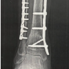
Plain radiography remains the most widely available tool to assess structural repair, and the Radiographic Union Score for Tibial fractures (RUST) – and its modified form RUST – provide standardized, validated metrics to quantify cortical bridging and loss of fracture line visibility on orthogonal radiographs. These scoring systems have demonstrated good inter- and intraobserver reliability and correlate with volumetric and biomechanical indices of callus mineralization in experimental and clinical studies [1,2].
Concurrently, systemic biochemical markers of bone turnover and mineral metabolism offer objective information on the biological processes underlying fracture repair. Markers of bone formation (notably alkaline phosphatase [ALP] and its bone-specific isoform) and indices of mineral homeostasis (serum calcium, phosphate, 25-hydroxyvitamin D, and parathyroid hormone [PTH]) change during the different phases of healing and have been investigated as potential adjuncts to imaging for early detection of delayed union [3,4].
Several clinical and experimental reports have documented a temporal rise in total and bone-specific ALP in association with callus formation, supporting its use as a dynamic biochemical indicator of osteoblastic activity during the reparative phase. Similarly, disturbances in Vitamin D status and perturbations of calcium–phosphate balance have been linked to slower or suboptimal consolidation in some cohorts, although interventional trials of Vitamin D supplementation report mixed effects on final union rates. PTH has also been examined for its complex, context-dependent role in bone remodeling and repair [5,6].
Given the complementary nature of radiographic scoring and circulating biochemical measurements, correlating serial biochemical profiles with validated radiographic healing scores could improve early identification of patients with impaired biological healing and refine prognostication. This study, therefore, aimed to evaluate the temporal changes in selected biochemical parameters and to determine their correlation with RUST at 24 weeks in adults with tibial shaft fractures.
Study design and setting
This hospital-based prospective observational study was conducted at a tertiary care teaching hospital in India. Written consent was obtained from all participants before enrolment.
Sample size and selection criteria
A total of 63 patients with radiographically confirmed tibial shaft fractures were included in the study. The sample size was determined based on previous literature indicating a moderate correlation between biochemical parameters and fracture healing scores, assuming a correlation coefficient (r) of 0.35, with 80% power and 5% level of significance.
Inclusion criteria
- Adults aged 18–60 years with fresh, closed tibial fractures treated conservatively or surgically
- Willingness to participate and comply with follow-up visits.
Exclusion criteria
- Pathological fractures or fractures associated with metabolic bone disease
- Chronic renal or hepatic disorders affecting calcium or Vitamin D metabolism
- Patients on corticosteroids, bisphosphonates, or Vitamin D/calcium supplementation before injury
- Open or compound fractures (Gustilo-Anderson type II or higher).
Clinical management
Patients underwent either closed reduction and internal fixation using intramedullary nailing or conservative management with casting, as deemed appropriate by the treating surgeon. Standard post-operative and rehabilitation protocols were followed in all cases.
Assessment schedule
Clinical and biochemical evaluations were performed at the time of injury (baseline) and subsequently at 6 weeks, 12 weeks, and 24 weeks post-fracture. Radiographic assessment was done concurrently using standard anteroposterior and lateral views of the leg.
Biochemical parameters
Venous blood samples were collected after overnight fasting and analyzed for:
- Serum calcium (mg/dL) – by o-cresolphthalein complexone method
- Serum phosphorus (mg/dL) – by ammonium molybdate ultraviolet method
- Serum ALP (U/L) – by kinetic colorimetric method
- Serum Vitamin D (ng/mL) – by chemiluminescent immunoassay
- Serum PTH (pg/mL) – by electrochemiluminescence immunoassay.
All analyses were performed in the hospital’s central biochemistry laboratory using standardized protocols and calibrated equipment.
Radiographic evaluation
Fracture healing was assessed radiographically using the RUSTs. Each cortex was scored from 1 to 3 (1 = fracture line visible with no callus, 2 = callus present but fracture line visible, 3 = bridging callus with no visible fracture line), with a total score ranging from 4 to 12. Radiographs were independently evaluated by two blinded orthopedic surgeons, and the mean score was used for analysis.
Statistical analysis
Data were compiled in Microsoft Excel and analyzed using the IBM Statistical Package for the Social Sciences Statistics version 26.0. Continuous variables were expressed as mean ± standard deviation, while categorical data were represented as frequencies and percentages.
Correlation between biochemical parameters and RUST scores was assessed using Pearson’s correlation. A P < 0.05 was considered statistically significant.
A total of 63 patients with tibial fractures were included in the study. The demographic and clinical characteristics are summarized in Table 1. The majority of participants were middle-aged males, and road traffic accidents represented the predominant cause of injury. Most fractures were closed and managed surgically with intramedullary nailing.

Table 1: Baseline demographic and clinical characteristics (n=63)
Progressive improvement was observed in the biochemical profile over the follow-up period (Table 2). Calcium and phosphorus levels demonstrated a steady upward trend throughout the 24-week duration, while serum ALP exhibited a characteristic rise during the intermediate phase of healing (6–12 weeks) followed by a decline toward baseline values at 24 weeks, consistent with the phase of callus remodeling. Vitamin D concentrations also showed gradual improvement during follow-up, accompanied by a concurrent decline in PTH levels, reflecting normalization of bone metabolism as healing progressed.

Table 2: Temporal changes in biochemical parameters during fracture healing
Radiographic assessment using the RUSTs revealed a consistent increase across all time points, indicating progressive cortical bridging and callus consolidation (Table 3). The improvement in mean RUST scores over successive follow-ups confirmed satisfactory union in the majority of patients by 24 weeks.

Table 3: Mean RUST at follow-up
Correlation analysis demonstrated statistically significant associations between biochemical parameters and radiographic healing scores at 24 weeks (Table 4). Serum ALP exhibited the strongest positive correlation with the RUST score, underscoring its utility as a dynamic biochemical marker of osteoblastic activity during fracture repair. Moderate positive correlations were also observed between calcium, phosphorus, and Vitamin D with RUST, suggesting their contributory role in bone mineralization and healing. In contrast, PTH levels showed a weak but significant negative correlation with radiographic union, indicating that persistently elevated PTH may reflect delayed mineralization or secondary hyperparathyroidism in slower healers.

Table 4: Correlation between biochemical parameters and RUST at 24 weeks
This study found that dynamic changes in routine biochemical indices of bone metabolism accompany radiographic progression of tibial fracture repair, and that serum ALP showed the strongest association with radiographic union at 24 weeks. These observations align with recent clinical case-series and cohort work that highlight a temporal relationship between ALP behavior and radiographic indices of consolidation, suggesting ALP as a useful, easily obtainable adjunct to imaging when interpreting biological healing activity [7].
The validity of radiographic scoring systems, such as the RUSTs as surrogates of mechanical and structural healing has been repeatedly demonstrated in preclinical and translational work; RUST correlates with callus mineral density, bone volume fraction and biomechanical properties, supporting its use as a quantitative radiographic end-point in human studies and justifying its use here as the comparator for circulating markers [8]. Against that background, the concordant rise in ALP during the reparative phase observed in our cohort – together with its positive correlation with RUST – supports the interpretation that rising ALP largely reflects osteoblastic/callus activity rather than non-specific systemic processes [7,9].
Changes in mineral indices (calcium and phosphate) and Vitamin D observed in our series echo prior clinical reports that document modest, time-dependent shifts in these analytes after long-bone fracture repair [9,10]. Several prospective studies of femoral/tibial fractures have described early post-operative increases in calcium and phosphate and variable ALP kinetics during the interval of callus formation and remodeling; such trends are biologically plausible given the mineralization demands of new bone and are compatible with our finding of moderate positive correlations between mineral markers and radiographic union [9,10].
The observed inverse relationship between PTH and radiographic union in our data is consistent with the complex role of PTH in bone biology. Pre-clinical work demonstrates that intermittent PTH administration can accelerate fracture repair and enhance callus formation in animal models, whereas chronically elevated endogenous PTH often reflects secondary disturbances in mineral metabolism (for example, Vitamin D deficiency or renal bone disease) and may associate with impaired bone microarchitecture [11]. Thus, the negative correlation we report likely reflects that higher circulating PTH in some patients was a marker of relative mineral insufficiency or perturbed remodeling rather than a direct inhibitory effect on healing [11].
Several recent clinical investigations have attempted to use circulating biomarkers to predict impaired healing early; results have been mixed. Some reports and systematic appraisals show promising associations between specific bone-turnover markers and healing trajectories, yet heterogeneity in marker selection, assay methods, timing, and endpoints has limited translation into routine practice [12,13]. Our results add to this body of evidence by demonstrating measurable, clinically accessible associations between widely available tests (total ALP, calcium, phosphate, Vitamin D, and PTH) and a validated radiographic score, but they also underscore the need for standardized assays and sampling protocols before biochemical monitoring can be recommended as a standalone predictor [13,14].
Strengths of our study include prospective serial sampling, use of a validated radiographic outcome (RUST), and analysis of multiple, complementary biochemical indices. Limitations deserve mention: This was a single-center observational study with a modest sample size, and total (rather than bone-specific) ALP was measured; systemic illness, nutritional status, and unmeasured local factors (fracture gap, soft-tissue injury) can confound circulating markers. Furthermore, while RUST is validated and correlates with structural metrics, radiographs are imperfect surrogates for mechanical strength [8,13].
In summary, our findings support a role for serial biochemical monitoring – particularly ALP, alongside mineral indices and Vitamin D – as an adjunct to radiographic assessment in tibial fracture follow-up. Future multicenter studies employing standardized BTM panels, bone-specific assays, and pre-specified thresholds are warranted to test whether early biochemical trajectories can prospectively identify patients at risk of delayed union and thereby guide timely interventions [13,14].
The present study demonstrates a significant relationship between biochemical markers of bone metabolism and radiographic evidence of fracture union in tibial fractures. Among the parameters evaluated, serum ALP showed the strongest positive correlation with radiographic healing scores, indicating its potential as a reliable biochemical indicator of osteoblastic activity. Improvements in calcium, phosphorus, and Vitamin D levels were also associated with progressive fracture consolidation, whereas elevated PTH levels were inversely related to healing. These findings suggest that periodic monitoring of biochemical parameters, alongside radiographic evaluation, can provide valuable insights into the biological progress of bone repair and may assist clinicians in predicting the course of fracture healing.
Monitoring biochemical markers, such as serum calcium, phosphorus, ALP, and Vitamin D can serve as valuable adjuncts in evaluating fracture healing progression. These parameters reflect underlying metabolic activity at the fracture site and correlate with radiographic healing outcomes. Incorporating biochemical assessment alongside imaging can enhance early detection of delayed union and guide timely therapeutic interventions.
References
- 1. Leow JM, Clement ND, Tawonsawatruk T, Simpson CJ, Simpson AH. The radiographic union scale in tibial (RUST) fractures: Reliability of the outcome measure at an independent centre. Bone Joint Res 2016;5:116-21. [Google Scholar] [PubMed]
- 2. Fiset S, Godbout C, Crookshank MC, Zdero R, Nauth A, Schemitsch EH. Experimental validation of the radiographic union score for tibial fractures (RUST) using micro-computed tomography scanning and biomechanical testing in an in-Vivo rat model. J Bone Joint Surg Am 2018;100:1871-8. [Google Scholar] [PubMed]
- 3. Perut F, Roncuzzi L, Gómez-Barrena E, Baldini N. Association between bone turnover markers and fracture healing in long bone non-union: A systematic review. J Clin Med 2024;13:2333. [Google Scholar] [PubMed]
- 4. Rathwa HS, Verma T, Chavali VH. Assessment of union in fractures: Role of serum alkaline phosphatase and ultrasonography. J Clin Orthop Trauma 2020;14:94-100. [Google Scholar] [PubMed]
- 5. Muljacić A, Poljak-Guberina R, Zivković O, Bilić V, Guberina M, Crvenković D. Course and rate of post-fracture bone healing in correlation with bone-specific alkaline phosphatase and bone callus formation. Coll Antropol 2013;37:1275-83. [Google Scholar] [PubMed]
- 6. Gatt T, Grech A, Arshad H. The effect of vitamin D supplementation for bone healing in fracture patients: A systematic review. Adv Orthop 2023;2023:6236045. [Google Scholar] [PubMed]
- 7. Ninomiya AF, Bertolucci V, Kaneko LO, Nonose N, Abreu LD, Harfuch GR, et al. Comparison of radiographic outcomes assessed via the radiographic union scale for tibial fractures and alkaline phosphatase levels during the tibial healing process: A series of case reports. Biology (Basel) 2024;13:407. [Google Scholar] [PubMed]
- 8. Cooke ME, Hussein AI, Lybrand KE, Wulff A, Simmons E, Choi JH, et al. Correlation between RUST assessments of fracture healing to structural and biomechanical properties. J Orthop Res 2018;36:945-53. [Google Scholar] [PubMed]
- 9. Sobhani Eraghi A, Saberi S, Molazemsanandaji B, Ghaznavi A. Investigating changes in calcium, phosphorus, alkaline phosphatase, and 25-hydroxy vitamin D after surgical repair of fractures of femur or tibia. Acta Biomed 2020;92:e2021019. [Google Scholar] [PubMed]
- 10. Chen Z, Xie L, Xu J, Lin X, Ye J, Shao R, et al. Changes in alkaline phosphatase, calcium, C-reactive protein, D-dimer, phosphorus and hemoglobin in elderly osteoporotic hip fracture patients. Ann Palliat Med 2021;10:1079-88. [Google Scholar] [PubMed]
- 11. Menger MM, Tobias AL, Bauer D, Bleimehl M, Scheuer C, Menger MD, et al. Parathyroid hormone stimulates bone regeneration in an atrophic non-union model in aged mice. J Transl Med 2023;21:844. Erratum in: J Transl Med 2024;22:828. [Google Scholar] [PubMed]
- 12. Ali S, Singh A, Yadav M, Siddiqui S, Pandey V, Mahdi AA, et al. Can impaired diaphyseal fracture healing be predicted early?: A cohort study of biomarkers. J Clin Orthop Trauma 2019;10 Suppl 1:S37-46. [Google Scholar] [PubMed]
- 13. Bhattoa HP, Vasikaran S, Trifonidi I, Kapoula G, Lombardi G, Jørgensen NR, et al. Update on the role of bone turnover markers in the diagnosis and management of osteoporosis: A consensus paper from the European Society for Clinical and Economic Aspects of Osteoporosis, Osteoarthritis and Musculoskeletal Diseases (ESCEO), International Osteoporosis Foundation (IOF), and International Federation of Clinical Chemistry and Laboratory Medicine (IFCC). Osteoporos Int 2025;36:579-608. [Google Scholar] [PubMed]
- 14. Minisola S, Cipriani C, Colangelo L, Labbadia G, Pepe J, Magnusson P. Diagnostic approach to abnormal alkaline phosphatase value. Mayo Clin Proc 2025;100:712-28. [Google Scholar] [PubMed]


Related Articles in Journal of Orthopaedic Case Reports
 February 1, 2026 Endoscopic Flexor Hallucis Longus Tenotomy for Post-traumatic Checkrein Deformity: A Case Report
 January 1, 2026 Correlation of Serum Vitamin D Levels and Incidence of Lateral Epicondylitis of the Elbow: An Observational Study in Eastern India
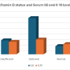 November 1, 2025 Relation of Serum Vitamin-D Level with Serum Interleukin-6 and Interleukin-10 in Patients with Primary Osteoarthritis of Knee
 November 1, 2025 Functional and Radiological Outcome of Ilizarov Ring Fixator versus Limb Reconstruction System in Compound Tibial Diaphyseal Fractures Grade II, IIIA, and IIIB