
Coronal tibiofemoral subluxation is common in varus knee osteoarthritis, is strongly associated with altered bone morphometry, is not reliably predicted by CPAK classification, and therefore warrants routine assessment on weight-bearing radiographs to optimize pre-operative planning, alignment correction, and soft-tissue balancing in total knee arthroplasty.
Dr. R Monish Kumar, Department of Orthopaedics, Vinayaka Mission’s Kirupananda Variyar Medical College and Hospital, Vinayaka Mission’s Research Foundation (Deemed to be University), Salem, Tamil Nadu, India. E-mail: moni97a@gmail.com
Abstract
Introduction: Coronal tibiofemoral subluxation (CTFS) is a frequently observed radiological feature in varus osteoarthritis (OA) of the knee and may reflect underlying biomechanical misalignment and osseous changes. Understanding its correlation with bone morphometry can help in surgical planning and risk stratification.
Aims: To analyze the coronal tibiofemoral (CTF) in varus OA knee and its correlation with bone morphometry in patients undergoing total knee arthroplasty.
Materials and Methods: This retrospective and prospective study was conducted between January 2024 and January 2025. A total of 98 patients with varus OA knees planned for total knee arthroplasty were included. Radiographic parameters, including arithmetic hip–knee–ankle (HKA) angle, joint line obliquity, CTF, lateral distal femoral angle (LDFA), medial proximal tibial angle (MPTA), coronal plane alignment of the knee (CPAK), and posterior condylar offset ratio (PCOR), were evaluated using standardized full-length scanograms.
Results: Among 98 patients, 60.2% were over 60 years old, and 61.2% were female. Body mass index classification showed that 42.9% were overweight and 25.5% were obese. Right knee involvement was observed in 53.1% of cases. CTF was present in 64 patients (65.3%) with a mean subluxation distance of 6.2 ± 1.5 mm. Most cases (75%) had subluxation distances between 0 and 6 mm. CPAK Type I was predominant (93.9%), whereas Type IV was seen in 6.1%. CTF prevalence was similar between CPAK Type I (65.2%) and Type IV (66.7%) (P = 0.887). Patients with CTF had higher LDFA (89.8° ± 2.1 vs. 87.9° ± 1.8), lower MPTA (84.5° ± 2.2 vs. 87.2° ± 1.9), higher PCOR (0.52 ± 0.06 vs. 0.48 ± 0.05), and more negative HKA angle (−7.2° ± 2.4 vs. −4.9° ± 2) than those without CTF (all P < 0.005). Correlation analysis showed a significant positive correlation of CTF with LDFA (r = 0.36, P = 0.004), PCOR (r = 0.3, P = 0.015), and HKA (r = 0.44, P = 0.003), and a negative correlation with MPTA (r = −0.42, P = 0.002).
Conclusion: CTF is highly prevalent in varus OA knees and is significantly associated with specific morphometric alterations, including increased LDFA and PCOR, decreased MPTA, and a more negative HKA angle. While CPAK Type I predominates in this population, the CPAK classification did not significantly correlate with subluxation presence. These findings underscore the importance of assessing CTFS and bone morphology preoperatively for individualized surgical planning and improved outcomes.
Keywords: Knee osteoarthritis, coronal tibiofemoral subluxation, tibiofemoral subluxation-ray.
Knee osteoarthritis (OA) is a major cause of pain and disability worldwide, particularly in aging populations. Varus malalignment, characterized by medial compartment overload, is the most common deformity associated with knee OA, often resulting in progressive joint space narrowing and mechanical imbalance. Coronal tibiofemoral subluxation (CTFS), a radiological phenomenon reflecting lateral displacement of the tibia relative to the femur, has emerged as an important biomechanical marker of disease severity in varus OA knees. Despite its potential clinical implications, CTFS remains underexplored in routine orthopedic evaluation. Recent studies have shown that tibiofemoral subluxation may influence surgical planning, post-operative alignment, and functional outcomes in total knee arthroplasty (TKA) [1]. In this context, evaluating the prevalence of CTFS in varus knees and understanding its anatomical correlates is essential for optimizing treatment strategies. Radiographic morphometry has become a reliable, non-invasive method for evaluating structural parameters contributing to OA progression. Parameters such as medial proximal tibial angle (MPTA), femoral condylar offset, and tibial plateau inclination are among the key factors that may influence tibiofemoral alignment and load distribution. A few reports have identified a relationship between altered bone morphology and the degree of subluxation in varus knees, suggesting a biomechanical interplay between osseous architecture and joint kinematics [2,3]. However, literature is scarce in systematically exploring this relationship in Indian populations, where anatomical variations and lifestyle factors such as squatting may further influence joint loading and morphometry. Therefore, there is a growing need for radiological studies that examine both subluxation prevalence and its morphological correlates, especially using standardized weight-bearing X-rays, which reflect functional joint positioning more accurately than non-weight-bearing modalities. Moreover, quantification of CTFS may have significant prognostic value. Studies have suggested that increased CTFS is associated with accelerated cartilage loss, greater mechanical instability, and poorer functional outcomes following conservative or surgical management [4,5]. Particularly in the context of TKA, persistent pre-operative subluxation can affect prosthetic alignment and may contribute to suboptimal post-operative outcomes or early failure. An understanding of whether subluxation correlates with bone angles or joint morphometry may also help in pre-operative templating and in deciding on the extent of bone cuts or ligament releases. Therefore, recognizing CTFS as more than just a radiographic finding, and instead as a potential indicator of altered biomechanics, may aid in tailoring personalized interventions for OA patients. Despite the growing recognition of CTFS in the global literature, regional data remains limited. Few Indian studies have evaluated the prevalence of coronal subluxation and its relationship with underlying bone morphology in varus knees, despite high OA burden in this population [6,7]. This study aims to bridge that gap using standard anteroposterior (AP) weight-bearing knee radiographs to determine the prevalence of CTFS in varus OA knees and assess its correlation with key radiographic bone parameters. Generating such data will not only enhance understanding of knee OA pathomechanics in the Indian context but may also contribute to improved clinical decision-making and surgical planning. Given the rising number of TKA procedures and the emphasis on alignment-based outcomes, this study holds practical relevance and clinical value in the management of knee OA.
Aim
To analyze the CTFS in varus OA knee and its correlation with bone morphometry in patients undergoing TKA.
Objectives
- To determine if there is a significant association between the prevalence of CTFS and specific CPAK types in patients with varus OA knee.
- To evaluate if there is a relationship between bone morphometry (MPTA, lateral distal femoral angle [LDFA], posterior condylar offset ratio [PCOR]) that influences subluxation.
Materials and Methods
Study design
This is a combined retrospective and prospective observational study aimed at analyzing the prevalence of CTFS in patients with varus OA of the knee and evaluating its correlation with various parameters of bone morphometry. The study is designed to assess pre-operative radiographic characteristics in patients scheduled for TKA.
Study setting and duration
The study will be conducted at the Department of Orthopedics, Vinayaka Missions Kirupanandha Variyar Medical College and Hospital, Salem. This study was approved by Vinayaka Missions Kirupanandha Variyar Medical College Ethical Committee with EC no:- CSP-MED/24/SEP/109/308, issued on February 12, 2024.
Sample Size
The study will include a total of 98 patients diagnosed with OA of the knee with varus deformity, who are planned for TKA during the study period.
Inclusion criteria
- Patients diagnosed as having OA of the knee and planned for TKA
- Patients with clinical and radiological evidence of varus deformity of the knee.
Exclusion criteria
- Patients with incomplete clinical or radiographic records
- Patients undergoing revision knee arthroplasty
- Patients with extra-articular deformities affecting the femur or tibia. Patients with non-standardized or poor-quality radiographs (e.g., non-weight-bearing, rotated, or misaligned scanograms)
- Patients with valgus deformity of the knee or arthritis due to other causes (e.g., post-traumatic, inflammatory, or infective).
Data collection
Radiological data will be collected from pre-operative full-length, weight-bearing AP scanograms of the lower limbs. Both retrospective and prospectively recruited patients will have radiographs assessed using standardized protocols.
Age distribution
Most patients (60.2%) were aged above 60 years, indicating a higher prevalence of varus knee OA in the elderly population. The largest age group was >70 years, accounting for 31.6% of cases. This suggests that CTFS and associated bony changes are more common in advanced age.
Gender distribution
A higher proportion of females (61.2%) was affected compared to males (38.8%), indicating a female predominance in varus OA cases. This trend may reflect hormonal, anatomical, or lifestyle-related factors contributing to disease prevalence.
Body mass index (BMI)
Most patients were either overweight (42.9%) or obese (25.5%), together accounting for 68.4% of the study population. Normal BMI was seen in 28.6% of patients, whereas only 3.1% were underweight. This highlights a strong association between higher BMI and varus OA.
Laterality
Right knee involvement was slightly more common (53.1%) compared to the left knee (46.9%) among the study population. The difference in laterality was minimal, indicating near-equal distribution. This suggests no strong lateral dominance in varus OA presentation.
Prevalence of coronal tibiofemoral (CTF) in study participants
CTFS was present in 65.3% of the study participants, indicating a high prevalence among patients with varus OA. The mean subluxation distance was 6.2 ± 1.5 mm. This underscores the significance of CTF as a common radiographic finding in such cases.
CTF characteristics
Most patients (75%) had a subluxation distance of 0–6 mm, indicating that mild-to-moderate CTFS is more common in varus OA knees. Only 25% of patients exhibited a subluxation distance >6 mm, suggestive of more advanced joint misalignment. The proposed detailed classification of CTFS reveals that most patients had subluxation distances between 2 and 6 mm, indicating that early to mild joint displacement is most common in varus OA knees. Higher-grade subluxation (>8 mm) was seen in a small proportion, suggestive of advanced disease. This stratification may aid in early detection, severity grading, and treatment planning. Grade II CTFS (5–10 mm) was the most common, seen in 59.4% of cases, indicating that moderate subluxation predominates in varus OA knees. Grade I (<5 mm) was present in 37.5%, whereas only 3.1% exhibited Grade III (>10 mm), suggesting severe subluxation is rare. Among the grades, Grade II showed the strongest correlation with morphometric alterations such as increased LDFA, decreased MPTA, and a more negative hip–knee–ankle (HKA) angle, highlighting its biomechanical significance (Table 1).
CPAK
Most patients (93.9%) belonged to CPAK Type I, indicating a strong predominance of this alignment pattern in varus knee OA. Type IV was seen in only 6.1% of cases, whereas Type VII was absent in the study population (Table 2).
Association
CTF was present in 65.2% of Type I and 66.7% of Type IV patients, showing no significant variation in subluxation prevalence between the two types.
The overall distribution of CTF across CPAK types was not statistically significant (P = 0.887). This suggests that CPAK classification may not strongly influence the presence of CTFS in varus knees (Table 3).
Measurement results with CTF
Patients with CTF had significantly higher mean LDFA (89.8° vs. 87.9°), lower MPTA (84.5° vs. 87.2°), higher PCOR (0.52 vs. 0.48), and more negative HKA angle (−7.2° vs. −4.9°) compared to those without CTF. All these differences were statistically significant (P < 0.005). This indicates a strong association between CTFS and altered bone morphometry in varus OA knees (Table 4).
Correlation
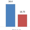
There was a statistically significant positive correlation of CTF with LDFA (r = 0.36) and PCOR (r = 0.3), and a strong positive correlation with HKA (r = 0.44). MPTA showed a significant negative correlation (r = −0.42), indicating that lower MPTA is associated with increased subluxation. These findings highlight that specific bone morphometric parameters are closely related to the degree of tibiofemoral subluxation (Table 5 and Fig. 1).
Age distribution
The predominance of patients over 60 years (60.2%) in this study underscores the increased susceptibility of the elderly to varus knee OA. This aligns with findings by Muraki et al., [8] who reported a higher prevalence of knee OA among individuals aged over 60 years in the Japanese population. Similarly, Zhang and Jordan noted that age is a significant risk factor for the development and progression of knee OA, with the incidence rising markedly in older age groups [9].
Gender distribution
The higher prevalence of varus knee OA in females (61.2%) observed in this study is consistent with global trends. Srikanth et al. [10] conducted a meta-analysis revealing that women are more likely to develop knee OA than men, potentially due to hormonal differences, biomechanics, and genetic factors. In addition, Sowers and Karvonen–Gutierrez highlighted that postmenopausal hormonal changes contribute to the increased risk of OA in women.
BMI
The association between higher BMI and varus knee OA, as evidenced by 68.4% of patients being overweight or obese, corroborates previous research. Felson et al. demonstrated that obesity is a significant risk factor for the development and progression of knee OA due to increased mechanical load and metabolic factors. Furthermore, Blagojevic et al. found that weight loss can reduce the risk of symptomatic knee OA, emphasizing the importance of weight management in prevention strategies [11,12,13].
Laterality
The near-equal distribution of right (53.1%) and left (46.9%) knee involvement suggests no significant lateral dominance in varus knee OA. This finding is in line with the study by Sharma et al., which reported symmetrical involvement of knees in OA patients, indicating that factors other than limb dominance may influence disease development [14].
Prevalence of CTF in study participants
The presence of CTF subluxation in 65.3% of patients highlights its commonality in varus knee OA. Schadler et al. reported similar findings, emphasizing that CTF subluxation is frequently observed in patients with moderate-to-severe varus OA. However, they also noted that CTF subluxation alone may not be an independent risk factor for the need for TKA [15].
CTF characteristics
The predominance of mild-to-moderate subluxation (2–6 mm) in 62.5% of patients (Grade I and II) suggests that early-stage joint misalignment is the most common presentation in varus knee OA. This finding is consistent with the observations of Tanaka et al., who reported that minor CTFSs are frequently seen in the early stages of OA and tend to worsen as the disease progresses. Their emphasis on early detection and timely intervention aligns with our results, underscoring the potential value of subluxation grading in guiding treatment decisions and slowing disease advancement [16].
CPAK
The overwhelming majority of patients (93.9%) classified as CPAK Type I indicates a strong association between this alignment pattern and varus knee OA. Yoon et al. observed a similar distribution, with CPAK Type I being predominant in patients with medial compartment OA. This suggests that CPAK classification can be a useful tool in understanding the alignment variations in OA patients [17].
Association
The similar prevalence of CTF in CPAK Type I (65.2%) and Type IV (66.7%) patients, with no significant statistical difference (P = 0. 887), suggests that CPAK classification may not strongly influence the presence of CTF subluxation. This finding is consistent with the study by Wang et al., who reported that while CPAK classification provides insight into knee alignment, it may not directly correlate with subluxation severity [18].
Measurement results with CTF
Patients with CTF exhibited significantly higher LDFA and PCOR, lower MPTA, and more negative HKA angles compared to those without CTF, indicating altered bone morphometry associated with subluxation. These findings agree with the study by Chang et al., who demonstrated that varus alignment and associated morphological changes are linked to increased mechanical stress and joint degeneration in knee OA [14].
Correlation
The significant correlations of CTF with LDFA (r = 0.36), MPTA (r = −0.42), PCOR (r = 0.3), and HKA (r = 0.44) underscore the relationship between bone morphometric parameters and subluxation severity. Sharma et al. highlighted that malalignment, particularly varus alignment, is a critical factor in the progression of knee OA, reinforcing the importance of these parameters in disease assessment [19].
This research assessed the frequency and clinical significance of CTF subluxation in patients with varus OA of the knee and its relation to some important bone morphometric parameters using plain radiographic measurements. The findings revealed that CTF subluxation is a prevalent radiological feature among individuals with varus knee deformities, noted in approximately 65.3% of the population studied. Importantly, a statistically significant association was found between CTF subluxation and several morphometric parameters. There was a moderate positive correlation with LDFA and PCOR, along with a strong positive correlation with HKA angle. Moreover, a significant negative correlation was noted with MPTA in the correlation, suggesting that less MPTA leads to more subluxation. These relationships illustrate how a change in bone structure greatly influences the scope of tibiofemoral subluxation. Despite most patients being classified to CPAK Type I in this study, the prevalence of CTF subluxation did not vary significantly among CPAK types. This indicates that while the CPAK classification system aids in understanding coronal alignment, it may not independently predict the presence or severity of subluxation. Age, gender, and BMI distributions in the study group were consistent with global epidemiological trends, supporting the multifactorial nature of knee OA. The relationship between CTF subluxation and morphometric changes suggests that early detection and structural analysis are essential. Understanding these changes increases insight into mechanical malalignment and its impact on the progression of disease. Routine radiographic assessments can be improved by integrating these measurements, which may increase precision in diagnosis, evaluation of disease severity, and tailored treatment strategies.
Strengths of the study
Focused radiographic analysis
The study utilizes standardized weight-bearing radiographs to assess both CTFS and bone morphometric parameters, ensuring reliable, objective, and reproducible measurements.
Homogeneous study population
By including only patients with varus OA knees, the study eliminates variability due to alignment differences (e.g., valgus knees), allowing for more accurate analysis of correlations specific to the varus subgroup.
Correlation with multiple morphometric parameters
The study comprehensively investigates multiple bone morphometry angles (LDFA, MPTA, PCOR, HKA), providing a detailed understanding of how various anatomical components influence subluxation.
Adequate sample size and statistical rigor
With 98 participants, the sample size is adequate for statistical analysis, and the study applies appropriate tests (e.g., Pearson correlation, Chi-square test) to establish the validity of its findings.
Limitations of the study
Cross-sectional design
The study’s cross-sectional nature limits its ability to establish causality or assess the progression of subluxation and morphometric changes over time.
Lack of functional outcome correlation
The study does not correlate radiographic findings with clinical or functional outcomes such as pain severity, joint function, or quality of life, which could enhance the clinical relevance.
Single-center study with potential selection bias
Conducted at a single institution, the findings may not be generalizable to broader populations due to possible demographic or regional variations in disease characteristics.
CTFS is a common and clinically relevant radiographic finding in varus knee OA. Pre-operative evaluation should extend beyond alignment classification to include detailed morphometric analysis of LDFA, MPTA, PCOR, and HKA angles. Integrating these parameters into routine radiographic assessment may aid surgical planning for TKA, guide soft-tissue balancing and bone resection strategies, and potentially improve post-operative outcomes.
References
- 1. Huang Y, Zhang W, Cheng J, Chen J, Chen H. Coronal tibiofemoral subluxation and its effect on total knee arthroplasty outcomes. J Orthop Surg Res 2021;16:121. [Google Scholar] [PubMed]
- 2. Tanaka Y, Takahashi M, Matsumoto T, Kuroda R. Radiographic analysis of tibiofemoral subluxation in osteoarthritic knees and its correlation with bone morphology. Knee Surg Sports Traumatol Arthrosc 2020;28:457-64. [Google Scholar] [PubMed]
- 3. Ahn S, Kim HJ, Kim HJ, Lee DH. Influence of tibial morphology on varus alignment and subluxation in osteoarthritic knees. Clin Orthop Surg 2022;14:155-61. [Google Scholar] [PubMed]
- 4. Gong Y, Li J, Zhang Y, Wang Z. Lateral tibiofemoral subluxation in varus knee OA: Prevalence and prognostic value. Int Orthop 2021;45:2091-8. [Google Scholar] [PubMed]
- 5. Kim TK, Chang CB, Kang YG, Kim SJ. Causes and patterns of coronal subluxation progression in medial knee osteoarthritis. Clin Orthop Relat Res 2019;477:2013-20. [Google Scholar] [PubMed]
- 6. Sharma A, Malhotra R, Bhandari A, Arora R. Radiographic prevalence and morphometric analysis of tibiofemoral subluxation in Indian varus osteoarthritic knees. Indian J Orthop 2023;57:84-90. [Google Scholar] [PubMed]
- 7. Patil N, Shetty GM, Patil S, Singh V. Association of knee joint morphometry with radiographic subluxation in Indian OA population. J Clin Orthop Trauma 2022;24:101732. [Google Scholar] [PubMed]
- 8. Muraki S, Akune T, Oka H. Prevalence and risk factors for knee osteoarthritis in elderly Japanese men and women. J Orthop Sci 2010;15:1-8. [Google Scholar] [PubMed]
- 9. Zhang Y, Jordan JM. Epidemiology of osteoarthritis. Clin Geriatr Med 2010;26:355-69. [Google Scholar] [PubMed]
- 10. Srikanth VK, Fryer JL, Zhai G. A meta -analysis of sex differences prevalence, incidence and severity of osteoarthritis. Osteoarthr Cartilage 2005;13:769-81. [Google Scholar] [PubMed]
- 11. Sowers MR, Karvonen- Gutierrez CA. The evolving role of obesity in knee osteoarthritis. Curr Opin Rheumatol 2010;22:533-7. [Google Scholar] [PubMed]
- 12. Felson DT, Zhang Y, Hannan MT, Naimark A, Weissman B, Aliabadi P, et al. Risk factors for incident radiographic knee osteoarthritis in the elderly: The Framingham study. Arthritis Rheum 1997;40:728-33. [Google Scholar] [PubMed]
- 13. Blagojevic M, Jinks C, Jeffery A, Jordan KP. Risk factors for onset of osteoarthritis of the knee in older adults: A systematic review and meta-analysis. Osteoarthritis Cartilage 2010;18:24-33. [Google Scholar] [PubMed]
- 14. Sharma L, Song J, Felson DT, Cahue S, Shamiyeh E, Dunlop DD. The role of knee alignment in disease progression and functional decline in knee osteoarthritis. JAMA 2001;286:188-95. [Google Scholar] [PubMed]
- 15. Schadler P, Kasparek M, Boettner F, Sgroi M, Faschingbauer M. Coronal tibiofemoral subluxation is not an independent risk factor for total knee arthroplasty in patients with moderate to severe varus-osteoarthritis: Data from the “osteoarthritis initiative”. Arch Orthop Trauma Surg 2017;137:1423-8. [Google Scholar] [PubMed]
- 16. Tanaka R, Ozaki T, Kuroda Y. Coronal tibiofemoral subluxation in patients with osteoarthritis was associated with knee pain and poor knee function. Med (Baltimore) 2022;101:e30532. [Google Scholar] [PubMed]
- 17. Yoon JR, Yang JH, Kim SH. Coronal plane alignment of the knee ( CPAK) type shifts toward constitutional varus in osteoarthritic knees. J Arthroplasty 2024;39:45-52. [Google Scholar] [PubMed]
- 18. Hunter DJ, Zhang Y, Niu J, Goggins J, Amin S, LaValley MP, et al. Increase in bone marrow lesions associated with cartilage loss: A prospective study of knee osteoarthritis. Arthritis Rheum 2006;54:1529-35. [Google Scholar] [PubMed]
- 19. Chang A, Moisio K, Chmiel JS. External knee adduction moment and varus thrust: What do these biomechanical variables reveal about medial knee osteoarthritis? Arthritis Rheum 2005;52:3535-9. [Google Scholar] [PubMed]


Related Articles in Journal of Orthopaedic Case Reports
 July 1, 2026 Efficacy of Intra-articular Growth Factor Concentrate in Knee Osteoarthritis: A Randomized Controlled Trial
 March 1, 2026 Photobiomodulation Therapy Modulates Inflammatory and Cartilage Biomarkers in Patients with Knee Osteoarthritis: A Pilot Case Series
 January 1, 2026 Comparative Outcomes of Arthroscopic Versus Open Meniscus Repair: An Observational Study
 September 1, 2025 Modern Perspectives on Unicompartmental Knee Arthroplasty: An Editorial Review