
Pull out suture technique for fixing coronoid fractures using a single lateral extensor digitorum communis split approach and Fiberwire Endobutton fixation by retrograde drilling will help us provide insights into the options for coronoid fracture fixation along with various associated compolex elbow injuries and help us guide to plan for the best treatment modalities for our patients.
Dr. Nihar Ranjan Mishra, Department of Orthopedics, Institute of Medical Sciences and SUM Hospital Campus II, Bhubaneswar, Odisha, India. E-mail: niharranjanmishra@soa.ac.in
Introduction: Coronoid fractures, while less common than other elbow injuries, present with significant challenges in maintaining elbow stability and function. Restoring the normal anatomy of the coronoid process (CP) is crucial for stabilizing the elbow joint, with surgical intervention often necessary for displaced or comminuted fractures. Suture button fixation offers advantages, such as ease of use, bone-sparing fixation, and reduced reliance on intraoperative X-rays. It is especially effective for small or fragmented fractures, with tools, such as aiming devices and repositioning pliers aiding in accurate fragment alignment. In this study, we present a novel technique that was used in six patients at our institute, involving the use of FiberWire and an Endobutton, approached through a single lateral split of the extensor digitorum communis.
Materials and Methods: Six patients from our institute, which is a tertiary care hospital, who underwent the procedure between May 2022 and July 2025, were followed for 1 year. Functional outcomes were assessed using the Mayo Elbow Performance Index and Disabilities of the Arm, Shoulder, and Hand score.
Results: Type 1 coronoid fractures fixed in this study resulted in early mobilization. The elbow joint was stable with a satisfactory functional arc. Repairing the anterior capsule and the damaged CP tip results in a more stable elbow, and fixing the associated fractured radial head and employing the suspensory action of an endobutton in type 1 and type 2 CP fractures produce positive results and a prompt return to activity. Comminuted fractures that are difficult to stabilize using a suture loop anchor can be treated with endobutton fixation.
Conclusion: Coronoid fracture fixation using this technique provides a reliable fixation for Type 1 and Type 2 fractures with the advantages of minimal soft tissue disruption, strong fixation, and early mobilization. It can be considered a promising option in the surgical management of coronoid fractures.
Keywords: Extensor digitorum communis, coronoid fractures, endobutton, disabilities of the arm, shoulder, and hand score.
Isolated fractures of the coronoid process (CP) are infrequent, with the majority occurring alongside other injuries, such as fractures of the radial head or proximal ulna, damage to the collateral ligaments, or concurrent joint dislocations. Coronoid fractures are present in 2–15% of all cases involving ulno-humeral dislocations [1,2]. The appropriate treatment strategy is determined by the fracture’s characteristics, such as its pattern and size, along with the extent of any associated bone or ligamentous damage [3]. One study found that CP fractures were present in 80% of elbow dislocations with radial head fractures and that patients with such injuries had the most frequent injury referred for delayed management [4].
Coronoid fractures are classified by the Regan-Morrey system based on the fracture’s height [4]. Type I involves small tip fractures, often from elbow dislocation. Type II involves <50% of the total coronoid height, and Type III involves more than 50%. The O’Driscoll classification describes coronoid fractures of the elbow with emphasis on their impact on stability. Type I involves fractures of the coronoid tip, usually linked with posterolateral rotatory instability. Type II affects the anteromedial facet, compromising varus and posteromedial stability. Type III represents basal fractures involving a larger portion of the coronoid, leading to marked anterior instability. This classification is particularly useful as it correlates fracture pattern with mechanism of injury and guides surgical planning. In this study, we present a novel technique that was used in six patients at our institute, involving the use of FiberWire and an Endobutton, approached through a single lateral split of the extensor digitorum communis (EDC).
Between May 2022 and July 2025, 4 males and 2 females presented with coronoid and radial head fracture associated with elbow dislocation who underwent this surgical technique were included. Ethical clearance was obtained from the Institutional Review Board for the study vide IEC no IEC/NEW/INST/2025/5044/007 approved on November 12th, 2025. The patient characteristics were analyzed and fracture classification was done based on the Regan-Morrey system and O’Driscoll classification. Routine pre-operative investigations, such as plain radiograph, stress X-ray, Magnetic Resonance Imaging, and 3D computed tomography scan were performed along with complete hemogram, serology, liver function test, and renal function test. Further post-operative assessment was done and functional scores were given at each follow-up for 1 year. Those with open fractures, associated shaft humerus or radius ulna shaft fractures were excluded from the study.
Surgical technique
After completing the pre-operative assessments, the patient was placed in a supine position on the operating table, under peripheral nerve block anesthesia. A tourniquet was positioned on the upper arm, just beneath the deltoid muscle, following the administration of a prophylactic antibiotic. Sterile surgical drapes were arranged and positioned behind the shoulder at the site of injury, ensuring the elbow could be properly positioned in both flexed and extended orientations. A 5–6 cm longitudinal oblique incision is made starting at the proximal margin of the lateral epicondyle and extending downward, crossing over the radial head and continuing toward the Lister tubercle (Fig. 1).

Figure 1: (a) Supine position on a hand table, (b) deep dissection after splitting the extensor digitorum communis tendon.
The EDC tendon is located and divided longitudinally, beginning at its origin on the lateral epicondyle and extending 20 mm downward from the radio capitellar joint. Next, the annular ligament and joint capsule are incised along the same line as the split in the EDC tendon, just anterior to the midpoint of the capitellum. The forearm is then pronated to shift the posterior interosseous nerve (PIN) away from the surgical incision. The coronoid was accessed from the lateral side after displacing the radial head fracture fragments. Upon noticing that the anterior portion of the capsule had been torn away from its attachment, we decided to repair the CP fracture by taking a capsular bite using FiberWire. Two parallel 2.0-mm K-wire holes were drilled in the posterior aspect of the proximal ulna. The sutures were then threaded through these holes using suture passers and were tightened to align the fracture fragments and secured by tying them to the subcutaneous posterior border of the ulna and fixed using Endo-button (Fig. 2).

Figure 2: (a) Repair of anterior portion of capsule by capsular bite using fiberwire, (b) Sutures threaded through K wire holes using suture passers, (c) endobutton passed through fiberwire and tied in posterior border of ulna.
The radial head was reduced and fixed using a Herbert screw or radial head plate, and with radial head prosthesis in one case (Fig. 3), depending upon the fracture pattern.

Figure 3: (a and b) radial head replacement with an appropriate-sized prosthesis.
The fixation was verified, and the wound was closed in multiple layers. Post-operatively, the limb was elevated, and swelling was closely monitored. A negative suction drain was not utilized [5]. (Fig. 4-6).

Figure 4: (a) Pre-operative X-ray of case 1 showing type 1A coronoid fracture and radial head fracture, (b) pre-operative X-ray of case 2 showing type IIA coronoid fracture with radial head fracture.

Figure 5: Pre-operative X-ray of case 6 showing bilateral elbow dislocation associated with olecranon fracture on left side and coronoid and radial head fracture on the right side.

Figure 6: (a) Sagittal computed tomography (CT) of case 1 showing type IA avulsion fracture of coronoid, (b) 3D CT of case 6 showing type IIB fracture of coronoid and grossly displaced radial head fracture.

Figure 7: (a) Intraoperative fluoroscopic image of case 2 post coronoid and radial head fixation with endobutton and radial head plate, (b) post fixation using endobutton and herbert screws in case 1.

Figure 8: Immediate post op X-ray of case 6 showing fixation of coronoid using the endobutton and radial head fracture managed with a prosthesis.
POST-OPERATIVE protocol
For the first 48 h after surgery, the elbow was kept elevated and immobilized in flexion using an above-elbow slab in a neutral rotation position. Wrist and shoulder exercises were encouraged without restriction. To avoid instability, the final 30° of elbow extension was restricted for the first 4 weeks. Indomethacin (100 mg daily) was administered during this period to manage pain, reduce swelling, and prevent heterotopic ossification. Sutures were removed approximately 14 days post-operatively. (Fig 7,8).
Clinical presentation
Between May 2022 and July 2025, six patients (four males and two females) underwent surgery using this technique at our institution. The mean age of the patients at the time of surgery was 41.6 years. All fractures were closed, with no neurovascular injuries. The distribution of fractures is depicted in Table 1.

The procedure was conducted under brachial block anesthesia, and a tourniquet was utilized for an average of 90 ± 10 min. The final elbow range of motion at the follow-up visit is shown in Table 2.

All elbows demonstrated stability with no evidence of instability. The mean Quick-disabilities of the arm, shoulder, and hand score was 8, and the Mayo Elbow Performance Score was 89.16. Full union was achieved in all coronoid and radial head fractures, and there were no signs of elbow dislocation or subluxation. Patients were able to return to work after an average of 4.5 months. No complications, such as hardware failure or infection were reported.
Josefsson et al. were the first to highlight the role of the CP in contributing to elbow instability in terrible triad injuries [6]. The stability of the elbow joint is heavily reliant on the integrity of the anterior capsule. According to Regan and Morrey’s classification, coronoid fractures of types 2 and 3 are associated with instability and should be managed surgically [4]. Conservative treatment for such fractures is typically ineffective and often leads to recurrent instability, making surgery the recommended approach. Even small coronoid fracture fragments are significant, as they may remain attached to the anterior articular capsule, playing a vital role in maintaining joint stability. Most CP fractures also involve disruption of the anterior articular capsule, further emphasizing their clinical importance. Coronoid insufficiency can result in posterolateral rotatory instability [7]. Research conducted by Han et al. demonstrated that the EDC splitting technique is an effective method for exposing the lateral elbow, offering a safe and dependable approach to access and visualize the coronoid and anterior part of the radial head. The study reported no major surgical complications, including inadvertent injury to the PIN or lateral ligaments [8]. Garrigues et al. demonstrated that the suture lasso fixation technique outperforms the suture anchor method for managing coronoid fractures, except in cases of comminuted fractures, where it proves ineffective [9]. For such complex fractures, we employed an endobutton to stabilize the fragments. The suture lasso method alone may not provide adequate coverage for all fragments, whereas a plate-like material can not only secure the pieces but also serve as a buttress [10]. Leveraging the attachment of the anterior capsule, we utilized the suspensory properties of the endobutton to enhance stability. A pre-shaped endobutton designed for optimal coverage further reinforced this effect, combining the benefits of its buttress-like support with its suspension capability [9]. By reducing the smaller coronoid fragments, partial repair of the anterior articular capsule was achieved, contributing to improved joint stability. In a study by Pai et al. concluded that suture anchors are suitable for managing small or multi-fragmented coronoid fractures, whereas larger coronoid fragments are best treated with open reduction and internal fixation using plates and screws [11]. The technique outlined by Salwan et al. demonstrated in multifragmentary fractures that an endobutton could be positioned in front of the coronoid fragments and used to suspend them through tunnels [12]. This approach allowed the fragments to remain securely held together, even with minimal attachment to the anterior capsule [13]. Our case series highlights that our technique provides a unique and innovative approach, offering improved visualization of fracture fragments and a straightforward yet stable fixation method for type 1 and type 2 coronoid fractures as classified by the Regan-Morrey system. Post-operative outcomes demonstrated significant improvement in functional scores with minimal to no notable complications.
Limitations
The small sample size limits statistical power and generalizability; however, this case series serves as a pilot study to establish technical feasibility and short-term outcomes. This is a single-center experience, which may limit external validity. Multicenter collaboration will be required to confirm reproducibility.
Our case series highlights that treating coronoid fractures with an endobutton and fiberwire using the EDC split approach is a safe, efficient, and dependable method. This technique provides superior visualization of the fracture fragments, enabling precise reduction and stable fixation. The suspensory function of the endobutton enhances the structural integrity of the repair, even in cases with minimal attachment to the anterior capsule. This approach has demonstrated excellent outcomes for both simple and complex coronoid fractures, with significant improvements in post-operative functional scores and minimal complications. Consistent with findings from Salwan et al., [12] the endobutton’s buttress effect reinforces fragment stabilization. In addition, as noted by Garrigues et al., [9] the technique is particularly advantageous when addressing comminuted fractures where traditional suture techniques may fall short. Furthermore, the EDC split approach minimizes soft tissue disruption, reduces the risk of iatrogenic nerve injury, and provides direct access to the surgical site. Combining these advantages with the durability of fiberwire fixation makes this method a promising addition to the surgical repertoire for managing coronoid fractures, ensuring better patient outcomes and reduced complication rates.
The treatment of coronoid fractures in complex elbow injuries is of paramount importance to provide satisfactory functional results. This article stresses on the importance of the fact that coronoid fractures can be managed through less invasive cost effective methods to provide optimal results.
References
- 1.[references_numbered] 1. Pugh DM, Wild LM, Schemitsch EH, King GJ, McKee MD. Standard surgical protocol to treat elbow dislocations with radial head and coronoid fractures. J Bone Joint Surg Am 2004;86:22-30. 2. O’Driscoll SW, Jupiter JB, Cohen MS, Ring D, McKee MD. Difficult elbow fractures: Pearls and pitfalls. Instr Course Lect 2003;52:113-34. 3. Doornberg JN, Ring D. Coronoid fracture patterns. J Hand Surg Am 2006;31:45-52. 4. Regan W, Morrey BF. Fractures of the coronoid process of the ulna. J Bone Joint Surg Am 1989;71:1348-54. 5. Berdusco R, Louati H, Desloges W, Papp SR, Pollock JW. Lateral elbow exposures: The extensor digitorum communis split compared with the Kocher approach. JBJS Essent Surg Tech 2015;5:e30. 6. Josefsson PO, Gentz CF, Johnell O, Wendeberg B. Surgical versus non-surgical treatment of ligamentous injuries following dislocation of the elbow joint. Clin Orthop Relat Res 1987;214:165-9. 7. O’Driscoll SW, Bell DF, Morrey BF. Posterolateral rotatory instability of the elbow. J Bone Joint Surg Am 1991;73:440-6. 8. Han F, Kong CH, Wang JQ, Dong JW, Yu T, Zhang J. Extensor digitorum communis splitting approach for exposure and fixation of coronoid process fractures. Orthop Surg 2020;12:234-42. 9. Garrigues GE, Wray WH 3rd, Lindenhovius AL, Ring DC, Ruch DS. Fixation of the coronoid process in elbow fracture-dislocations. J Bone Joint Surg Am 2011;93:1873-81. 10. Ring D, Doornberg JN, Jupiter JB. Surgical techniques for coronoid fractures. J Hand Surg Am 2006;31:1679-89. 11. Pai V, Pai V. Use of suture anchors for coronoid fractures in the terrible triad of the elbow. J Orthop Surg (Hong Kong) 2009;17:31-5. 12. Salwan A, Saoji A, Pisulkar G, Awasthi AA, Taywade S. Use of endobutton for small avulsion fracture of coronoid in the terrible triad of the elbow: A case report. Cureus 2023;15:e38119. 13. Terada N, Yamada H, Seki T, Urabe T, Takayama S. The importance of reducing small fractures of the coronoid process in the treatment of unstable elbow dislocation. J Shoulder Elbow Surg 2000;9:344-6. [/references_numbered] [Google Scholar | PubMed]


Related Articles in Journal of Orthopaedic Case Reports
 May 1, 2025 Functional Outcome of Distal Radius Fractures Managed by Minimally Invasive Plate Osteosynthesis: A Prospective Study of 20 Patients
 October 1, 2024 A Study of Functional Outcome and Assessment of Role of Proximal Humerus Internal Locking System (Philos) Plating in Elderly Population with Proximal Humerus Fracture: A Case Series
 March 1, 2026 Clinical outcome of Intramedullary Screw Fixation in Metacarpal and Phalangeal Fractures
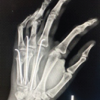 November 1, 2025 Outcome of Non-thumb Metacarpal Shaft Fractures Treated by Two Different Techniques – K Wire and Herbert Screw Fixation: A Comparative Single Centre Study