
The PENG block can be safely performed intraoperatively by orthopedic surgeons under direct vision during a posterior approach in total hip arthroplasty.
Dr. Alejandro Zylberberg, Department of Orthopaedic Surgery, Clínica Universidad de los Andes, Av. La Plaza 2501, Las Condes, Santiago 7620001, Chile. E-mail: alezylber@gmail.com
Abstract
Introduction: The pericapsular nerve group (PENG) block is an emerging technique for perioperative analgesia in hip surgery, offering effective pain control while preserving motor function. Its use has been associated with reduced opioid consumption and improved early mobilization compared to conventional blocks. Despite these advantages, practical implementation of the technique remains variable across surgical settings. This study explores the anatomical feasibility of performing the PENG block intraoperatively by the surgeon, providing a potential alternative delivery route integrated within the surgical procedure.
Materials and Methods: A cadaveric feasibility study was conducted on a fresh-frozen right lower limb from an 83-year-old male. An ultrasound-guided PENG block was first performed by an anesthesiologist using standard technique. Subsequently, a surgeon placed a second needle through a posterolateral approach under direct visualization of the anterior hip capsule. Needle positions were compared through surgical exposure through a Hueter-type anterior approach.
Results: Both needles were located in the same myofascial plane between the iliopsoas tendon and pubic ramus, with the intraoperative needle positioned <1 cm proximal to the ultrasound-guided one. No neurovascular structures were compromised.
Conclusion: This cadaveric study demonstrates that intraoperative surgeon-performed PENG block placement through a posterior approach is anatomically feasible and accurately replicates the needle position of the conventional ultrasound-guided technique. These findings support the anatomical validity of this approach; however, further in vivo studies are required to evaluate its clinical efficacy and safety.
Keywords: Pericapsular nerve group block, regional anesthesia, intraoperative anesthesia, total hip replacement, ultrasound-guided nerve block, surgeon-performed block, cadaveric study.
Total hip replacement (THR) is one of the most frequently performed orthopedic procedures, but optimal perioperative pain management remains a challenge [1]. Inadequate analgesia may lead to delayed mobilization and increased risk of thromboembolic events [2,3]. Evidence shows that multimodal analgesia, including peripheral nerve blocks (PNB), allows better control of post-operative pain in THR [1,4,5]. Femoral nerve block (FNB) is one of the most frequently performed PNBs, allowing adequate post-operative pain control and reduction in opioid consumption [6,7,8]. Its main disadvantages are the risk of neurovascular injury and quadriceps weakness due to motor blockade, which may delay ambulation [6,9,10]. In 2018, a new regional anesthesia technique for hip fractures was described [11], known as the pericapsular nerve group (PENG) block. This block targets the anterior capsule of the hip joint by anesthetizing the articular branches of the femoral, obturator, and accessory obturator nerves. This concept is supported by anatomical studies demonstrating predominant sensory innervation of the anterior and superolateral hip capsule [12,13]. The original technique is performed with an injection directed toward the anterior hip capsule under ultrasound guidance [11]. The PENG block allows rapid motor recovery, reducing the risk of falls related to quadriceps motor blockade observed with FNB [14,15]. Other studies have shown that this block may be superior to other PNBs in terms of post-operative pain reduction after THR and lower opioid consumption [14,15,16,17]. Despite its increasing clinical use, the need for ultrasound guidance limits its availability in resource-constrained settings or during high-volume surgical sessions. We hypothesize that the PENG block can be effectively performed intraoperatively under direct visualization by the surgeon, without ultrasound, using anatomical landmarks. This cadaveric study aims to compare the anatomical accuracy of a surgeon-performed intraoperative PENG block with the conventional ultrasound-guided technique. To our knowledge, this technique has not been previously described.
Institutional Review Board consent was obtained for this study. A fresh-frozen right lower extremity cadaveric specimen from an 83-year-old male, placed in the lateral decubitus position was used. First, following the technique described for the PENG block [11], a trained anesthesiologist placed a low-frequency ultrasound probe (Phillips Xperius®, 2–5 MHz) in a transverse plane over the anterior superior iliac spine and then rotated it approximately 45° clockwise. The iliopubic eminence, iliopsoas tendon, and femoral artery were identified. A first needle was inserted from lateral to medial into the myofascial plane between the iliopsoas tendon (anterior) and the pubic ramus (posterior) (Fig. 1). In a second step, a posterolateral approach to the hip was performed, and a non-cemented THR was implanted according to standard technique. The anteroinferior quadrant of the articular capsule was directly visualized, and a second needle was inserted above the shoulder of the femoral stem, oriented medially and approximately 15° distally, penetrating the articular capsule by approximately 1 cm (Fig. 2). No tracers or contrast media were injected through either needle. Subsequently, a Hueter-type anterior approach to the hip was performed to evaluate and compare the position of both needles (Fig. 3).
The ultrasound-guided needle was found to be placed as described in the original technique, in the myofascial plane between the iliopsoas tendon (anterior) and the pubic ramus (posterior). The needle placed under direct vision was placed in the same tissue layer, <1 cm proximal to the ultrasound placed needle (Fig. 3). There was no damage to any neurovascular structure by the needles.
This cadaveric study tries to answer if a PENG block intraoperatively performed from a posterolateral approach to the hip is feasible, precise, and safe. The results show that, from an anatomic point of view, it is possible and safe. Both needles were placed <1 cm away from each other. The original PENG block technique recommends a 20 cc volume injection. Diffusion of the anesthetic fluid on the myofascial plane makes this small distance irrelevant. This block may be performed by the surgeon during a THR operation. Small size clinical reports have shown the benefits of PENG block regarding post-operative pain above other PNB, such as FNB or fascia iliaca compartment block (FICB). A recent systematic review and meta-analysis, which included 15 randomized clinical trials, most of them comparing PENG block with FICB, demonstrated better results of the former in pain relief in the 1st post-operative hours and lower opioid consumption [15]. Another meta-analysis by Pai et al showed that PENG block after hip replacement surgery was associated with less pain in the first 24 h, when compared with placebo, FICB and periarticular infiltration [18]. Intraoperative blocks are frequently performed for knee surgery, especially adductor canal block, and are less frequently used in hip surgery [19]. There are few reports on intraoperative hip blocks. One of these is the psoas compartment block, which targets the branches of the lumbar plexus within the psoas muscle: The femoral nerve, the lateral femoral cutaneous nerve, and the obturator nerve. Its usefulness has been demonstrated in the direct anterior approach and in the anterolateral approach, for which it was originally described [20]. The proposed block of this study may have a wider clinical application, as the posterior approach is much more frequently used for THR than anterolateral approach. From a clinical perspective, the ability of the surgeon to perform the PENG block intraoperatively under direct visualization may offer practical advantages. These include reduced reliance on ultrasound equipment and trained anesthesiologists, streamlined operating room workflow, and the potential to facilitate effective analgesia without delaying surgery. This technique could be particularly valuable in low-resource settings or during high-volume surgical lists where time and equipment availability are constrained. This study has several limitations. Although unlikely, the extrapolation of ex vivo findings to in vivo conditions may be influenced by differences in tissue properties. Furthermore, as this was a single-specimen study, broad generalization of the findings is limited, and potential anatomical variability between individuals should be taken into account. While the anatomical landmarks targeted by the PENG block are relatively consistent, patient-specific factors, such as soft-tissue thickness or underlying hip pathology may affect intraoperative access and needle trajectory in vivo. Although needle placement was anatomically consistent with the ultrasound-guided technique in this specimen, these findings should be interpreted as a proof of concept demonstrating anatomical feasibility rather than clinical generalizability. Confirmation in larger cadaveric series and future in vivo studies is required to assess variability, reproducibility, safety, and potential clinical efficacy.
This cadaveric study demonstrates the anatomical feasibility and precision of an intraoperative PENG block performed through a posterior approach during THR. The needle placement under direct surgical visualization closely approximated that of the ultrasound-guided technique, without injury to neurovascular structures. These findings support the potential for surgeons to safely integrate intraoperative PENG block into the surgical workflow, especially in settings where ultrasound guidance is unavailable. Further studies are warranted to evaluate the clinical efficacy and reproducibility of this technique in vivo.
This cadaveric study supports the feasibility of an intraoperative PENG block performed by orthopedic surgeons during total hip arthroplasty through a posterior approach. Incorporating this technique into routine surgical practice may optimize perioperative pain management without relying on preoperative anesthesiologist-administered blocks.
References
- 1. Karam JA, Schwenk ES, Parvizi J. An update on multimodal pain management after total joint arthroplasty. J Bone Joint Surg Am 2021;103:1652-62. [Google Scholar] [PubMed]
- 2. Min BW, Kim Y, Cho HM, Park KS, Yoon PW, Nho JH, et al. Perioperative pain management in total hip arthroplasty: Korean hip society guidelines. Hip Pelvis 2016;28:15-23. [Google Scholar] [PubMed]
- 3. Tsinaslanidis G, Tsinaslanidis P, Mahajan RH. Perioperative pain management in patients undergoing total hip arthroplasty: Where do we currently stand? Cureus 2020;12:e9049. [Google Scholar] [PubMed]
- 4. Kim E, Shin WC, Lee SM, Choi MJ, Moon NH. Efficacy of pericapsular nerve group block for pain reduction and opioid consumption after total hip arthroplasty: A meta-analysis of randomized controlled trials. Hip Pelvis 2023;35:63-72. [Google Scholar] [PubMed]
- 5. Park HJ, Park KK, Park JY, Lee B, Choi YS, Kwon HM. Peripheral nerve block for pain management after total hip arthroplasty: A retrospective study with propensity score matching. J Clin Med 2022;11:5456. [Google Scholar] [PubMed]
- 6. Ilfeld BM, Mariano ER, Madison SJ, Loland VJ, Sandhu NS, Suresh PJ, et al. Continuous femoral versus posterior lumbar plexus nerve blocks for analgesia after hip arthroplasty: A randomized, controlled study. Anesth Analg 2011;113:897-903. [Google Scholar] [PubMed]
- 7. Aoyama Y, Sakura S, Abe S, Tadenuma S, Saito Y. Continuous quadratus lumborum block and femoral nerve block for total hip arthroplasty: A randomized study. J Anesth 2020;34:413-20. [Google Scholar] [PubMed]
- 8. Guay J, Johnson RL, Kopp S. Nerve blocks or no nerve blocks for pain control after elective hip replacement (arthroplasty) surgery in adults. Cochrane Database Syst Rev 2017;10:CD011608. [Google Scholar] [PubMed]
- 9. Kwofie MK, Shastri UD, Gadsden JC, Sinha SK, Abrams JH, Xu D, et al. The effects of ultrasound-guided adductor canal block versus femoral nerve block on quadriceps strength and fall risk: A blinded, randomized trial of volunteers. Reg Anesth Pain Med 2013;38:321-5. [Google Scholar] [PubMed]
- 10. Kuchálik J, Magnuson A, Lundin A, Gupta A. Local infiltration analgesia or femoral nerve block for postoperative pain management in patients undergoing total hip arthroplasty. A randomized, double-blind study. Scand J Pain 2017;16:223-30. [Google Scholar] [PubMed]
- 11. Girón-Arango L, Peng PW, Chin KJ, Brull R, Perlas A. Pericapsular nerve group (PENG) block for hip fracture. Reg Anesth Pain Med 2018;43:859-63. [Google Scholar] [PubMed]
- 12. Short AJ, Barnett JJ, Gofeld M, Baig E, Lam K, Agur AM, et al. Anatomic study of innervation of the anterior hip capsule: Implication for image-guided intervention. Reg Anesth Pain Med 2018;43:186-92. [Google Scholar] [PubMed]
- 13. Gerhardt M, Johnson K, Atkinson R, Snow B, Shaw C, Brown A, et al. Characterisation and classification of the neural anatomy in the human hip joint. Hip Int 2012;22:75-81. [Google Scholar] [PubMed]
- 14. Huda AU, Ghafoor H. The use of pericapsular nerve group (PENG) block in hip surgeries is associated with a reduction in opioid consumption, less motor block, and better patient satisfaction: A meta-analysis. Cureus 2022;14:e28872. [Google Scholar] [PubMed]
- 15. Chaudhary K, Bose N, Tanna D, Chandnani A. Ultrasound-guided pericapsular nerve group (PENG) block versus femoral nerve block for positioning during spinal anaesthesia in proximal femur fractures: A randomised comparative study. Indian J Anaesth 2023;67:913-9. [Google Scholar] [PubMed]
- 16. Farag A, Hendi NI, Diab RA. Does pericapsular nerve group block have limited analgesia at the initial post-operative period? Systematic review and meta-analysis. J Anesth 2023;37:138-53. [Google Scholar] [PubMed]
- 17. Wang Y, Wen H, Wang M, Lu M. The efficiency of ultrasound-guided pericapsular nerve group block for pain management after hip surgery: A meta-analysis. Pain Ther 2023;12:81-92. [Google Scholar] [PubMed]
- 18. Pai P, Amor D, Lai YH, Echevarria GC. Use and clinical relevancy of pericapsular nerve block (PENG) in total hip arthroplasty: A systematic review and meta-analysis. Clin J Pain 2024;40:320-32. [Google Scholar] [PubMed]
- 19. Drapeau-Zgoralski V, Bourget-Murray J, Hall B, Horton I, Dervin G, Duncan K, et al. Surgeon-performed intraoperative peripheral nerve blocks and periarticular infiltration during total hip and knee arthroplasty: A critical analysis review. JBJS Rev 2022;10:e22.00105. [Google Scholar] [PubMed]
- 20. Green C, Flannery O, Crotty J, Felle P, Harmon D, Masterson E, et al. A cadaveric study of injectate spread in the psoas compartment with a direct iliopsoas injection suggested for use during surgery. Clin Anat 2011;24:763-7. [Google Scholar] [PubMed]


Related Articles in Journal of Orthopaedic Case Reports
 June 1, 2026 Determining the Accuracy of Acetabular Cup Size using Acetate Templates on Digital Radiographs in Patients Undergoing Total Hip Replacement
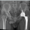 February 1, 2026 Fungal Infection of Native Hip Joint Presenting as Secondary Arthritis in 52-Year-Old Male – A Rare Case Report
 January 1, 2026 Total Hip Replacement in Fracture of Hip with Unicameral Bone Cyst – A Rare Case Report
 January 1, 2026 Harnessing 3D Printing Technology for Complex Acetabular Reconstruction in Revision Total Hip Arthroplasty: From Childhood Hip Trauma to Customized Modern Solutions