
Pre-operative radiological parameters – particularly facet angle, disc height, and lateral listhesis – are important predictors of clinical outcomes in patients undergoing lumbar interbody fusion for degenerative lumbar spondylolisthesis. Identifying these factors before surgery helps in better patient selection, surgical planning, and prediction of recovery, ultimately improving post-operative functional outcomes.
Dr. Karthik Devaraj, Department of Orthopaedics, ESIC Medical College and Hospital, Chennai, Tamil Nadu, India. E-mail: medicothedoc@gmail.com
Introduction: Degenerative lumbar spondylolisthesis (DLS) is a common spinal disorder in the ageing population, characterised by the forward displacement of a vertebral body due to degenerative changes. Lumbar interbody fusion (LIF) is the standard surgical treatment, yet outcomes remain variable, with a significant proportion of patients experiencing unfavourable results. Identifying predictive factors influencing these outcomes is critical for improving surgical planning, patient selection, and post-operative care.
Materials and Methods: An observational cohort study included 52 patients aged 45–75 years diagnosed with single-level degenerative spondylolisthesis. Patients underwent LIF, and clinical outcomes were measured using the Visual Analogue Scale (VAS) and Oswestry Disability Index (ODI). Radiological parameters, including facet angle (FA), disc height (DH), and lateral listhesis (LLS), were assessed preoperatively and postoperatively. Logistic regression and multivariate analysis were conducted to evaluate predictors of unfavourable outcomes, with significance set at P < 0.05.
Results: FA (P = 0.039), LLS (P = 0.021), and DH (P = 0.032) were significant pre-operative predictors of unfavourable outcomes. Patients with inadequate postoperative DH restoration exhibited slower recovery, while sustained DH (P = 0.012) was critical for long-term success. Favourable outcomes demonstrated significant reductions in VAS (7.0 to 2.5) and ODI (28 to 8) scores at 6 months compared to the unfavourable cohort.
Conclusion: FA, LLS, and DH are pivotal predictors of clinical outcomes in LIF for DLS. Monitoring these parameters enables personalised surgical planning and improved patient outcomes. Future studies should validate these findings in multicentre settings and explore machine learning for enhanced predictive modelling.
Keywords: Degenerative lumbar spondylolisthesis, lumbar interbody fusion, Oswestry Disability Index, facet angle, lateral listhesis.
Degenerative lumbar spondylolisthesis (DLS) is a prevalent condition in the ageing population, characterised by the forward slippage of one vertebral body over another due to degeneration of the intervertebral disc and facet joints. This condition often leads to spinal instability, nerve root compression, and associated symptoms such as chronic low back pain, radiating leg pain, and functional impairment. As the global population continues to age, the healthcare burden of DLS is expected to rise, making effective treatment strategies crucial. While lumbar interbody fusion (LIF) combined with decompression has emerged as the standard surgical approach, outcomes remain variable, with a significant proportion of patients experiencing suboptimal results [1,2]. One of the primary challenges in managing DLS is the variability in patient-reported outcomes following surgery. Despite advancements in surgical techniques, unfavourable outcomes, such as persistent pain, limited functional recovery, and dissatisfaction, remain common [3,4]. These poor outcomes not only impact the quality of life for patients but also contribute to increased healthcare costs, frequent reoperations, and strained doctor–patient relationships [5]. Understanding the factors that influence these outcomes is critical for improving patient selection, optimising surgical techniques, and tailoring postoperative care [6,7]. Tools such as the Visual Analogue Scale (VAS) for pain assessment and the Oswestry Disability Index (ODI) for evaluating functional disability have been widely adopted to quantify surgical outcomes [8,9]. However, their application has primarily focused on outcome measurement rather than outcome prediction. Identifying pre-operative and intraoperative predictors that influence unfavourable outcomes could enable clinicians to develop individualised treatment plans and set realistic expectations for patients. This study aims to address the gaps in understanding the predictors of unfavourable clinical outcomes in DLS treatment. By analysing pre-operative and post-operative parameters, including radiological and functional metrics, we seek to identify key factors contributing to poor short-term (2-week) and long-term (6-month) outcomes. Ultimately, the goal is to develop a predictive framework that can guide clinical decision-making and improve patient satisfaction, paving the way for evidence-based strategies to enhance the effectiveness of surgical interventions for DLS.
Aim:
The aim of the study is to investigate pre-operative and post-operative predictors of unfavourable outcomes in DLS surgery and assess their impact on short-term (2-week) and long-term (6-month) recovery.
Study design:
This study employed an observational cohort design to investigate the predictors of unfavourable clinical outcomes in patients undergoing LIF for DLS. The study was conducted at the Department of Orthopaedics, ESIC Medical College and PGIMSR, and Institutional Ethics Committee approval was obtained (IEC/2022/2/39); Chennai. In a total of 52 patients, the sample size was calculated using an estimated prevalence of unfavourable outcomes of 30%, with a confidence level of 95% and precision of 10%, using the formula n = Z2pq/d2. The calculated sample size was approximately 50 patients, and hence, 52 patients aged 45–75 years were included in the study based on predefined inclusion and exclusion criteria.
Inclusion criteria:
- Patients aged 45–75 years
- Diagnosis of single-level DLS (Grades I–III, Meyerding scale)
- Patients who had failed conservative management (e.g., physical therapy, non-steroidal anti-inflammatory drugs)
- Availability of complete personal data, measured parameters, and operative details
- A minimum follow-up period of 6 months postoperatively.
Exclusion criteria:
- Patients with high-grade spondylolisthesis (Grades IV and V)
- Previous invasive spinal procedures in the lumbar region
- Cases involving implant failure, multi-level decompression, or secondary surgery
- Non-degenerative causes of spondylolisthesis, including isthmic, traumatic, pathological, or dysplastic etiologies
- Cases with spinal tumors, deformities, fractures, infections, or other unrelated spine diseases
- Patients with significant systemic comorbidities classified as American Society of Anaesthesiologists III or higher.
Procedure:
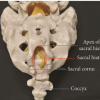
The surgical procedure for LIF was conducted under standardised conditions by the same experienced surgeon to ensure consistency. After obtaining informed consent and performing pre-operative evaluations, including radiological and laboratory assessments, patients were placed under general anaesthesia in a prone position on a radiolucent table as shown in Fig. 1. A midline incision was made to expose the spinous processes, lamina, and facet joints. Posterior decompression, including laminectomy and foraminotomy, was performed to relieve neural compression. Pedicle screws were inserted bilaterally using the intersection technique and confirmed with fluoroscopy. Following the removal of the intervertebral disc and preparation of endplates, an interbody cage (bean or bullet cage) packed with autologous bone graft was inserted into the disc space. Transforaminal LIF (TLIF) was the preferred technique to minimise neural injury. Rods were secured to the pedicle screws, and compression was applied to restore disc height (DH) and alignment. After securing stability, the wound was closed in layers to ensure a watertight seal. Postoperatively, patients received antibiotics and DVT prophylaxis, with mobilisation starting on day 1 and assisted walking by day 4 as shown in Fig. 2. Follow-ups were conducted at 2 weeks, 3 months, and 6 months to assess clinical and radiological outcomes, including VAS and ODI scores and fusion status. No intraoperative complications were observed, and postoperative care was tailored to facilitate recovery and prevent complications.
Post-operative outcomes were evaluated at two key time points:
- Short-term outcomes (2 weeks postoperatively)
- Long-term outcomes (6 months postoperatively).
Radiological evaluations were performed at the same intervals to measure changes in facet angle (FA), DH, and lateral listhesis (LLS). Surgical details, such as the fusion method (PLIF/TLIF), pedicle screw size, and bone graft type, were documented for all cases. Osteoporosis was diagnosed using dual-energy X-ray absorptiometry. A T-score of ≤ −2.5 at the lumbar spine or femoral neck was considered diagnostic of osteoporosis.
Statistical analysis:
Statistical analyses were performed using the Statistical Package for the Social Sciences (SPSS) (SPSS, version 17.0). Continuous variables were summarised as mean ± standard deviation, while categorical variables were presented as frequencies and percentages. Comparisons between groups: Independent sample t-tests or Mann–Whitney U-tests were used for comparing continuous variables between favourable and unfavourable outcome groups. Paired t-tests or Wilcoxon signed-rank tests were applied for within-group comparisons. Chi-square tests were used for categorical variables. Predictive modelling: Logistic regression analysis was performed to identify significant predictors of unfavourable outcomes. Key parameters such as pre-operative FA, DH, and LLS were assessed as independent variables. Multivariate regression analysis was used to evaluate their combined impact on outcomes. Threshold for significance: a P < 0.05 was considered statistically significant for all tests.
The study included 52 patients diagnosed with DLS, aged between 45 and 75 years. The average age was 53.25 years, with the highest proportion of patients in the 55–65 age range. Females were the predominant demographic, with 40 female patients compared to 12 males, reflecting a 3:1 female-to-male ratio. Osteoporosis was a significant comorbidity, observed in 63.5% of patients, further highlighting the vulnerability of older female patients to degenerative spine conditions (Fig. 3). A histogram showing the age distribution of patients reveals a clustering of cases in the middle-aged and elderly population, emphasising the progressive nature of degenerative spinal diseases. A pie chart illustrates the female predominance in the cohort, aligning with known epidemiological patterns for DLS.
Predictive factors:
FA: Pre-operative FA emerged as a critical predictor of unfavourable short-term outcomes (p = 0.039). Patients with higher FAs showed greater axial load-bearing, leading to prolonged recovery times. LLS: Lateral displacement of vertebral bodies (LLS) was another significant predictor. Patients with pre-operative LLS experienced more instability and degenerative changes, affecting both short-term and long-term outcomes. DH: Reduced pre-operative DH correlated with worse clinical outcomes, likely due to limited intervertebral space and increased compressive forces (Table 1).
Short-term versus long-term predictors:
Short-term (2-week) outcomes were heavily influenced by immediate post-operative factors, including post-operative DH and early restoration of spinal alignment. Long-term (6-month) outcomes, however, were more dependent on sustained DH (P = 0.012) and stabilisation of FAs (Table 2).
Outcomes: VAS and ODI scores:
Patients were grouped into favourable and unfavourable cohorts based on improvements in the VAS and ODI scores. The unfavourable group exhibited slower improvement in pain and functional disability at both 2-week and 6-month follow-ups. Favourable patients demonstrated significant reductions in VAS (from 7.0 preoperatively to 2.5 at 6 months) and ODI (from 28 preoperatively to 8 at 6 months), highlighting the importance of identifying predictive factors early in treatment planning (Figs. 4 and 5).
The study demonstrates a strong correlation between pre-operative predictors and clinical outcomes in patients undergoing LIF for DLS. Pre-operative FA, DH, and LLS emerged as critical predictors of unfavourable outcomes. A significant pre-operative FA (P = 0.039) was associated with increased axial loads on the lumbar spine, contributing to delayed recovery and persistent symptoms in some patients [1]. Similarly, reduced DH (P = 0.032) highlighted the degenerative changes affecting intervertebral spaces, which are crucial for load-bearing and spinal stability [4]. LLS (P = 0.021), indicative of vertebral instability, was another important factor linked to both short-term and long-term unfavourable outcomes [8]. Collectively, these predictors provided valuable insights into the structural and biomechanical factors influencing surgical outcomes and long-term patient recovery. Short-term (2-week) outcomes were primarily influenced by immediate postoperative parameters, such as postoperative DH and spinal alignment restoration. Patients who achieved adequate DH restoration post-surgery experienced faster reductions in pain and improved functionality, as measured by the VAS and ODI [5]. Importantly, sustained improvements in DH at 6 months (P = 0.012) emerged as a critical factor for long-term success, further validating the importance of optimising this parameter during surgery [2]. Early identification of these predictors allows clinicians to plan personalised interventions and closely monitor high-risk patients, potentially mitigating unfavourable outcomes. Emerging studies utilising deep learning and predictive modelling techniques have reinforced the importance of radiological parameters, such as DH and FA, in determining clinical outcomes [7]. Dong et al. highlighted the value of machine learning algorithms in identifying predictors for unfavourable outcomes in spinal fusion, with DH and axial load distribution emerging as pivotal factors [10]. These findings align with the results of this study, where multivariate regression analysis identified pre-operative DH and FA as statistically significant predictors of recovery. Similar to the findings of Ghogawala et al., the significance of the pre-operative FA was underscored as a determinant of spinal stability and short-term recovery [5]. However, unlike previous studies, this research specifically links higher FAs with delayed functional improvements, emphasising its importance as a biomechanical factor influencing axial load-bearing capacity. This study provides unique insights by demonstrating the combined impact of preoperative LLS and DH on long-term outcomes. While previous research has largely focused on individual predictors, this study offers a holistic view by integrating multiple radiological and clinical parameters into a predictive framework. In addition, the inclusion of 2-week outcomes as a benchmark for early recovery distinguishes this study from others, offering a practical timeline for assessing initial surgical success. The identification of pre-operative predictors has direct implications for patient selection and counselling. Patients with high FAs, significant LLS, or reduced DH can be flagged as high-risk candidates for unfavourable outcomes [10]. Clinicians can use this information to set realistic expectations, discuss potential risks, and emphasise the importance of adherence to postoperative rehabilitation protocols. This proactive approach enhances patient satisfaction and trust. The findings highlight the importance of achieving optimal restoration of DH and spinal alignment during surgery. Surgeons can use pre-operative imaging to identify structural abnormalities and tailor their surgical techniques accordingly [10]. For instance, patients with significant LLS may benefit from additional stabilisation measures, such as pedicle screw fixation and larger interbody cages [11]. Close monitoring of high-risk patients during the early post-operative period (2 weeks) is crucial for identifying signs of unfavourable outcomes. Regular imaging and functional assessments can help clinicians evaluate progress and implement timely interventions, such as physical therapy or additional surgical adjustments, if necessary [12]. As a single-centre study, the findings may not be generalisable to broader patient populations. Variations in surgical techniques, patient demographics, and healthcare infrastructure could influence the applicability of these results in other settings. Future studies involving multicentre collaborations are essential to validate these findings across diverse populations [13]. The relatively small sample size (52 patients) limits the statistical power of the study. Although significant correlations were identified, larger sample sizes are necessary to confirm the robustness of these predictors and to explore additional factors that may influence outcomes [14]. The study’s 6-month follow-up period provides valuable insights into short-term and intermediate outcomes but does not capture long-term complications, such as adjacent segment disease or implant failure. Future studies should include extended follow-up periods to assess the durability of surgical outcomes and the development of late-stage complications [15]. The integration of advanced imaging techniques and machine learning algorithms in future research could further enhance the predictive accuracy of clinical and radiological parameters. For instance, automated segmentation of magnetic resonance imaging and computed tomography images could streamline the identification of predictors, while deep learning models could offer personalised risk stratification for patients [16].
Pre-operative radiological parameters – FA, DH, and LLS – are significant predictors of unfavourable outcomes in patients undergoing LIF for DLS. These factors influence both short-term and long-term recovery, highlighting their importance in optimising surgical planning and improving clinical outcomes.
Careful pre-operative evaluation of facet angle, disc height, and lateral lysthesis is essential in patients undergoing lumbar interbody fusion for degenerative spondylolisthesis. Identifying these factors can help predict unfavourable outcomes and guide surgical planning to achieve optimal disc height restoration and spinal stability, thereby improving patient recovery.
References
- 1. </p> [Google Scholar] [PubMed]
- 2. <ol> [Google Scholar] [PubMed]
- 3. <li>Hammerberg KW. New concepts on the pathogenesis and classification of spondylolisthesis. Spine (Phila Pa 1976) 2005;30 6 Suppl:S4-11.</li> [Google Scholar] [PubMed]
- 4. <li>Pan W, Zhao JL, Xu J, Zhang M, Fang T, Yan J, <em>et al</em>. Lumbar alignment and patient-reported outcomes after single-level transforaminal lumbar interbody fusion for degenerative lumbar spondylolisthesis with and without local coronal imbalance. J Neurosurg Spine 2020;34:464-70.</li> [Google Scholar] [PubMed]
- 5. <li>Zhong W, Liang X, Luo X, Huang T, Quan Z. Complications rate of and risk factors for the unplanned reoperation of degenerative lumbar spondylolisthesis in elderly patients: A retrospective single-centre cohort study of 33 patients. BMC Geriatr 2020;20:301.</li> [Google Scholar] [PubMed]
- 6. <li>Försth P, Ólafsson G, Carlsson T, Frost A, Borgström F, Fritzell P, <em>et al</em>. A randomized, controlled trial of fusion surgery for lumbar spinal stenosis. N Engl J Med 2016;374:1413-23.</li> [Google Scholar] [PubMed]
- 7. <li>Ghogawala Z, Dziura J, Butler WE, Dai F, Terrin N, Magge SN, <em>et al</em>. Laminectomy plus fusion versus laminectomy alone for lumbar spondylolisthesis. N Engl J Med 2016;374:1424-34.</li> [Google Scholar] [PubMed]
- 8. <li>Xi X, Zeng Z, Li F, Wang C, Ma B, Xie N, <em>et al</em>. Caudad insertion of pedicle screws facilitates interbody distraction during spondylolisthetic vertebrae restoration: A retrospective study. Pain Ther 2021;10:1537-50.</li> [Google Scholar] [PubMed]
- 9. <li>Shengtao D<em>.</em> Predictive modeling for poor clinical outcomes in spinal fusion using machine learning. Spine J 2020;10:1544-58.</li> [Google Scholar] [PubMed]
- 10. <li>Blumenthal C, Curran J, Benzel EC, Potter R, Magge SN, Harrington JF Jr., <em>et al</em>. Radiographic predictors of delayed instability following decompression without fusion for degenerative grade I lumbar spondylolisthesis. J Neurosurg Spine 2013;18:340-6.</li> [Google Scholar] [PubMed]
- 11. <li>Zhou Q, Zhang JX, Zheng YF, Teng Y, Yang HL, Liu H, <em>et al</em>. Effects of different pedicle screw insertion depths on sagittal balance of lumbar degenerative spondylolisthesis, A retrospective comparative study. BMC Musculoskelet Disord 2021;22:850.</li> [Google Scholar] [PubMed]
- 12. <li>Dong F<em>.</em> Deep learning-assisted risk stratification in spinal surgery. J Orthop Res 2021;39:689-97.</li> [Google Scholar] [PubMed]
- 13. <li>Mummaneni PV, Whitmore RG, Curran J Degenerative lumbar spondylolisthesis: Trends in surgery and patient outcomes. Neurosurg Focus 2013;35:E2.</li> [Google Scholar] [PubMed]
- 14. <li>Lee CH, Chung CK, Kim CH, Kwon JW. Health care burden of spinal diseases in the elderly in South Korea: An analysis of the National Database 2008-2012. Acta Neurochir (Wien) 2016;158:989-96.</li> [Google Scholar] [PubMed]
- 15. <li>Auerbach JD, Tamai K, Chan D. Adjacent segment degeneration and disease following lumbar fusion. J Am Acad Orthop Surg 2016;24:365-74.</li> [Google Scholar] [PubMed]
- 16. <li>Radcliff KE, Kepler CK, Jakoi A, Sidhu GS, Rihn J, Vaccaro AR, <em>et al</em>. Adjacent segment disease in the lumbar spine following different treatment interventions. Spine J 2013;13:1339-49.</li> [Google Scholar] [PubMed]
- 17. <li>Di Martino A, Russo F, Denaro V. Adjacent segment disease after lumbar fusion: A systematic review. Int J Spine Surg 2021;15:9-17.</li> [Google Scholar] [PubMed]
- 18. <li>Sharma A, Vaccaro AR, Lim MR. Emerging technologies for lumbar spinal surgery: Robotics and navigation. Spine J 2020;20:1325-33.</li> [Google Scholar] [PubMed]
- 19. </ol> [Google Scholar] [PubMed]
- 20. <p> [Google Scholar] [PubMed]


Related Articles in Journal of Orthopaedic Case Reports
 March 1, 2026 Caudal Epidural Steroid Injection in adults with Chronic Lower Backache: Comparison of Landmark-Guided Technique and Ultrasonography-Guided Technique
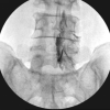 October 1, 2025 Effectiveness of Epidural Methylprednisolone Injection in Management of Lumbar Prolapsed Intervertebral Disc: A Comparison of Caudal, Transforaminal and Interlaminar Routes
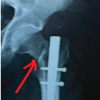 March 1, 2026 Management of a Missed Iatrogenic Fracture Neck of Femur in an Operated Case of Fracture Shaft Femur- Case Report
 April 1, 2025 Beyond Hemostasis: Exploring Intra-articular Tranexamic Acid’s Analgesic Effect in Post-operative Care in Total Knee Arthroplasty – Original Article