


Introduction
Popliteal artery aneurysms (PAAs) presents as the most frequently encountered peripheral arterial aneurysms, accounting for approximately 67% of all such cases in clinical practice [1]. The typical demographic for this pathology mainly consists of males over the age of 65, with systemic atherosclerosis acting as the primary underlying etiology [2]. Due to this gradual atherosclerotic progression, true PAAs are exceptionally rare among younger populations. When an aneurysm or pseudoaneurysm presents in a patient under the age of 40, it universally indicates an atypical etiology, such as localized vascular trauma, congenital connective tissue disorders, or mycotic vessel manifestations [3].
In terms of recent years, the clinical profile of young patients presenting with severe, acute vascular complications has shifted significantly. This change is largely driven by the rising incidence of intravenous (IV) drug abuse, a trend increasingly documented in specific geographic regions, including Northern India [4]. In this demographic, repeated and inadvertent arterial injections of illicit drugs and their diluents can lead to direct endothelial trauma, periarterial extravasation, and hematoma formation. Later bacterial contamination of these hematomas rapidly erodes the arterial wall, resulting in the formation of infected mycotic pseudoaneurysms [5]. While the femoral artery stays the most common site of injury due to its accessibility, peripheral vessels like the popliteal artery are frequently implicated as proximal veins become sclerosed from chronic abuse [6].
The rupture of a popliteal aneurysm is a profound clinical emergency characterized by limb-threatening ischemia and potential exsanguination. While the stable PAAs are primarily known for causing distal thromboembolism, an acute rupture can alters the patient’s prognosis; literature indicates that delayed or missed diagnoses of ruptured PAAs can lead to amputation rates as high as 100% [7]. In the context of IV drug abuse, the presence of localized tissue necrosis, soft-tissue infection, and systemic bacteremia can severely complicate the standard revascularization efforts [8].
This case report describes the successful surgical management of a ruptured PAA in a 35-year-old male with a history of chronic IV drug use. It highlights the need for a high index of clinical suspicion, rapid diagnostic evaluation, and decisive operative intervention to prevent ischemia and achieve limb salvage in this high risk demographic.
Case Report
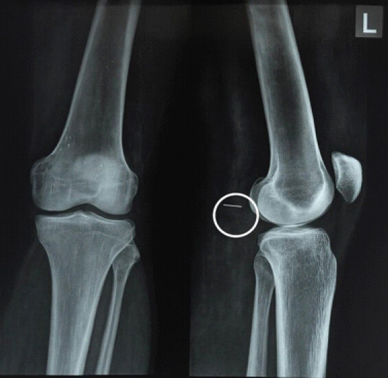
A 35-year-old male presented to the emergency department with a 1-week history of progressive pain and swelling in the left popliteal fossa. The patient had a known history of chronic IV heroin use, specifically reporting repeated self-injections into the left popliteal region. He denied any recent acute trauma or constitutional symptoms such as fever or weight loss. Physical examination revealed a firm, tender, and pulsatile mass in the left popliteal fossa. Distal pulses (dorsalis pedis and posterior tibial) were significantly diminished compared to the contralateral limb, and the leg was cool to the touch distal to the knee. Sensory and motor functions were intact, and there were no signs of compartment syndrome. Initial radiographic evaluation included anteroposterior and lateral X-ray views of the left knee. Notably, the lateral view revealed a retained foreign body appearing to be a needle in the soft tissues of the popliteal region, correlating with the patient’s history of self-injection (Fig. 1).
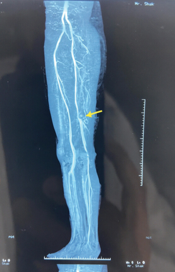
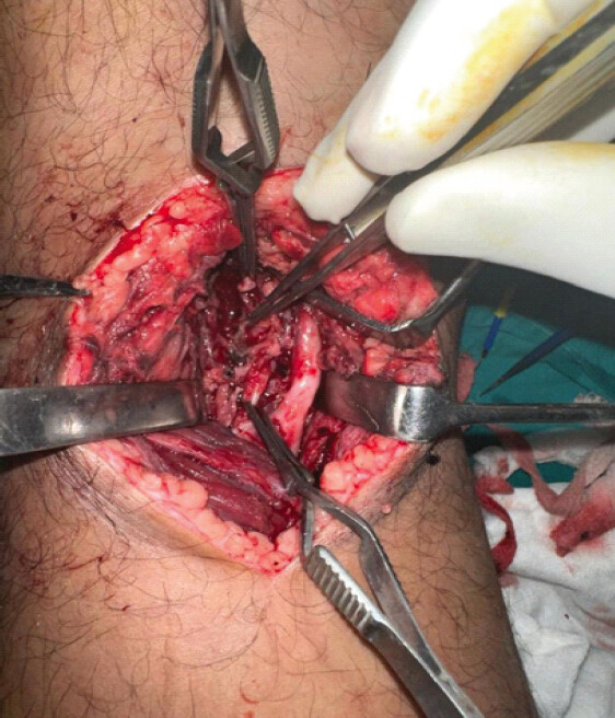
Color Doppler ultrasonography of the left lower extremity showed normal venous flow but revealed dampened, mono-phasic arterial waveforms in the posterior tibial artery and biphasic flow with reduced velocity in the popliteal artery. These findings indicated a significant proximal arterial lesion. Subsequent contrast-enhanced computed tomography angiography (CTA) confirmed a large PAA measuring 5.8 × 3.0 cm (Fig. 2). The CTA demonstrated active contrast extravasation into a surrounding hematoma, confirming a rupture and significant luminal compression. The patient was taken for urgent surgical intervention. Utilizing a posterior approach, the popliteal artery was exposed. Intraoperative findings confirmed a true aneurysmal sac with a focal wall rupture and an associated large, compressive hematoma (Fig. 3). Following systemic heparinization, the diseased aneurysmal segment was resected. Due to the length of the resulting arterial defect, a primary anastomosis was not feasible. Therefore, a reverse saphenous vein graft (RSVG) was harvested from the ipsilateral limb to reconstruct the popliteal artery. To protect the vascular repair and prevent mechanical stress on the graft during the early healing phase, an across knee external fixator was applied with the knee maintained in 30° of flexion. Local debridement of the inflamed tissue, hematoma, and foreign body (needle) was completed. Total tourniquet time was 45 min. The post-operative recovery was uneventful. The patient received a brief course of heparin and was transitioned to enoxaparin. The external fixator provided stable immobilization, and distal pulses remained palpable and symmetric throughout the admission. At the 6-week follow-up, a repeat angiography confirmed excellent potency of the RSVG with no signs of stenosis or leakage. Consequently, the external fixator was removed, and the patient began a structured physical therapy program for knee range of motion. He was further advised to cease the IV drug to use to prevent recurrence or other vascular insults. Relevant substance use treatment was started as a part of his care plan.
Discussion
PAAs are predominantly degenerative and atherosclerotic, typically occurring in males over the age of 60. Their manifestation in a 35-year-old is exceptionally rare, as this demographic lacks the systemic atherosclerotic risk profile associated with traditional aneurysmal disease [9]. While trauma and mycotic infections are known causes in younger populations, this case highlights a distinct etiology repetitive arterial wall trauma from chronic IV heroin injection. Clinically, PAAs often remain asymptomatic until complications such as thrombosis or rupture occur. In our patient, the pulsatile mass and diminished distal pulses were classic signs of a vascular emergency. While Duplex ultrasound serves as an excellent initial screening tool, contrast-enhanced CTA remains the gold standard for surgical planning [10]. It accurately delineates the aneurysm’s dimensions, the site of rupture, and the status of the distal run off vessels [10]. The management of a ruptured PAA is strictly surgical. In this case, the extent of the arterial wall destruction required resection rather than simple repair. While primary end-to-end anastomosis is ideal, the resulting gap often requires interposition grafting. We used an autologous RSVG, which remains the gold standard for peripheral arterial reconstruction, especially in potentially contaminated fields common in IV drug users. Unlike prosthetic grafts, autologous veins offer superior potency rates and a higher resistance to infection [11]. Furthermore, the application of an across knee external fixator in 30 degrees of flexion was a critical adjunct. In the popliteal region, extreme extension or unpredictable joint movement can jeopardize the integrity of a fresh venous graft. Temporary rigid immobilization ensures a tension-free environment for the anastomosis to mature. Our protocol of maintaining fixation until patency was confirmed through angiography at 6 weeks allowed for a safe transition to mobilization without compromising the vascular reconstruction.
Conclusion
PAAs in young adults are rare and should prompt immediate investigation into atypical etiologies, such as localized trauma from IV drug abuse. Early clinical suspicion and advanced imaging (CTA) are essential for definitive surgical planning. Prompt surgical resection followed by RSVG and temporary stabilization with an across-knee external fixator is an effective strategy for limb salvage. This comprehensive approach restores distal perfusion while protecting the vascular repair in the high-mobility popliteal zone.
Clinical Message
A pulsatile popliteal mass with diminished distal pulses in a young intravenous drug user must be regarded as a vascular emergency until proven otherwise, as a ruptured popliteal artery aneurysm carries a limb threatening and also potentially life threatening prognosis. It is necessary to do a prompt contrast- enhanced CT angiography which is essential to define the lesion and plan reconstruction accordingly. In the contaminated high mobility popliteal zone, aneurysmal resection with an autologous reverse saphenous vein graft, protected by across knee external fixator provides a durable & infection resistance limb salvage. Additionally, addressing the underlying substance use is integral to prevent recurrence of such events.
Conflict of Interest:
Source of Support:
Nil
Consent:
The authors confirm that informed consent was obtained from the patient for publication of this article
References
- Dawson I, Sie RB, Van Bockel JH.Atherosclerotic popliteal aneurysm. Br J Surg 1997;84:293-9. [Google Scholar] | [PubMed]
- Michaels JA, Galland RB.Management of asymptomatic popliteal aneurysms: The use of a Markov decision tree to determine the criteria for a conservative approach. Eur J Vasc Surg 1993;7:136-43. [Google Scholar] | [PubMed]
- Vogel TR, Dombrovskiy VY, Carson JL, Haser PB, Lowry SF, Graham AM.Infectious complications after elective vascular surgical procedures. J Vasc Surg 2010;51:122-9. [Google Scholar] | [PubMed]
- Saini NS, Luther A, Mahajan A, Joseph A.Infected pseudoaneurysms in intravenous drug abusers: Ligation or reconstruction?. Int J Appl Basic Med Res 2014;4 Suppl 1:S23-6. [Google Scholar] | [PubMed]
- Padberg F Jr, Hobson R 2nd, Lee B, Anderson R, Manno JE, Breitbart G.Femoral pseudoaneurysm from drugs of abuse: Ligation or reconstruction?. J Vasc Surg 1992;15:642-8. [Google Scholar] | [PubMed]
- Barbetta I, Van Den Berg JC.Access and hemostasis: Femoral and popliteal approaches and closure devices-why, what, when, and how?. Semin Intervent Radiol 2014;31:353-60. [Google Scholar] | [PubMed]
- Sie RB, Dawson I, van Baalen JM, Schultze Kool LJ, Van Bockel JH.Ruptured popliteal artery aneurysm. An insidious complication. Eur J Vasc Endovasc Surg 1997;13:432-8. [Google Scholar] | [PubMed]
- Samarakoon LB, Ho DC, Tan YK, Kum SW, Lim DM.Infected femoral pseudoaneurysms in intravenous drug abusers: A decade of experience from a Singapore tertiary centre. Singapore Med J 2021;62:135-8. [Google Scholar] | [PubMed]
- Jadhav S, Sanagar S, Kaushik S, Munde H.Giant popliteal artery aneurysm in a teenager: An unusual occurrence. Korean J Thorac Cardiovasc Surg 2019;52:116-8. [Google Scholar] | [PubMed]
- Rief M, Rief A, Bornemann-Cimenti H, Rief P.Idiopathic pseudoaneurysm of the popliteal artery with endovascular treatment: A case report. Radiol Case Rep 2023;18:3336-40. [Google Scholar] | [PubMed]
- Varothayan S, Vinojan S, Dhadchayini R, Gobinath S, Shathana P.Case report: Retrogenicular popliteal artery pseudoaneurysm following trivial knee hyperextension. Int J Surg Case Rep 2024;124:110439. [Google Scholar] | [PubMed]
© 2026 Journal of Orthopaedic Case Reports - Published by Indian Orthopaedic Research Group
About the Authors
-

Dr. Tushar Jethi
-

Dr. Shikhar Bindal
-

Dr. Rishabh Guliani
-

Dr. Rajnish Garg
-

Dr. Tarkik Thami
Related Articles in Journal of Orthopaedic Case Reports
 May 1, 2026 Functional Outcomes and Complications of Megaprosthetic Reconstruction in Limb-Salvage Surgery for Musculoskeletal Tumors: A Scoping Review
 June 10, 2022 Cutaneous Actinomycosis of Popliteal Fossa: A Rare Case Report
 June 19, 2021 Outcomes of Tumor Resection and Reconstruction by Megaprosthesis around Knee: Our Experience
 February 10, 2021 Secondary Erosive Arthritis in a Young Lady – A Rare Manifestation of Primary Antiphospholipid Antibody Syndrome