


Introduction
Angiomatoid fibrous histiocytoma (AFH) is a soft-tissue neoplasm of low malignant potential, typically involving the superficial soft-tissue of the extremities in children [1]. The World Health Organisation has classified it as a tumor of uncertain differentiation with moderate biological potential and an incidence of 0.3% of all soft-tissue neoplasms [2]. It is a spindle cell or epithelioid tumor surrounded by sleeves of lymphoid tissue in the following areas: Antecubital fossa, axilla, inguinal region, and supraclavicular region [3,4]. It is considered to arise from pluripotent stem cells involving the fusion of the Ewing Sarcoma breakpoint Region 1 gene (EWSR1) and either the cAMP Response Element Binding protein 1 (CREB1) or Activating Transcription Factor 1 (ATF1) gene. An association with other diseases such as human immunodeficiency virus, neuroblastoma, testicular tumors, and Hodgkin’s lymphoma has been reported.
The appearance of AFH on plain radiograph is non-specific: A mass with cystic areas, pseudocapsule, and internal fluid-fluid levels [5]. However, the occurrence of fluid-fluid levels is non-specific, as it may occur in other lesions, such as soft-tissue hemangioma, hematoma, and malignant fibrous histiocytoma. In the reported cases of AFH with soft-tissue involvement, these lesions were indistinguishable from other bone lesions merely on the basis of radiological appearance.
Hereditary spherocytosis (HS) is a common type of hemolytic anemia that can present as intermittent jaundice and splenomegaly. The soft-tissue lymph node swelling in HS can mimic extramedullary hematopoiesis after splenectomy [6]. There are reports of the occurrence of non-Hodgkin lymphoma and chronic myeloid myelocytic leukemia with HS [7,8]. This is a rare case of AFH involving the soft tissue of the shoulder in a post-splenectomy patient with HS; no direct association has been documented previously. Parental consent was taken for the use of patient’s data, radiographic images, and clinical details of the patient.
Case Report
A male child in early adolescence presented with a 2-month history of right shoulder pain and swelling. The swelling progressed from pea-sized to the size of a cricket ball and was painful, with pain exacerbated by shoulder movement. He had a low-grade fever for 5 days, which resolved with medication. There were no systemic symptoms such as loss of appetite, weight loss, trauma, generalized joint involvement, or morning stiffness.
On examination, a single, firm, tender, fluctuant, lobulated swelling measuring 10 × 6 cm was noted in the right periscapular region. It had well-defined margins, raised overlying skin temperature, and no neurovascular deficit.
The patient was born full-term through cesarean section, with a birth weight of 2.6 kg, and was treated in the Pediatric Intensive Care Unit for 7 days for neonatal jaundice. He had a history of multiple transfusions from age four for undiagnosed anemia. Eight months prior, he presented with pallor, jaundice, generalised weakness, intermittent breathlessness, and abdominal distension. He was diagnosed with HS and underwent splenectomy for massive splenomegaly.
Investigations
-
-> Laboratory studies revealed leukocytosis (15,500/mm3) and elevated C-reactive protein (152 mg/L)
-
-> A plain radiograph did not reveal any lytic or sclerotic lesion
-
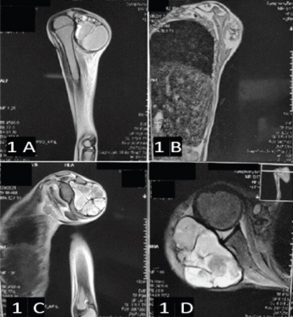
-> Magnetic resonance imaging (MRI) revealed a multiloculated lesion with T1 hypointensity and T2 hyperintensity, surrounded by a pseudocapsule – features suggestive of a complex cystic neoplasm or hematoma (Fig. 1)
-
-> A bone scan ruled out an infectious pathology
-
-> Fine-needle aspiration was inconclusive. No organisms grew on the culture.
Differential diagnosis
-
-> AFH
-
-> Synovial sarcoma
-
-> Malignant peripheral nerve sheath tumor (MPNST)
-
-> Hydatid cyst
-
-> Pseudotumor
-
-> Extramedullary hematopoiesis.
Table 1 compares the features of the differential diagnosis.
Features differential diagnosis of AFH
| Feature | Angiomatoid fibrous histiocytoma | Synovial sarcoma | Malignant peripheral nerve sheath tumor | Hydatid cyst |
|---|---|---|---|---|
| Etiology | Neoplastic Ewing sarcoma breakpoint region 1- Element Binding protein 1 gene fusion | Malignant–t(X;18) translocation (SYT-SSX fusion) | Malignant-often associated with Neurofibromatosis Type 1 (NF1) | Parasitic infection (Echinococcus) |
| Clinical features | Slow-growing, painless mass in children and adolescents in extremities. | Slow-growing mass in adolescent and adults near large joints, initially painless later can present with pain. | Rapidly growing painful mass near nerve trunks in adults, associated with numbness and parasthesia | Any age patient living in endemic region of echinococcus±allergic reactions secondary to rupture of cyst. |
| MRI Findings | Well-circumscribed, multiloculated, T1 hypointense, T2 hyperintense with fluid-fluid levels, thick pseudocapsule, internal septations | Heterogeneous with cystic/solid areas and septations; may calcify; possible hemorrhage | Ill-defined, heterogeneous; necrosis; fusiform shape along nerve trajectory | Multiloculated cyst with daughter cysts, T2 hyperintense with hypointense rim (“hydatid sand”). |
| Histopathology | Pseudoangiomatous blood-filled cystic spaces, fibrous pseudocapsule, spindle-epithelioid cells, lymphoid cuff, hemosiderin | Monophasic (spindle cells) or biphasic (spindle+epithelial); glandular or cystic patterns possible | Hypercellular spindle cells, high mitoses, necrosis, and alternating hypo- and hypercellular areas | Laminated ectocyst and germinal endocyst; surrounding inflammatory cells with eosinophils |
Treatment
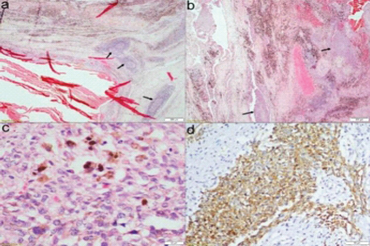
An open excisional biopsy with wide resection was performed. Intraoperatively, a well-encapsulated mass was identified and excised in total. Histopathology revealed multiple blood-filled cystic spaces lined by spindle and epithelioid cells with hemosiderin and lymphocytic infiltrate, with tumor-free margins, suggestive of AFH. Immunohistochemistry showed epithelial membrane antigen (EMA+), desmin+, and cluster of differentiation (CD)99 (focal)+, consistent with AFH (Fig. 2).
Outcome and follow-up
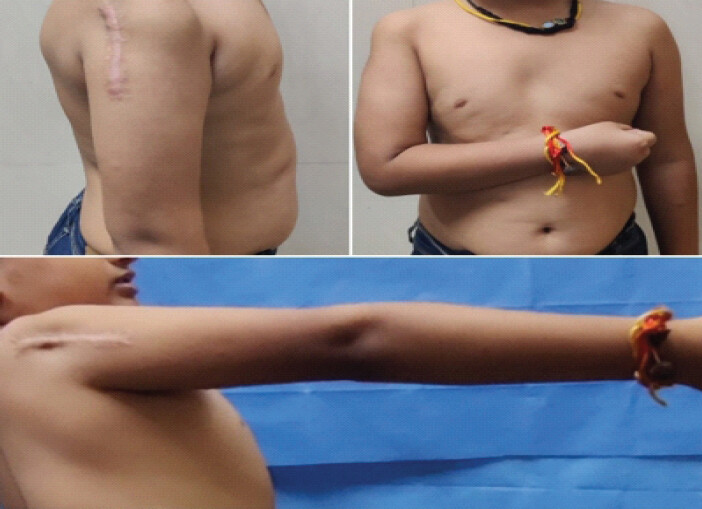
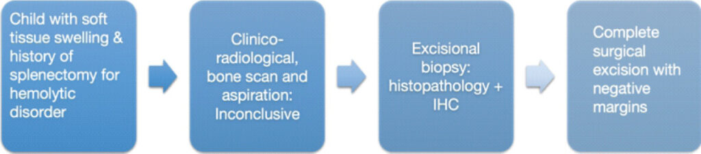
At a 3-year follow-up, the patient had no recurrence and regained moderate shoulder mobility. He had the following range of motion in his shoulder: Forward flexion 70°, extension 30°, abduction 90°, adduction 20°, external rotation 80°, and internal rotation 90° (Fig. 3). He had a Musculoskeletal Tumor Society score-93 of 66.67 % and a Quick Disability of Arm Shoulder and Hand score of 20.5% at the 3 years follow-up. An algorithm for the evaluation and management of soft-tissue swelling in post-splenectomy patients with hemolytic anemia is presented in Flow Chart 1.
Discussion
AFH is a rare soft-tissue neoplasm first described by Enzinger in 1979 while studying 42 cases of unusual fibro-histiocytic sarcoma, in which the median age was 13 years [9]. It is a unique tumor that blends clinical, radiologic, and histologic ambiguity. Its imaging appearance often suggests hematomas, benign vascular lesions, cystic-necrotic lesions, and calcifying lesions [10]. Radiological mimicry of an infectious etiology frequently confuses the diagnosis, emphasizing the need for tissue biopsy [11]. In our case, we considered infective foci as differential diagnosis due to raised inflammatory markers after splenectomy, while MRI suggested a cystic collection or pseudotumor.
Histologically, AFH is characterized by pseudoangiomatous spaces, fibrous pseudocapsule, chronic inflammatory infiltrate, and hemosiderin-laden macrophages [12]. Immunohistochemistry typically demonstrates EMA and desmin positivity, with variable CD99, and is negative for S100 and cytokeratin, helping differentiate it from MPNST, rhabdomyosarcoma, and synovial sarcoma [3]. In our case, aspiration was inconclusive, but histopathological features of spindle and epithelioid cells with hemosiderin and lymphocytic infiltrate were suggestive of AFH. Immunohistochemistry staining supplemented the diagnosis by EMA+, desmin+, and CD99 (focal)+. Gene fusion testing can provide diagnostic confirmation, especially in ambiguous cases, though it was not available in our setting. Recent reviews emphasize the utility of next-generation sequencing or reverse transcription polymerase chain reaction for EWSR1-CREB1 or ATF1 fusions [13]. Surgery is the mainstay of management, with an excellent prognosis when primary complete excision is achieved [14]. While AFH is mostly indolent, it carries a local recurrence risk of 15–20% and a rare metastatic potential of 1–3% [15]. Adjuvant radiotherapy or chemotherapy may be considered when excision is incomplete or in the presence of distant metastasis [16,17]. Wide local excision with negative margins remains the cornerstone of treatment. We did not observe local or distant recurrence at the 3 years of follow-up.
HS is a common cause of hemolytic anemia in children and results from disruption of the red blood cell membrane. The development of a swelling could be due to an extramedullary hematopoiesis in a splenectomized patient with HS [18,19]. The prolonged erythropoietin stimulation might play an etiological role in some of these patients. These tumors contain a more copious fat component than usual hematopoietic tumors. Extramedullary hematopoiesis was reported in various parts of the body in HS, ranging from lymph nodes, pleura, and mediastinum to adrenal glands and liver. Other tumors have also been reported in HS, including B-cell lymphoma, acute lymphoblastic leukemia (ALL), and pancreatic schwannoma [20,21,22]. Our case was unique because the soft-tissue mass adjacent to the scapula that could be confused with extramedullary hematopoiesis. It could be postulated that erythropoietin played some role in the development of the tumor in this patient. Our case is unique for its co-occurrence with HS and post-splenectomy. While previous reports describe HS patients developing hematologic malignancies such as acute myeloid leukemia (AML) or chronic myeloid leukemia (CML) [8,23], no association with AFH has been described to date. The patient’s chronic erythroid stress and post-splenectomy immune modulation could theoretically create a permissive microenvironment for mesenchymal transformation, though this remains speculative.
There have been reports suggesting an association between AFH and leukemia, lymphoma, and platelet disorders [8,23,24,25]. Given that a soft-tissue tumor has been linked to two blood cell lineages, it is worth considering whether a similar association could exist with the third lineage- erythrocytes. In our case, we noted a connection between AFH and a condition involving red blood cells, though this observation remains preliminary and requires further investigation. This case thus expands the clinical spectrum of AFH and raises the possibility of a link between chronic hematologic stress and mesenchymal tumorigenesis, warranting further research.
Conclusion
AFH can mimic benign or infective lesions radiologically, making histopathology essential for diagnosis. Immunohistochemistry (EMA+, desmin+, focal CD99+) is critical in differentiating AFH from sarcomas. This is the first reported case of AFH in a child with HS after splenectomy. The development of AFH in this setting suggests a possible but as yet unproven link between chronic hematologic stress, immune modulation, and mesenchymal tumorigenesis. Wide local excision with close surveillance may provide acceptable outcomes, given the recognized risk of local recurrence associated with AFH.
Clinical Message
This is the first reported case linking AFH to hereditary spherocytosis, expanding its clinical spectrum, and immunohistochemistry plays a critical role in confirming the diagnosis. Therefore, AFH should be considered in the differential diagnosis of soft-tissue swelling in post-splenectomy patients with HS.
Conflict of Interest:
Nil
Source of Support:
Nil
Consent
The authors confirm that informed consent was obtained from the patient for publication of this article
How to Cite this Article
Sehrawat S, Kumar VS, Kapoor L, Mridha AR. Angiomatoid Fibrous Histiocytoma in a Child with Hereditary Spherocytosis: A Rare Co-occurrence. Journal of Orthopaedic Case Reports 2026 October;16(10): 63-68.
References
- Ponmar M, Badrinath T, Ramachandran A, Kurian JJ, Gaikwad P, Thomas BP. Case series of angiomatoid fibrous histiocytoma (AFH)-a clinico-radiological and pathological conundrum. Indian J Surg Oncol 2025;16:19-30. [Google Scholar] | [PubMed]
- Bansal A, Goyal S, Goyal A, Jana M. WHO classification of soft tissue tumours 2020: An update and simplified approach for radiologists. Eur J Radiol 2021;143:109937. [Google Scholar] | [PubMed]
- Zeng Q, Li JZ, Li GP, Chen YP, Song FL, Gao F. Clinical and pathological analyses of 14 cases of angiomatoid fibrous histiocytoma. Med Mol Morphol 2024;57:299-305. [Google Scholar] | [PubMed]
- Mocellin S. Angiomatoid fibrous histiocytoma. In: Soft Tissue Tumors: A Practical and Comprehensive Guide to Sarcomas and Benign Neoplasms. Cham: Springer International Publishing; 2020 89-91. [Google Scholar] | [PubMed]
- Khader M, Alyafei T, Ibrahim S, Elaiwy O. Angiomatoid fibrous histiocytoma (AFH) unusual clinical presentation and unique radiological findings. BJR Case Rep 2021;7:20190069. [Google Scholar] | [PubMed]
- Calhoun SK, Murphy RC, Shariati N, Jacir N, Bergman K. Extramedullary hematopoiesis in a child with hereditary spherocytosis: An uncommon cause of an adrenal mass. Pediatr Radiol 2001;31:879-81. [Google Scholar] | [PubMed]
- Alhaddad M, Elsabaawy M, Wahb A, Hendy O. Hereditary spherocytosis evolving to non-hodgkin lymphoma. Arch Cancer Res 2017;5:159-62. [Google Scholar] | [PubMed]
- Xiaoqiu W, Bingmu F, Jinhong J, Zhigang Q, Guangli M, Yonghua L. A case report of hereditary spherocytosis with concomitant chronic myelocytic leukemia. Open Med (Wars) 2016;11:152-4. [Google Scholar] | [PubMed]
- Enzinger FM. Angiomatoid malignant fibrous histiocytoma: A distinct fibrohistiocytic tumor of children and young adults simulating a vascular neoplasm. Cancer 1979;44:2147-57. [Google Scholar] | [PubMed]
- Martinez SJ, Moreno CC, Vinson EN, Dodd LG, Brigman BE. Angiomatoid fibrous histiocytoma: Novel MR imaging findings. Skeletal Radiol 2016;45:661-70. [Google Scholar] | [PubMed]
- Cheng M, Chen Y. Disseminated lymph node tuberculosis after splenectomy: An unusual case report in an adolescent. BMC Infect Dis 2021;21:1181. [Google Scholar] | [PubMed]
- Shi H, Li H, Zhen T, Zhang F, Dong Y, Zhang W. Clinicopathological features of angiomatoid fibrous histiocytoma: A series of 21 cases with variant morphology. Int J Clin Exp Pathol 2015;8:772-8. [Google Scholar] | [PubMed]
- Kobayashi H, Makise N, Shinozaki-Ushiku A, Ishibashi Y, Ikegami M, Kohsaka S. Scapular angiomatoid fibrous histiocytoma with EWSR1-CREB1 fusion in an adult patient. Case Rep Orthop 2021;2021:9434222. [Google Scholar] | [PubMed]
- Maher OM, Prieto VG, Stewart J, Herzog CE. Characterization of metastatic angiomatoid fibrous histiocytoma. J Pediatr Hematol Oncol 2015;37:e268-71. [Google Scholar] | [PubMed]
- Saito K, Kobayashi E, Yoshida A, Araki Y, Kubota D, Tanzawa Y. Angiomatoid fibrous histiocytoma: A series of seven cases including genetically confirmed aggressive cases and a literature review. BMC Musculoskelet Disord 2017;18:31. [Google Scholar] | [PubMed]
- Demand A, Barber S, Powell S, Britz G. Angiomatoid fibrous histiocytoma: Primary intracranial lesion with thoracic spine metastasis and a malignant course. Illustrative case. J Neurosurg Case Lessons 2024;7:CASE23535. [Google Scholar] | [PubMed]
- Corley EA, Pace E, Barnacle AM, Patel PA, Thway K, Chisholm JC. Evidence of chemoresponsiveness in unresectable metastatic angiomatoid fibrous histiocytoma. J Pediatr Hematol Oncol 2023;45:e279-84. [Google Scholar] | [PubMed]
- Roberts AS, Shetty AS, Mellnick VM, Pickhardt PJ, Bhalla S, Menias CO. Extramedullary haematopoiesis: Radiological imaging features. Clin Radiol 2016;71:807-14. [Google Scholar] | [PubMed]
- Chaves SA, Le Guellec S, Pugnet G, Astudillo L, Sailler L, Moulis G. Development of sarcoma on a ectopic extramedullary hematopoiesis secondary to hereditary spherocytosis: Case report and literature review. La Rev Med Int 2020;41:559-61. [Google Scholar] | [PubMed]
- Valentina C, Cristina G, Manuel C, Simona D, Luca N, Benedetta S. Feasibility of intensive chemotherapy in hereditary spherocytosis. Hematol Rep 2025;17:11. [Google Scholar] | [PubMed]
- Martinez-Climent JA, López-Andreu JA, Ferris-Tortajada J, Pérez-Sirvent ML, Castel-Sanchez V. Acute lymphoblastic leukaemia in a child with hereditary spherocytosis. Eur J Pediatr 1995;154:753-4. [Google Scholar] | [PubMed]
- Saito A, Shimada N, Niiya M, Yamane T, Fujise K, Kobayashi M. A case of pancreatic schwannoma with hereditary spherocytosis. Nihon Shokakibyo Gakkai Zasshi 2005;102:605-11. [Google Scholar] | [PubMed]
- Qian J, Shen Q, Yin H, Shi WY, Yang L, Zhang YP. Development of myelofibrosis and acute monocytic leukemia in a patient with hereditary spherocytosis: A case report. Medicine (Baltimore) 2019;98:e18266. [Google Scholar] | [PubMed]
- Morgan LM, Miller ER, Raj AB, Coventry SC, Elster JD. Angiomatoid fibrous histiocytoma with paraneoplastic platelet storage pool deficiency. Pediatrics. 2018;141(3):e20162065. [Google Scholar] | [PubMed]
- Lee JT, Manzie T, Fielder T. Lurking in the lymphoid cuff: dual diagnosis of angiomatoid fibrous histiocytoma and small lymphocytic lymphoma in the mandible. Pathology. 2026 Feb 1;58:S11. [Google Scholar] | [PubMed]
© 2026 Journal of Orthopaedic Case Reports - Published by Indian Orthopaedic Research Group
About the Authors
-

Sandeep Sehrawat
-

Venkatesan Sampath Kumar
-

Love Kapoor
-

Asit Ranjan Mridha
-

Dheeraj Yadav
-

Shah Alam Khan
Related Articles in Journal of Orthopaedic Case Reports
 March 12, 2017 Complex Regional Pain Syndrome following an Episode of Herpes Zoster: A Case Report
 July 11, 2014 Klippel Feil Syndrome: A Rare Case Report
 September 10, 2023 Common Peroneal Nerve Splitting in Proximal Fibular Osteochondroma: A Rare Presentation
 April 1, 2025 Clinicoradiological Outcome of Variable Angle Volar Locking Plate in the Management of Distal Radius Fractures