


Introduction
The treatment of pincer and cam lesions of the hip joint depends on whether it’s a focal or global lesion with the global form having more extreme acetabular over coverage. The main challenge of using arthroscopic techniques in trimming these lesions is due to difficulties in hip distraction, central compartment access, instrument navigation, acetabuloplasty, and chondral surgery of the posterior acetabulum [1]. The peripheral compartment first technique can overcome these difficulties in access to the hip through arthroscopy [2]. The peripheral compartment first technique allows both CAM deformity correction with limited capsulotomies, helping preserve iliofemoral ligament function with a decreased chance of iatrogenic chondral and labral injury [3]. In the peripheral compartment, first technique portals are created with minimal capsulotomies to preserve fluid pressure, and this helps in the ballooning of the capsule and also in increased exposure in the peripheral compartment. To increase the maneuverability of the arthroscopic instruments and for improved visualization, a synovectomy and capsular thinning can be performed, mainly in the zona orbicularis with the help of a shaver and radiofrequency wand [3].
Case Report
A 54-year-old male who is an accountant by profession presented to us with complaints of bilateral hip pain for the past 1 month with a history of similar complaint and severe restriction of movements of both hip joints, which had evolved over a few years.
The patient has history of pain and stiffness of the neck and lower back.
The patient is a known case of type 2 diabetes mellitus on oral hypoglycemic agents.
On examination, there was severe restriction of rotation and extension of hip joint with stiffness of spine. Right hip joint had 90° of flexion with no extension and no internal rotation and 25° of external rotation. Left hip joint had 85° of flexion with no extension and no internal rotation and 20° of external rotation. No localized tenderness was elicited in both the hip joints. The hip impingement test was positive bilaterally. On checking his hip movements, internal rotation and external rotation were restricted bilaterally.
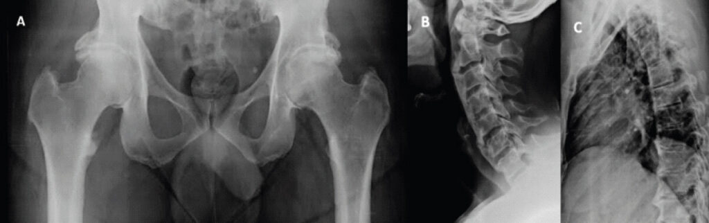
Metabolic evaluation was carried out for diffuse idiopathic skeletal hyperostosis. X-ray of both hip joints showed global pincer and CAM lesions involving both sides Fig. 1a. X-ray of the cervical and thoracolumbar spine showed spinal fusion which correlated with the limited movements of the thoracic and lumbar spine Fig. 1b with magnetic resonance imaging (MRI) of the cervical and lumbar spine correlating with the same. Blood investigations were negative for human leucocyte antigen B27 and ACCP which ruled out the possibility of ankylosing spondylosis.
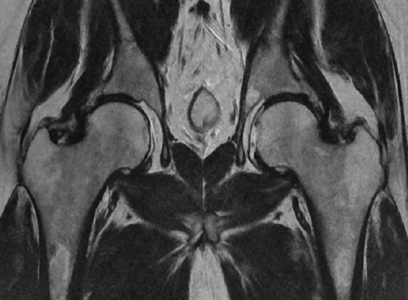
MRI of both hip joints showed a bilateral mixed type of femoroacetabular impingement with a predominant pincer component, non-visualized anterosuperior labrum, and partial thinning of the articular cartilage surrounding the fovea, as shown in Fig. 2.
The patient was diagnosed with bilateral femoroacetabular impingement with predominant pincer lesions with seronegative spondyloarthropathy.
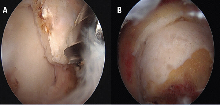
The patient underwent a planned arthroscopic pincer and CAM osteoplasty with the hip in 15-° flexion and slight internal rotation after fluoroscopic confirmation of hip joint distraction. Anterior superior iliac spine and greater trochanter were marked and an anterolateral portal was created under fluoroscopic guidance. Anterior and posterolateral portals were made and diagnostic arthroscopy was performed showing Grade 2 cartilage lesions 8 × 5 mm and 5 × 5 mm were noted over the weight-bearing dome of the head. The hip was repositioned with a flexion of 60° and the CAM lesion over the anterosuperior region was identified along with the pincer lesion Fig. 3a. Pincer and CAM osteoplasty was performed under fluoroscopic guidance Fig. 3b. It was done as a staged procedure by peripheral compartment first technique with the right hip followed 3 months later left hip. The movements were checked in the immediate post-operative period and found to have significant improvement with 45° of external rotation and 20° of internal rotation. Post-operative physiotherapy included non-weight-bearing for 6 weeks followed by partial weight-bearing from 6 to 12 weeks.
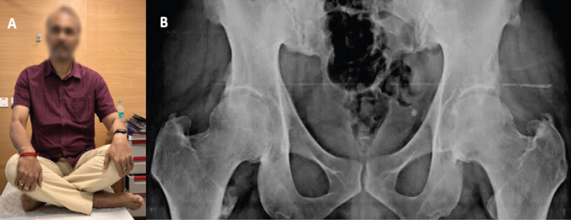
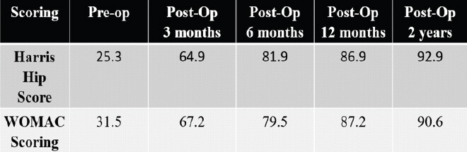
He achieved complete painless range of motion at 6 months follow-up after the 1st hip surgery Fig. 4a correlating with the post-operative X-ray Fig. 4b and the patient being able to have complete painless weight-bearing. The Harris hip score improved from a pre-operative average of 25.3 to a post-operative average of 88.9 at 2 years and the Western Ontario McMaster Universities Osteoarthritis Index score improving from a pre-operative average of 31.5 to a post-operative average of 87.2 at 2 years after the second surgery Fig. 5.
Discussion
A pincer lesion is a bony overhang that occupies the anterolateral acetabular rim and it causes over-coverage of the femoral head causing impingement and pain. CAM lesion is a bony bump or a bony prominence at the junction of the femoral head and neck. When a cam and pincer lesion causes impingement, it produces a mechanical collision of the femoral CAM with the acetabular pincer lesion resulting in pinching of the labrum and the cartilage [4]. The feasibility of arthroscopic global pincer excision is proven over the open technique which requires surgical dislocation of the hip. The common challenges that can occur include difficulty in the distraction of the hip for visualization, instrument navigation, acetabuloplasty, approach to the posterior labrum, and another one being the requirement of an experienced arthroscopic surgeon [5]. Arthroscopic labral repair is found to be superior to labral debridement. The arthroscopic technique also overcomes the need for surgical dislocation of the hip which can predispose to avascular necrosis of the femoral head [6]. The arthroscopic pincer and cam debridement with peripheral compartment first technique is protective against iatrogenic chondral and labral damage. It also helps in being conservative to the joint capsule [7]. The ease of arthroscopic surgery depends on the area of the hip that we need access to. A lateral CAM lesion can be addressed with the hip in extension whereas for a large posterolateral CAM deformity, traction might be required for better proximal exposure [2]. The surgical treatment focuses on better hip motion and prevention of abutment against the acetabular rim with an early surgical intervention other than giving a relief of symptoms might also reduce the pace of progression of the degenerative process, especially in the younger population [8]. In the case of bilateral femoroacetabular impingement, the surgeon should take adequate pre-operative imaging, proper history, patient lab tests, and operative fitness before deciding on a single sitting surgery or a staged procedure for both hips and to decide on the timing between each procedure in case of staged procedure [9]. Recent studies have found 3 extra articular types of hip impingement that is not familiar for majority of hip surgeons. These include low anterior inferior iliac spine (AIIS) impingement, also known as sub-spine impingement, ischio-femoral impingement (IFI) and pelvi-trochanteric impingement. Considering these atypical varieties of impingement is important while evaluating patient for hip or groin pain for which no specific cause can be found which may require further evaluation [10].
Conclusion
Arthroscopic rim trimming of global pincer and cam lesions through peripheral compartment first technique gives satisfactory results when followed up with aggressive physiotherapy as it does not require surgical dislocation of the hip and is also conservative to the hip joint capsule.
Clinical Message
Arthroscopic rim trimming or debridement is an excellent alternative for open debridement in a patient with global pincer and cam lesions with a faster recovery due to minimal soft tissue violation.
Conflict of Interest:
Nil
Source of Support:
Nil
Consent:
The authors confirm that informed consent was obtained from the patient for publication of this article
How to Cite this Article
Jos S, Kunnamkottu SJ, Paul P, Jayaram R, Nambiyath MJ. Bilateral Global Pincer-Type Femoroacetabular Impingement with CAM Lesions: Arthroscopic Management Using a Peripheral First A pproach. Journal of Or thopaedic Case R epor ts 2026 October;16(10): 69-72.
References
- Matsuda DK, Gupta N, Hanami D. Hip arthroscopy for challenging deformities: Global pincer femoroacetabular impingement. Arthrosc Tech 2014;3:e197-204. [Google Scholar] | [PubMed]
- Dantas P, Gonçalves S, Mascarenhas V, Camporese A, Marin-Peña O. Hip arthroscopy with initial access to the peripheral compartment provides significant improvement in FAI patients. Knee Surg Sports Traumatol Arthrosc 2021;29:1453-60. [Google Scholar] | [PubMed]
- Dantas P, Gonçalves S, Mascarenhas V, Barreira M, Marin-Peña O. Hip arthroscopy with initial access to the peripheral compartment: A detailed step-by-step technique description. Arthrosc Tech 2020;9:e1651-5. [Google Scholar] | [PubMed]
- O'Rourke RJ, El Bitar Y. Femoroacetabular impingement. In: StatPearls. Treasure Island, FL:StatPearls Publishing 2023. [Google Scholar] | [PubMed]
- Matsuda DK, Gupta N, Hanami D. Hip arthroscopy for challenging deformities: Global pincer femoroacetabular impingement. Arthrosc Tech 2014;3:e197-204. [Google Scholar] | [PubMed]
- Pun S, Kumar D, Lane NE. Femoroacetabular impingement. Arthritis Rheumatol 2015;67:17-27. [Google Scholar] | [PubMed]
- Larson CM, LaPrade RF, Floyd ER, McGaver RS, Bedi A. Acetabular rim disorders/pincer-type femoroacetabular impingement and hip arthroscopy. Sports Med Arthrosc Rev 2021;29:35-43. [Google Scholar] | [PubMed]
- Ganz R, Parvizi J, Beck M, Leunig M, Nötzli H, Siebenrock KA. Femoroacetabular impingement: A cause for osteoarthritis of the hip. Clin Orthop Relat Res 2003;417:112-20. [Google Scholar] | [PubMed]
- Kumar MV, Shanmugaraj A, Kay J, Simunovic N, Huang MJ, Wuerz TH . Bilateral hip arthroscopy for treating femoroacetabular impingement: A systematic review. Knee Surg Sports Traumatol Arthrosc 2022;30:1095-108. [Google Scholar] | [PubMed]
- Bech NH, Haverkamp D. Impingement around the hip: beyond cam and pincer. EFORT Open Rev. 2018 Feb 21;3 2:30-38. doi: 10.1302/2058-5241.3.160068.. PMID: 29657843; PMCID: PMC5890128.. [Google Scholar] | [PubMed] | [CrossRef]
© 2026 Journal of Orthopaedic Case Reports - Published by Indian Orthopaedic Research Group
About the Authors
-

Sujit Jos
-

Shobit Joseph Kunnamkottu
-

Prannoy Paul
-

Rakshit Jayaram
-

Mohammed Jishar Nambiyath
Related Articles in Journal of Orthopaedic Case Reports
 December 10, 2021 Early Breakage of All Proximal Locking Compression Screws under Non-weight-bearing Conditions after Derotational Femoral Osteotomy in a Child with Cerebral Palsy: A Case Report
 April 1, 2026 Cedell’s Fracture of the Talus Managed with Herbert Screw Fixation through Posteromedial Approach – A Case Report
 August 1, 2025 Comparative Analysis of Dorsal Distraction Plating Versus External Fixation in the Management of Intra-Articular Distal Radius Fractures
 May 23, 2018 Why there are so many submission, why the rush to publish?