


Introduction
Fractures of the distal fibula are among the most common lower limb injuries in adolescent athletes [1,2]. They usually occur after identifiable trauma, such as ankle inversion, direct contact, or high-energy rotational forces during sports activities. They are associated with typical clinical findings such as pain, swelling, and functional limitation, facilitating early diagnosis and management [1,2].
The Danis–Weber classification categorizes fibular fractures according to their relationship to the distal tibiofibular syndesmosis and remains widely used in clinical practice for ankle fractures. It is described in orthopedic references such as the AO surgery references for trauma guidelines [3].
We report the case of a 15-year-old presenting with a suprasyndesmotic distal fibular fracture consistent with a Weber C pattern following a non-contact mechanism at a football practice. This incident was characterized by the absence of spontaneous pain and minimal clinical findings on examination. This case highlights the potential discrepancy between the mechanism of injury, clinical symptoms, and radiological findings in adolescent patients.
Case Report
Patient information
A 15-year-old amateur football player presented to the emergency department after an incident during training. While attempting a shot, he missed the ball during a kicking motion and immediately felt a crack in his left leg. He reported no direct trauma, collision, or fall and was able to continue playing. He later sought medical evaluation for a delayed, unusual sensation after the game.
Clinical findings
At presentation, the patient was able to ambulate with mild discomfort but reported no spontaneous pain in the ankle or leg (Visual Analog Scale 0/10 at rest, 2/10 on deep palpation). Physical examination revealed:
-
Absence of swelling or deformity of the ankle
-
Strictly painless palpation of both malleoli
-
Painless varus stress testing
-
Isolated elective tenderness at the distal third of the fibula
-
Preserved ankle mobility
-
Absence of neurovascular deficit.
The clinical presentation was therefore remarkably poor in symptoms compared with the diagnosed fracture injury. Standardized pain scoring was not prospectively logged throughout subsequent follow-up visits, though the patient consistently reported an absence of pain during routine weight-bearing.
Diagnostic assessment
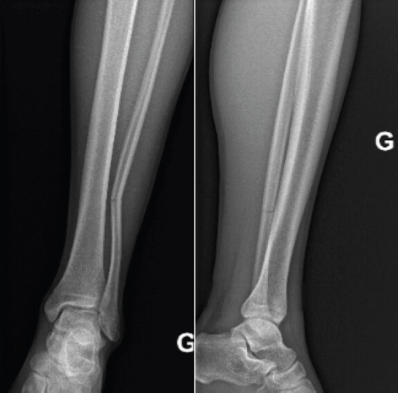
Standard radiographs of the left leg were nevertheless taken and revealed a fracture located between the middle and distal thirds of the left fibula (Fig. 1). The fracture line was situated above the distal tibiofibular syndesmosis, corresponding to a suprasyndesmotic fibular fracture consistent with a Weber C pattern. Initial plain radiographs were interpreted as a non-displaced, greenstick-type suprasyndesmotic fracture. However, advanced acute imaging (such as magnetic resonance imaging [MRI]) was not obtained at presentation, leaving open the possibility of occult syndesmotic or ligamentous strain. No associated tibial fracture or ankle joint abnormality was detected. No systemic bone evaluation was performed to assess potential metabolic or bone mineral density abnormalities, given the absence of fragility fracture history or endocrine disorders and the lack of any suspicious osseous lesion on radiographs.
Initial management
Conservative treatment was initiated [4] with a below-knee plaster splint, as recommended by the AO surgery reference [3] in the case of a non-displaced, stable fracture. Treatment also included non-weight-bearing using crutches and functional rest. No analgesic medication was required. The patient was discharged with orthopedic follow-up.
Follow-up
At the orthopedic follow-up consultation, 9 days after initial discharge, the patient reported no pain. Clinical examination showed painless mobilization of the knee and ankle. Control radiographs confirmed the non-displaced fracture without secondary displacement.
At 1 month, the patient remained completely asymptomatic. Radiographs demonstrated the appearance of bone callus formation at the fracture site, allowing progressive return to practice under supervision.
At 2 months, the patient continued to report full functional recovery, although mild tenderness on palpation of the distal fibula persisted. There was no edema, instability, or ankle pain. Radiographs still demonstrated a visible fracture line with periosteal apposition. An ultrasound examination was performed, revealing minimal inflammatory changes and periosteal reaction without Doppler hyperemia.
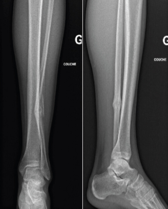
4 months after the initial injury, radiographs continued to show a persistence of the fracture line (Fig. 2).
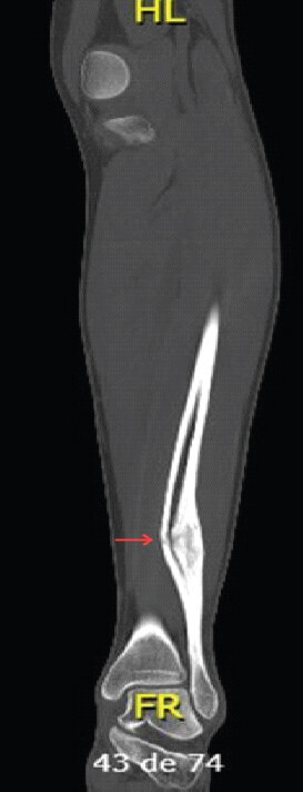
At 8 months, due to the persistent radiographic radiolucency, a computed tomography (CT) scan was ordered to definitively characterize the healing process and rule out pseudarthrosis, revealing robust hypertrophic bridging callus. CT imaging demonstrated a large hypertrophic callus surrounding the fracture site without evidence of bone lysis, instability, or pathological features (Fig. 3). These findings confirmed an active healing process and excluded pseudarthrosis. Return-to-sport clearance at 4 months was determined via clinical stability, absence of focal tenderness, and full range of motion; however, formal validated patient-reported outcome measures (e.g., American Orthopaedic Foot and Ankle Society, foot and ankle outcome score [FAOS]) and functional sport-specific discharge testing were not recorded. The patient progressively resumed full sport activity without limitation or pain.
Discussion
This case illustrates several unusual aspects of distal fibular fractures in adolescent athletes. In the Danis–Weber classification, Weber-C fractures occur above the syndesmosis and are often associated with a rupture of the interosseous ligament between the distal tibia and fibula, and the interosseous membrane up to the level of the fracture, frequently implying syndesmotic disruption and potential ankle instability [3,5]. These injuries are typically related to high-energy mechanisms such as external rotation forces or contact trauma, and usually present with marked clinical symptoms [5, 6]. In unstable syndesmotic injuries, surgical stabilization using screw or suture-button fixation may be required to restore ankle stability and maintain reduction [7]. However, in the present case, the absence of clinical and radiological signs of instability supported a conservative approach.
An unusual feature of this case was the minimal clinical presentation at initial evaluation, with only localized tenderness of the fibular shaft and absence of traumatic contact or significant mechanical stress. Most fibular fractures in sports occur following ankle inversion injuries, direct impact, or high rotational forces. In contrast, the present injury occurred during a simple kicking motion. This atypical mechanism and clinical paucity may lead to underdiagnosis or delayed diagnosis if imaging is not performed, particularly because syndesmotic injuries may be difficult to identify clinically [6].
In adolescents, fracture patterns and mechanisms may differ due to the biomechanical properties of the immature skeleton. Increased bone elasticity and the presence of open physes may result in incomplete or greenstick-type fractures without associated ligamentous disruption [8, 9].
Finally, this case highlights the persistence of radiological abnormalities despite complete clinical recovery. At 4 months, the fracture line remained visible on radiographs, raising concern for delayed union or pseudarthrosis. However, CT imaging demonstrated hypertrophic callus formation without pathological features, confirming active bone healing and illustrating the utility of advanced imaging when persistent radiographic abnormalities raise concern for instability or non-union [10]. Such findings are consistent with the high osteogenic potential observed in children and adolescents and may explain the discrepancy between imaging and clinical outcomes [8,11] and emphasize the importance of correlating these aspects when managing sports injuries in adolescents.
Non-contact fibular fractures during kicking are highly uncommon. Biomechanically, missing the ball during a maximal-velocity kicking motion can cause an uncontrolled deceleration phase, generating sudden violent eccentric contraction of the peroneal and ankle musculature, or abrupt torsional stresses transmitted across the fibula. While this mechanism appears to have induced the suprasyndesmotic failure in this patient, a single observational case report cannot definitively establish a causal relationship or define universal biomechanical thresholds. This case illustrates that significant fibular fractures may present with minimal symptoms in adolescent athletes. A Weber C fibular fracture may exceptionally occur without direct contact and with minimal clinical manifestations, possibly delaying diagnosis. Persistent radiological abnormalities should be interpreted cautiously, as they may reflect ongoing healing rather than pathology. Clinical evaluation remains central to management. Conservative treatment allowed complete recovery and safe return to sport in this young athlete.
Study limitations
Several limitations of this report must be acknowledged:
-
Generalizability and design: As an isolated case report without a comparative cohort, these findings cannot be broadly generalized to all adolescent athletic populations
-
Diagnostic and metabolic workup: Acute MRI was not performed, which limits definitive exclusion of concomitant occult chondral or syndesmotic micro-injuries. Furthermore, metabolic bone parameters were not investigated
-
Outcome measures and follow-up: Recovery was tracked clinically rather than with validated instruments (such as FAOS or the Foot and Ankle Ability Measure) or objective kinetic return-to-sport testing. Patient compliance and formal rehabilitation progression were not prospectively logged
-
Long-term monitoring: Follow-up concluded at 8 months. Long-term surveillance would be required to rule out late-onset micro-instability or post-traumatic degenerative remodeling.
Conclusion
This case describes a Weber C distal fibular fracture occurring without direct contact during a kicking motion in an adolescent athlete, with only minimal symptoms at presentation. In the absence of clinical and radiological signs of syndesmotic instability, conservative management led to complete recovery and a safe return to sport. Radiographic abnormalities persisted at 4 months, but CT showed hypertrophic callus consistent with ongoing healing rather than non-union. Clinicians should keep a low threshold for imaging in adolescent athletes with localized fibular tenderness, even after a low-energy, non-contact mechanism. Persistent radiographic findings should be interpreted alongside clinical recovery, with CT reserved for cases where instability or non-union is suspected.
Clinical Message
A Weber C fibular fracture can occur in an adolescent athlete without direct contact and with minimal symptoms, so imaging should be considered despite a benign presentation. A fracture line that persists on radiographs in a clinically recovered adolescent may reflect ongoing healing, so correlate it with the clinical course (and CT if needed) before diagnosing non-union.
Conflict of Interest:
Nil
Source of Support:
Nil
Consent:
The authors confirm that informed consent was obtained from the patient for publication of this article
How to Cite this Article
Delgado H, Bossière A, Marchal C. Atypical Suprasyndesmotic Distal Fibular Fracture in an Adolescent Athlete with Minimal Symptoms: A Case Report. J Orthop Case Rep. 2026 November;16(11): 121-125. doi: 10.13107/jocr.2026.v16.i11.8410.
References
- Beck JJ, VandenBerg C, Cruz AI, Ellis HBJr. Low energy, lateral ankle injuries in pediatric and adolescent patients: A systematic review of ankle sprains and nondisplaced distal fibula fractures. J Pediatr Orthop 2020;40:283-7. [Google Scholar] | [PubMed]
- Rougereau G, Noailles T, Khoury GE, Bauer T, Langlais T, Hardy A. Is lateral ankle sprain of the child and adolescent a myth or a reality? A systematic review of the literature. Foot Ankle Surg 2022;28:294-9. [Google Scholar] | [PubMed]
- AO Foundation. AO Surgery Reference. Davos, CH:AO Foundation. Available from: https://surgeryreference.aofoundation.org. [Last accessed on 2026 May 04]. [Google Scholar] | [PubMed]
- Schmittenbecher PP, Slongo TF. Techniques of lower limb immobilization in children and adolescents. Oper Orthop Traumatol 2025;37:213-27. [Google Scholar] | [PubMed]
- Shore BJ, Kramer DE.. Management of syndesmotic ankle injuries in children and adolescents. J Pediatr Orthop 2016;36 Suppl 1:S11-4. [Google Scholar] | [PubMed]
- Chang AL, Mandell JC.. Syndesmotic ligaments of the ankle: Anatomy, multimodality imaging, and patterns of injury. Curr Probl Diagn Radiol 2020;49:452-9. [Google Scholar] | [PubMed]
- Lurie BM, Paez CJ, Howitt SR, Pennock AT.. Suture-button versus screw fixation in adolescent syndesmotic injuries: Functional outcomes and maintenance of reduction. J Pediatr Orthop 2021;41:e427-32. [Google Scholar] | [PubMed]
- Canton G, Sborgia A, Maritan G, Fattori R, Roman F, Tomic M. Fibula fractures management. World J Orthop 2021;12:254-69. [Google Scholar] | [PubMed]
- Alexanian AA, Loveland K, Friebele JC.. Pediatric ankle syndesmosis injuries: Current concept review. J Pediatr Orthop Soc North Am 2022;4:553. [Google Scholar] | [PubMed]
- Silva LN, Morimoto LR, Watanabe GG, Rhodes NG, Cruz IA, Arruda PH. Distal tibiofibular syndesmosis: Anatomy, biomechanics, imaging approach, and postoperative evaluation. Radiographics 2026;46:e250065. [Google Scholar] | [PubMed]
- Weber B, Kalbitz M, Baur M, Braun CK, Zwingmann J, Pressmar J.. Lower leg fractures in children and adolescents-comparison of conservative vs. ECMES treatment. Front Pediatr 2021;9:597870. [Google Scholar] | [PubMed]
© 2026 Journal of Orthopaedic Case Reports - Published by Indian Orthopaedic Research Group
About the Authors
-

Hebe Delgado
-

Anaèle Bossière
-

Christophe Marchal
Related Articles in Journal of Orthopaedic Case Reports
 June 1, 2026 Clinicoradiological Outcomes after Percutaneous Radiofrequency Ablation of Benign Bone Tumors: A Prospective Observational Study
 March 1, 2026 Assessment of Lumbar Foraminal Stenosis: Diagnostic Value and Clinical Correlation: A Systematic Review
 November 1, 2025 Incidence and Functional Impact of Malrotation after Intramedullary Nailing of Femoral Shaft Fractures: A Prospective Computed Tomography-based Observational Study
 October 1, 2026 Osteogenic Potential of Bone Callus: A Cross-sectional Study on Microscopic Structure and Healing Stages