


Introduction
Normal coronal knee alignment is commonly described with reference to the mechanical axis of the lower limb. Valgus knee deformity is defined when this axis passes lateral to the center, and may be graded by the anatomic tibiofemoral angle as mild (<15°), moderate (15–30°) and severe (>30°) [1,2,3]. Common causes include rheumatoid arthritis (RA), post-traumatic arthritis, rickets [3]. Total knee arthroplasty for valgus knee can provide reliable pain relief and good functional outcomes but often presents technical challenges related to bone loss, contracted lateral soft tissues, ligament balancing, patellar tracking, and peroneal nerve protection [4,5,6,7,8,9,10,11]. Stress and insufficiency fractures after total knee arthroplasty (TKA) have been reported in the femoral neck, tibia, calcaneus, and metatarsals [12,13,14,15,16,17,18]. Intraoperative proximal fibula fracture during TKA is uncommon, with the available literature largely limited to delayed stress fractures, fibular head avulsion, and cases involving patients with RA [17,18,19,20,21]. We report an undisplaced proximal fibula fracture identified fluoroscopically during primary TKA for a moderate valgus deformity with clinical and radiologic follow-up at 1 year. The purpose of this report is to describe the presentation and management of this uncommon event rather than to infer causation or treatment superiority.
Case Report
A 73-year-old female presented with progressive left knee pain over several years, which was refractory to conservative therapy. She had a history of right sided TKA done 8 months prior. Her medical history included gout, osteoporosis, chronic kidney disease, coronary artery disease, and congestive heart failure. She used a walker for ambulation.
Clinical findings
Physical examination and diagnostic assessment
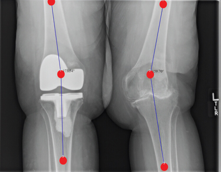
The left knee demonstrated a correctable valgus deformity, stable ligamentous examination, intact distal neurovascular status. Her range of motion (ROM) from 5° to 110° with a pre-operative Knee Injury and Osteoarthritis Outcome Score for Joint Replacement (KOOS-JR) score of 54.8. Knee radiographs showed Kellgren-Lawrence grade 4 osteoarthritis and an anatomic tibiofemoral angle of approximately 159°, representing approximately 15° of valgus relative to the contralateral post-operative alignment of approximately 174° (Fig. 1). Pre-operative dual-energy X-ray absorptiometry (DEXA) measurements documented T-scores of −2.5 at the left femoral neck, −3.0 at the left total hip, −2.9 at the right femoral neck, and −2.9 at the right total hip, confirming pre-existing osteoporosis.
Therapeutic intervention
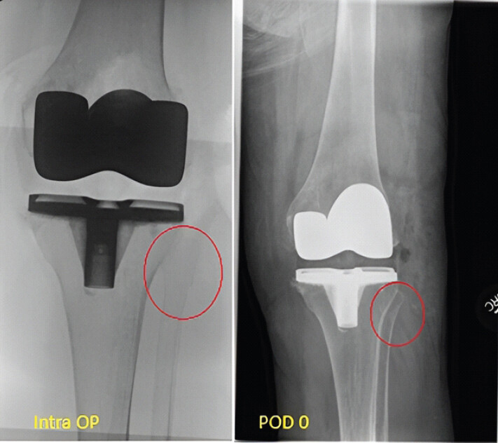
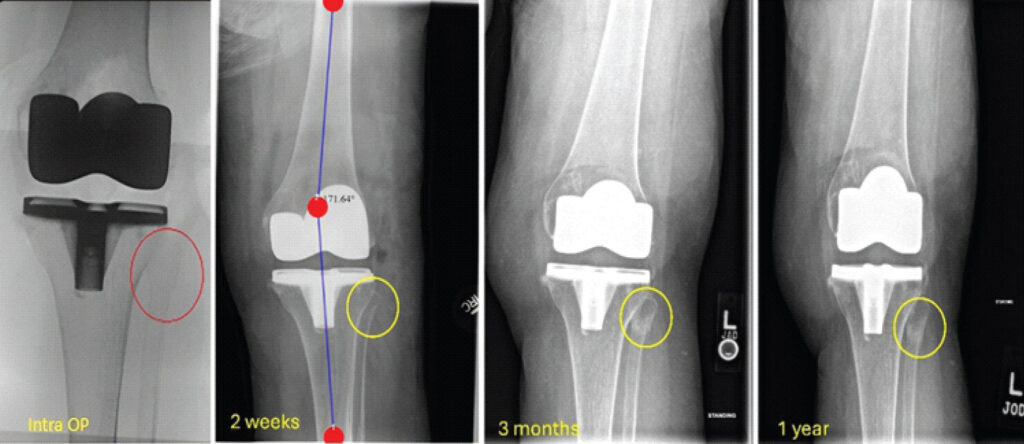
We performed a primary left TKA using a standard medial parapatellar approach and a Zimmer Biomet Persona medial-congruent bearing prosthesis. Distal femoral cut was made in 3° of valgus to address the deformity. The correction and lateral balancing sequence consisted of osteophyte removal, release of the posterolateral capsule, and pie-crusting of the iliotibial band. No additional lateral soft-tissue release was performed. After trialing and cementation, the knee was stable to varus-valgus and anteroposterior stress throughout the ROM. Fluoroscopy after component placement revealed an undisplaced fracture at the proximal fibular head-neck junction (Fig. 2). As the fracture was undisplaced and both component position and knee stability were preserved, non-operative management was selected. The patient was allowed to bear weight as tolerated immediately, perform active knee range-of-motion exercises, and mobilize in a knee brace. The brace was discontinued after 2 weeks, and no additional rehabilitation restrictions were imposed. Post-operative distal neurovascular examination was normal.
Follow-up and outcomes
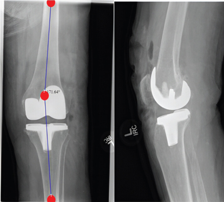
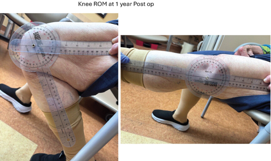
Serial radiographs demonstrated progressive callus formation without displacement, and the fracture was considered radiographically healed by 3 months. The early post-operative alignment was maintained, with an anatomic tibiofemoral angle of approximately 172° on the 2-week standing radiograph (Fig. 3). At the 1-year follow-up, the patient reported no proximal fibular pain, fracture-related symptoms, or pain during activities of daily living. She was very satisfied with the result and had a KOOS-JR score of 84.6/100. Examination showed knee ROM from 0° to 117° and no instability to varus-valgus or anteroposterior stress (Fig. 4 and Video 1). Peroneal motor and sensory function were intact. No instrumented or dynamometric strength testing was performed. She walked without an assistive device at home and continued to use a walker outdoors. She could negotiate the steps required when traveling to the hospital. Standing anteroposterior and lateral radiographs at 1 year showed a healed proximal fibula fracture, maintained TKA component position and alignment, and no evident interval fracture displacement (Fig. 5). No computed tomography or magnetic resonance imaging was obtained because clinical and serial radiographic assessments were considered sufficient for routine follow-up. Complete timeline of the patient’s clinical history and follow-up is summarized in Table 1.
Clinical history and follow-up
| Time | Finding/Intervention |
|---|---|
| Preoperative | Progressive left knee pain, History of right TKA, correctable approximately 15° valgus deformity; ROM 5–110°; KOOS-JR 54.8/100, documented osteoporosis on DEXA. |
| Day 0 | Left TKA, Intraoperative proximal fibula fracture identified fluoroscopically, satisfactory component placement and stable knee |
| Day 1 | Weight-bearing as tolerated; active, unrestricted knee ROM in a brace was permitted |
| 2 weeks | Brace discontinued, standing alignment approximately 172°; no additional rehabilitation restrictions |
| 3 months | Radiographic union without displacement or fracture-related complication. |
| 1 year | ROM 0–117°; stable knee; intact peroneal motor and sensory function; no fibular symptoms; KOOS-JR 84.6/100; radiographs showed maintained union and component position. |
KOOS-JR: Knee injury and osteoarthritis outcome score for joint replacement, TKA: Total knee arthroplasty, ROM: Range of motion, DEXA: Dual-energy X-ray absorptiometry
Video 1: Clinical video demonstrating active knee range of motion and functional recovery at 3-month follow-up.
Patient perspective
The patient initially expressed concern when informed of the fracture but understood the rationale for non-operative treatment after the stability of the fracture and knee were explained. At approximately 1 year, she reported no fibular symptoms or pain during daily activities was very satisfied with the outcome, walked without assistance at home, and continued to use a walker outdoors.
Discussion
Proximal fibula fracture after TKA is rarely reported. Vaish et al. described a fatigue fracture 14 years after TKA in a patient with RA; Takai et al. reported a stress fracture 7 weeks after TKA in a patient with severe valgus deformity and RA; Robati et al. described an acute presentation 5 weeks after TKA; and Phadnis et al. reported perioperative fibular head avulsion [18,19,20,22]. More recently, Chappidi et al. reported a proximal fibula fracture diagnosed intraoperatively during TKA for a valgus knee after an audible pop raised suspicion of the injury. Their report discussed the potential mechanism and management of this uncommon complication [18,19,20,21,22].
The present case adds to the limited literature on intraoperative proximal fibula fractures during TKA. Although valgus correction, transient hyperextension, cementation, or force transmission through the lateral collateral ligament, biceps femoris, popliteus, and other proximal fibular attachments are biomechanically plausible contributors, the mechanism cannot be established from this observation alone [23]. Bone fragility may also have contributed, as prior DEXA measurements confirmed osteoporosis, while chronic kidney disease, gout, advanced age, and limited baseline mobility may have further affected bone health or loading [24,25,26,27].
More broadly, intraoperative periprosthetic fractures during primary TKA are uncommon, with reported incidences ranging from approximately 0.2–4.4%; tibial intraoperative fractures have been reported in approximately 0.1–0.5% of cases. Management depends on fracture location and morphology, implant stability, and associated soft-tissue or ligamentous injury. Stable, non-displaced fractures with well-fixed components may be managed non-operatively with protected weight-bearing and progressive ROM, whereas unstable or displaced fractures generally require fixation or modification of the reconstruction [28,29,30].
In this present case, non-operative management with short-term bracing, weight-bearing as tolerated, and active motion resulted in successful radiographic union and a satisfactory 1-year clinical result. This outcome supports non-operative care as a reasonable option in a carefully selected, stable fracture; however, it does not demonstrate equivalence or superiority to surgical management. Displacement, implant instability, ligamentous instability, progressive fracture propagation, neurologic deficit, or inability to mobilize would require individualized reassessment and may alter management.
This report has several limitations. This single case lacks a control group and cannot establish generalizability, causation, independent risk factors, or the comparative effectiveness of conservative treatment. Multiple comorbidities and osteoporosis precluded identification of the principal contributor. The use of a single implant system, surgeon-specific technique, and limited follow-up further restricts broader and long-term conclusions.
Conclusion
An undisplaced proximal fibula fracture can occur and be recognized intraoperatively during TKA for a valgus knee. In this patient, preserved component and knee stability supported non-operative management, followed by radiographic union, good Knee ROM, and a favorable KOOS-JR score at approximately 1 year. The report principally emphasizes recognition, assessment of displacement and stability, and individualized treatment.
Clinical Message
When an intraoperative proximal fibula fracture is identified during TKA, management should be based on fracture displacement, implant position, knee stability, neurologic findings, bone quality, and the patient’s ability to mobilize. Short-term bracing with weight-bearing as tolerated was successful in this stable, undisplaced fracture, but broader preventive or treatment recommendations require larger studies.
Conflict of Interest:
Nil
Source of Support:
Nil
Consent
The authors confirm that informed consent was obtained from the patient for publication of this article
How to Cite this Article
Khadilkar S, Mahatme RJ, Moore S, Gangavaram A, Silva M, Ashfaq K. Intraoperative Proximal Fibula Fracture during Total Knee Arthroplasty for Moderate Valgus Knee Deformity: A Case Report. J Orthop Case Rep 2026 November;16(11): 61-66. doi: 10.13107/jocr.2026.v16.i11.8388.
References
- Ranawat AS, Ranawat CS, Elkus M, Rasquinha VJ, Rossi R, Babhulkar S. Total knee arthroplasty for severe valgus deformity. J Bone Joint Surg Am 2005;87(Suppl 1)271-84. [Google Scholar] | [PubMed]
- Keblish PA. The lateral approach to the valgus knee. Surgical technique and analysis of 53 cases with over two-year follow-up evaluation. Clin Orthop Relat Res 1991;271:52-62. [Google Scholar] | [PubMed]
- Favorito PJ, Mihalko WM, Krackow KA. Total knee arthroplasty in the valgus knee. J Am Acad Orthop Surg 2002;10:16-24. [Google Scholar] | [PubMed]
- Karachalios T, Sarangi PP, Newman JH. Severe varus and valgus deformities treated by total knee arthroplasty. J Bone Joint Surg Br 1994;76:938-42. [Google Scholar] | [PubMed]
- Krackow KA, Jones MM, Teeny SM, Hungerford DS. Primary total knee arthroplasty in patients with fixed valgus deformity. Clin Orthop Relat Res 1991;273:9-18. [Google Scholar] | [PubMed]
- Stern SH, Moeckel BH, Insall JN. Total knee arthroplasty in valgus knees. Clin Orthop Relat Res 1991;273:5-8. [Google Scholar] | [PubMed]
- Gebhardt EM, Laurencin CT. Total knee replacement in severe valgus deformity. Am J Knee Surg 1992;5:135-9. [Google Scholar] | [PubMed]
- Whiteside LA. Correction of ligament and bone defects in total arthroplasty of the severely valgus knee. Clin Orthop Relat Res 1993;288:234-45. [Google Scholar] | [PubMed]
- Healy WL, Iorio R, Lemos DW. Medial reconstruction during total knee arthroplasty for severe valgus deformity. Clin Orthop Relat Res 1998;356:161-9. [Google Scholar] | [PubMed]
- Miyasaka KC, Ranawat CS, Mullaji A. 10- to 20-year followup of total knee arthroplasty for valgus deformities. Clin Orthop Relat Res 1997;345:29-37. [Google Scholar] | [PubMed]
- Idusuyi OB, Morrey BF. Peroneal nerve palsy after total knee arthroplasty. Assessment of predisposing and prognostic factors. J Bone Joint Surg Am 1996;78:177-84. [Google Scholar] | [PubMed]
- Hardy DC, Delince PE, Yasik E, Lafontaine MA. Stress fracture of the hip. An unusual complication of total knee arthroplasty. Clin Orthop Relat Res 1992;281:140-4. [Google Scholar] | [PubMed]
- Pankaj A, Malhotra R, Logani V, Bhan S. Bilateral femoral neck stress fractures following total knee arthroplasty: A case report and review of literature. Arch Orthop Trauma Surg 2007;127:549-52. [Google Scholar] | [PubMed]
- Kumm DA, Rack C, Rütt J. Subtrochanteric stress fracture of the femur following total knee arthroplasty. J Arthroplasty 1997;12:580-3. [Google Scholar] | [PubMed]
- Ozdemir G, Azboy I, Yilmaz B. Bilateral periprosthetic tibial stress fracture after total knee arthroplasty: A case report. Int J Surg Case Rep 2016;24:175-8. [Google Scholar] | [PubMed]
- Takai H, Kiyota K, Nakane N, Takahashi T. Charcot fracture in the calcaneus after total knee arthroplasty: A case report. J Orthop Case Rep 2016;6:92-5. [Google Scholar] | [PubMed]
- Harato K, Ozaki M, Sakurai A, Kudo Y, Otani T. Stress fracture of the first metatarsal after total knee arthroplasty: Two case reports using gait analysis. Knee 2014;21:328-31. [Google Scholar] | [PubMed]
- Vaish A, Vaishya R, Agarwal AK, Vijay V. Stress fracture of the proximal fibula after total knee arthroplasty. BMJ Case Rep 2016;2016:bcr2016214886. [Google Scholar] | [PubMed]
- Robati S, Kazzam M, McIntyre D, Wood DG. Acute presentation of a proximal fibular stress fracture after a total knee arthroplasty. BMJ Case Rep 2021;14:e234954. [Google Scholar] | [PubMed]
- Phadnis A, Johnston P, Glasgow M. Avulsion of the fibular head post-total knee replacement. Knee Surg Sports Traumatol Arthrosc 2007;15:1340-2. [Google Scholar] | [PubMed]
- Chappidi VS, Abhilash P, Jain M. Iatrogenic fibula head avulsion fracture in fixed valgus arthritic knee during total knee arthroplasty. J Orthop Case Rep 2026;16:251-4. [Google Scholar] | [PubMed]
- Takai H, Kii S, Murayama M, Nakane N, Takahashi T. Ipsilateral stress fracture of the proximal fibula after total knee arthroplasty in a patient with severe valgus knee deformity on a background of rheumatoid arthritis. Int J Surg Case Rep 2018;45:17-21. [Google Scholar] | [PubMed]
- Seebacher JR, Inglis AE, Marshall JL, Warren RF. The structure of the posterolateral aspect of the knee. J Bone Joint Surg Am 1982;64:536-41. [Google Scholar] | [PubMed]
- Coughlan T, Dockery F. Osteoporosis and fracture risk in older people. Clin Med (Lond) 2014;14:187-91. [Google Scholar] | [PubMed]
- Gembillo G, Sessa C, Morale W, Zanoli L, Catalano A, Silipigni S. Fracture risk in chronic kidney disease: Addressing an overlooked complication. Metabolites 2025;15:460. [Google Scholar] | [PubMed]
- Tzeng HE, Lin CC, Wang IK, Huang PH, Tsai CH. Gout increases risk of fracture: A nationwide population-based cohort study. Medicine (Baltimore) 2016;95:e4669. [Google Scholar] | [PubMed]
- Ji MX, Yu Q. Primary osteoporosis in postmenopausal women. Chronic Dis Transl Med 2015;1:9-13. [Google Scholar] | [PubMed]
- Smitterberg CW, Hoveidaei AH, Delanois RE, Mont MA. Intraoperative tibial periprosthetic fractures during total knee arthroplasty: Incidence, risk factors, and early outcomes. J Knee Surg 2026;39:645-8. [Google Scholar] | [PubMed]
- Siddiqi A, Tozzi D, Springer BD, Rosen A. Diagnosis and management of intraoperative periprosthetic fractures in primary total knee arthroplasty. J Am Acad Orthop Surg 2023;31:e760-8. [Google Scholar] | [PubMed]
- Siddiqi A, Ahmed A, Pasqualini I, Molloy RM, Krebs VE, Piuzzi NS. Intraoperative fractures sustained during total knee arthroplasty: A critical analysis review. JBJS Rev 2023;11((6). doi: 10.2106/JBJS.RVW.23.00010. [Google Scholar] | [PubMed] | [CrossRef]
© 2026 Journal of Orthopaedic Case Reports - Published by Indian Orthopaedic Research Group
About the Authors
-

Siddharth Khadilkar
-

Ronak J Mahatme
-

Shawn Moore
-

Anish Gangavaram
-

Maduwilla Silva
-

Kashif Ashfaq
Related Articles in Journal of Orthopaedic Case Reports
 June 1, 2025 Tracking Pin Site Fractures and Measures to Overcome During Robotic-Assisted Total Knee Arthroplasty: Two Case Reports
 June 1, 2025 A Case Report on the Management of Peri-Prosthetic Fracture in a Post-TKA Patient with Rheumatoid Arthritis: Surgical Strategy and Treatment Outcome
 November 1, 2026 Management of Periprosthetic Total Knee Infection with Partial Hardware Removal: A Case Report
 November 1, 2026 First Reported Case of Periprosthetic Fungal Knee Joint Infection Caused by Candida viswanathii: A Case Report