


Introduction
The management of patients with localized articular cartilage defects presents an ongoing challenge to orthopedic surgeons. Hyaline cartilage heals poorly in response to partial thickness injury with minimal regenerative capabilities [1]. Full-thickness injuries lead to the formation of fibrocartilage, which is mechanically inferior to the native cartilage [2]. Inadequate repair of this cartilaginous defect can lead to continued cartilage degradation and may progress to osteoarthritis [2].
Both conservative and surgical techniques have been explored to identify the optimum management strategies when managing articular cartilage damage. Autologous matrix-induced chondrogenesis (AMIC) was designed to improve on microfracture alone by utilizing a collagen bilayer seeded with chondrocytes as a scaffold, similar to the principles used in matrix-associated chondrocyte transplantation [3]. AMIC is a single-step minimally invasive procedure in which microfracture is followed by the application of a biodegradable collagen I/III membrane to cause clot stabilization and allow the differentiation of the mesenchymal stem cells (MSCs) present within the clot to differentiate into chondrocytes [4].
Present evidence of AMIC has demonstrated efficacy in the treatment of chondral and osteochondral defects throughout multiple joints.
The aim of this review is to identify the strengths and limitations of AMIC to treat chondral defects in the knee and compare it to other available cartilage repair procedures.
Background
The presence of articular cartilage is key in the normal function of synovial joints. It covers the opposing articular bony surfaces, providing a smooth surface with low friction. It also forms a protective cushion that is resistant to compressive loads and deformation [5], thus allowing for pain-free joint movement. In utero and throughout early life, chondrocytes build articular cartilage. In early adulthood, they switch to cartilage-maintaining activity, leading to lower anabolic and proliferative capacity [2]. These features, combined with the avascular nature of articular cartilage led to its limited self-healing ability [1]. Due to this, superficial injuries where there is no hemorrhage or inflammatory response undergo little to no healing. In full-thickness injuries where there is also injury to the subchondral bone, hemorrhage and blood clot formation can aid in the healing process [6]. Acute cartilaginous injuries can lead to symptoms of locking, pain, swelling and functional impairment [7] whilst more generalized degeneration causes stiffness, immobility and loss of quality of life [8]. Full thickness injuries lead to the formation of fibrocartilage rather than hyaline cartilage [9], which is lacks the mechanical properties of native cartilage and bonds poorly with adjacent normal cartilage [2]. This fibrocartilage replacement has been demonstrated to breakdown under normal joint loading [6]. Regardless of the depth or origin of articular cartilage defect, there is an increased risk of the development of osteoarthritis [10].
Chondral and osteochondral defects can be managed conservatively or surgically. Conservative management strategies include the restriction of physical activities, and physiotherapy has yielded mixed results. Poorer prognosis was also identified in those with larger lesions >12 mm2, unstable lesions, and in those with open physis [11]. Therefore, these patients, along with those with mechanical symptoms, should be offered surgical treatment.
A variety of surgical techniques are used in the management of chondral and osteochondral defects. These can largely be divided into repair, bone marrow stimulating techniques, and transplantation. The foundation of bone marrow stimulation (BMS) for the management of osteochondral defects was initially described by Pridie in 1959 [12]. This was further developed by Steadman et al. in the 1990s into the microfracture procedure [13], which remains a popular surgical technique. Microfracture involves the penetration of subchondral bone to access the bone marrow. This allows the formation of a blood clot that contains MSCs, bioactive molecules, and fibrin [14], which can aid in the healing process. Microfracture provides good mid-term results; however, unsatisfactory results have been reported in long-term results, especially in those with larger lesions [15].
Transplantation techniques can be performed with either implantation of osteochondral plugs into the defect with autograft or allograft tissue. Autografts are commonly done by osteochondral autograft transplant (OAT), mosaicplasty, or by the introduction of autologous chondrocytes as in autologous chondrocyte implantation (ACI). Both OAT and mosaicplasty involve autograft transplantation of articular cartilage from less important anatomical locations, usually away from the articular surface. Whereas OAT utilises a single osteochondral plug, mosaicplasty uses multiple smaller autografts to fill the defect [16]. ACI was initially described by Brittberg et al. in 1994 [17] and is a two-step procedure in which chondrocytes are harvested from a non-weight-bearing surface, expanded in a laboratory before reimplantation. This technique has been further developed into collagen-covered ACI (ACI-C) and matrix-assisted chondrocyte implantation (MACI).
Osteochondral allograft transplantation (OCAT) was first reported as a treatment method for chondral and osteochondral defects in the 1980s by Meyer’s et al. [18] and involves the transplantation of surviving hyaline cartilage and subchondral bone into the cartilage defect. Since then, the technique has continued to develop and remains an effective treatment, particularly for larger cartilage defects, with graft survival reported up to 59.4% at 25 years post-operatively [19].
AMIC was described by Behrens in 2005 [20], which combines microfracturing of the subchondral bone after cartilage debridement with the fixation of a collagen I/III matrix. This covers the blood clot providing additional structure and protection against shearing forces during joint motion, as well as allowing the MSCs to develop into chondrocytes [21]. AMIC has been demonstrated to be a safe and effective treatment for the management of chondral and osteochondral defects [22,23] in the short and medium-term; however, longer-term outcomes are required to establish this technique as the optimal surgical choice.
Principles
AMIC was designed to expand on BMS techniques, such as subchondral drilling and microfracture. BMS techniques work on the principle that accessing the highly vascular bone marrow allows the formation of a clot within the defect. This clot contains a variety of active substrates and MSCs that eventually form fibrocartilage [4]. Microfracture is a simple, cost-effective single-stage procedure that can be completed arthroscopically and, if required, in conjunction with additional procedures, such as meniscus surgery or ligament repair [24]. Fibrocartilage is not an ideal replacement for hyaline cartilage as it lacks the viscoelastic properties and therefore is unable to distribute force or absorb shock as effectively [2,24] and deteriorates more quickly under normal joint loading [6]. These factors may contribute to the relatively high failure rates seen in microfracture [25].
AMIC aims to improve on standard BMS techniques by the application of a 3D matrix to act as a scaffold for clot formation and durability. The scaffold material should ideally mimic the biological, architectural, and structural properties of the native tissue whilst allowing cell infiltration, attachment, proliferation, and differentiation [16]. A variety of different materials have been used, including natural protein- or carbohydrate-based scaffolds and synthetic scaffolds [26]. Three matrices are commonly used: Chondro-Gide (Geistich Biomaterials), Chondrotissue (BioTissue), and Hyalofast (Fida Advanced Biopolymers). The most common matrix used in AMIC is Chondro-Gide, a porcine-derived collagen I/III bilayer membrane that functions as both a porous cell adhesive and a smooth occlusive layer. The porous layer allows MSCs to attach, proliferate, and differentiate, while the occlusive layer aims to keep the clot within the defect. Gille et al. [27] demonstrated that collagen I/III matrices showed better retention of their chondrocyte morphology and properties for chondrogenesis than collagen II matrices.
Chondrotissue is an absorbable non-woven polyglycolic acid textile treated with hyaluronic acid [28]. This works as a sponge to hold the blood clot and progenitor cells within the defect, protecting the area and inducing hemostasis [28]. Hyaluronic acid has also been demonstrated to cause differentiation of MSCs along the chondrogenic lineage [28]. The feasibility of chondrotissue was demonstrated by Erggelet et al. [29] in ovine models where its use resulted in increased type II collagen formation when compared to microfracture alone.
Hyalofast is a 3D, semi-synthetic, non-woven matrix made of a proprietary material called Hyaff, which is a chemically modified form of hyaluronic acid [30]. It was designed on the principle of hyaluronic acid causing the differentiation of MSCs into chondrocytes and was confirmed as a viable material by Facchini et al. [31] in laboratory studies. As with other matrices, it is designed to be a scaffold for clot formation as well as a protective layer.
Surgical Technique and variations
The standard AMIC technique is described by Benthien and Behrens [4] and is indicated for symptomatic full-thickness cartilage lesions (International Cartilage Repair Society [ICRS] grade 4) and in <1.5 cm2 in size. It can be completed under spinal or general anesthesia and begins with an arthroscopic examination of the affected joint. The size and location of the defect are identified, and additional lesions and ligamentous injuries are excluded. A minimally invasive arthrotomy is completed, and the defect is visualized openly. A curette is used to remove damaged cartilage and subchondral bone. A template can be measured at this stage. If an osteochondral defect is identified, cancellous bone can subsequently be harvested from the head of the tibia through the same incision. Microfracture is then carried out. This was initially described using microfracture awls; however, the authors subsequently changed their recommendation to the use of a 1.1 mm K-wire following studies demonstrating its improved effectiveness at reaching the subchondral stroma. Following microfracture, the matrix is hydrated and then attached. The matrices require different hydration fluids, and guidance is available from the manufacturer. Attachment of the matrix is commonly done with fibrin glue, sutures, or a combination of both. Concerns have been raised that suturing of the matrix may lead to cartilage damage [32], and fibrin glue may reduce chondrocyte proliferation and migration [33]; however, follow-up studies have not demonstrated any difference in these techniques [34]. The collagen matrix is placed below the level of the cartilage to ensure it is not displaced when the joint moves. The joint should then be cycled gently and reassessed to ensure there has been no displacement of the membrane. Post-operatively, the authors recommend limited weight bearing, with flexion limited to 30° for 2 weeks post-op followed by a further 2 weeks each at 60° and 90°. More recent studies exploring early full weight bearing and full range of motion have been published with good results [35].
Nanofractured AMIC (NAMIC) is a variation of AMIC where nanofracture is used in place of microfracture. It was described by Benthien and Behrens in 2015 [36] as a further advancement of standard AMIC. Using nanofracture allows smaller holes (<1 mm diameter) to be created that penetrate deeper into the bone (9 mm). This leads to reduced trabecular fragmentation and compaction and subsequently better restoration of normal bone architecture [37]. This allowed the authors to update their rehabilitation recommendations in line with more recent publications, with a shorter period of reduced weight bearing and earlier physiotherapy than following a standard AMIC procedure. Initial results for NAMIC are promising [38], but long-term data are still required.
An all-arthroscopic variation of AMIC has also been described [39]. Following initial debridement, the knee is emptied of irrigation fluid and dry arthroscopy is performed. This facilitates drilling of the subchondral layer with a 1.1 mm K-wire. The Chondro-glide membrane is then inserted and clamped into place before attachment with fibrin glue. In larger lesions, multiple pieces of Chondro-glide are inserted to fill the defect. This is left to set before the knee is cycled. Here the authors describe the use of these flexion-extension cycles to identify the safe range of motion for use in rehabilitation post-operatively. More recently, an all arthroscopic nanofracture AMIC technique has been described [40]. This is similar to the technique described by Piontek et al. [39], but nanofracture is used for subchondral bone penetration in place of K-wire drilling.
Clinical Outcomes
The AMIC procedure has now been in regular clinical use for more than 20 years. Its use has been described for the treatment of chondral and osteochondral defects in various joints, though most of the literature is related to the knee and talus.
Knee
There is a growing body of literature to support the use of AMIC within the knee. Early data published by Gille et al. [41] followed up 27 patients who underwent AMIC procedures for chondral defects within the knee. They were followed up for a mean of 37 months. Post-operatively, these patients demonstrated increased Myer, Lysholm, Tegner, ICRS, and Cincinnati scores at 12, 24, and 36 months compared to pre-operative scores, with the highest scores being achieved at 24 months. Of the patients who underwent a questionnaire, 87% were subjectively highly satisfied with the results. Peras et al. [42] added this data with a retrospective multicenter study including 101 patients with a mean follow-up of 2.5-years. The mean knee injury and osteoarthritis score (KOOS) increased from 45% to 73%, the mean International Knee Documentation Committee (IKDC) score increased from 43% to 74%, and the mean SF-36 score increased by 20%. Magnetic resonance imaging (MRI) scans were completed at 2 years in 58 patients who had a mean magnetic resonance observation of cartilage repair tissue (MOCART) 2.0 score of 75, though there was no correlation between these radiological outcomes and the functional outcomes.
Kaiser et al. [43] demonstrated good long-term outcomes for 26 patients following AMIC procedures. Their data showed a sustained improvement in both Lysholm and Visual Analog Scale (VAS) for pain scores between 2- and 9-years post-procedure. However, patients in this study underwent leg axis correction when malalignment was identified, which may have influenced the outcomes. Tradati et al. [44] assessed the outcomes of AMIC on patellar chondral and osteochondral defects. They demonstrated improvements in IKDC scores and VAS pain scores in line with other literature at 2- and 5- years. The specificity of this, however, led to a reduced participant number, with only 14 meeting inclusion criteria.
Whilst AMIC was initially described for use in defects <1.5 cm2, present literature exists that reports good outcomes in much larger defects [41,42,43,44]. The present data demonstrate both good short- and medium-term outcomes for knee AMIC. Longer-term data are still required. Present published literature rarely discriminates between patellar and femoral lesions or chondral and osteochondral defects. Often these outcomes are reported together. Further research investigating these individually will allow for more robust analysis of outcomes.
Ankle
Good post-operative outcomes have been reported for ankle AMIC. Weigelt et al. [45] followed 33 patients for a mean of 4.7-years post-operatively following AMIC procedures to the talar dome in patients with osteochondral defects. The demonstrated significant improvement in VAS pain scores (6.4–1.4) and Tegner score (3.5–5.2), with a mean American Orthopedic Foot and Ankle Society (AOFAS) score of 93 sustained at final follow-up. This study included radiological follow-up with MRI and MOCART score. The mean MOCART score was 60.6, which is similar to other literature. As with Peras et al. [42] study, the MOCART score did not correlate clinically.
Longer-term follow-up of talar dome AMIC by Deiss et al. [46] also demonstrated good outcomes. 18 of the included patients were followed up 10-years post-operatively. Functional foot index D scores had a statistically significant reduction that was sustained at 10-years (56–21). Additional research with larger patient numbers is needed to improve on present literature.
Hip
The use of AMIC in the hip is less common than in the knee and ankle, and the available literature reflects this. An all-arthroscopic technique for treatment of hip cartilage defects was described by Fontana in 2012 [47]. This technique was used by the same author in a series of 201 patients with a mean defect size of 2.9 cm2, who were followed up for 5-years [48]. Post-operatively, their outcome was assessed with the modified Harris Hip Score (mHHS). A sustained improvement was seen from baseline (44.9) from 6 months post-operatively through to final follow-up. Maximum benefit was reported at 3 years post-operatively, with a mean mHHS score of 85.5, with no patients reporting a poor score (<60). Further studies with longer follow-up are required for hip AMIC.
Upper limb
There is currently limited literature related to the use of AMIC in the upper limb. Cuéllar et al. [49] described the use of AMIC in the glenohumeral joint and reported stability of the implanted membranes at 2- and 7-months, though no functional or VAS pain scores were completed. AMIC has also been explored in the hand and wrist by López-Osornio De Vega et al. [50], who completed a 3-case series with patients involved describing improved pain and function post-operatively. This is an emerging field of research and much more data will be required before this is an established practice.
Comparative Studies
AMIC versus microfracture
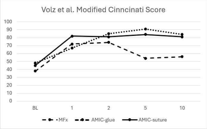
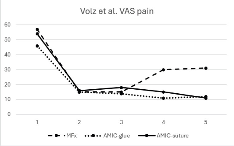
There is a large volume of publications comparing AMIC and microfracture outcomes in multiple joints. Volz et al. [22] completed a randomized controlled trial for knee AMIC (glued and sutured groups) versus microfracture. Patients were assessed with the modified Cincinnati score. Mean scores in all groups improved at 1- and 2-year post-operatively, though whilst the scores were maintained or improved in the AMIC groups at 5 years, there was a decrease in the microfracture group. This was also seen in both subjective and objective functional scoring, where all groups saw improved at 1 year post-operatively, but at 5 years, 66% of the microfracture group had abnormal knees compared to 6–7% in the AMIC groups. MRI assessment of the defects was completed at 2 years post-operatively, which demonstrated that two-thirds of the defect was filled in 60% of the AMIC groups, whereas this degree of filling was present only in 25% of the microfracture group. Volz et al. [34] subsequently published 10-year follow-up data for the same patients. There was no statistically significant change in the modified Cincinnati (Fig. 1) or VAS pain scores (Fig. 2) between the 5- and 10-year in either the AMIC or microfracture group; thus, at 10 years the AMIC outcomes remain superior to microfracture. Both the AMIC and microfracture groups underwent little change in their modified Cincinnati and VAS pain scores between the 5- and 10-year follow-up period.
Similar results were reported by Migliorini et al. [51], who compared AMIC versus microfracture in patellar lesions. The AMIC group saw improvements in IKDC, Lysholm, Tegner, and VAS pain scores when compared to the microfracture group at a mean follow-up of 45 months. Lower failure rates were seen in the AMIC group, and revision or progression to arthroplasty was the same in both groups.
Migliorini et al. [52] published data on talar shoulder AMIC versus microfracture and followed patients up for a mean of 43 months. They demonstrated an improvement in AOFAS, VAS pain, and Tegner scores in the AMIC group compared to the microfracture group (Table 1). There were also greater rates of revision surgery in the microfracture group, with 39% undergoing further surgery for ongoing pain at 4 years post-operatively compared to 10% in the AMIC group. Becher et al. [53], however, reported no statistically significant difference between microfracture and AMIC in the talus, comparing the Hannover Scoring System, VAS pain, function, and satisfaction scores at 5 years post-operatively. This paper limited the size of included defects to < 2 cm2, whereas Migliorini et al. [52] patients had a mean defect size of 2.7 cm2, possibly suggesting increased effectiveness in larger defects.
Results of functional and radiographical scores comparing AMIC and microfracture (MFx) in the talus, Migliorini et al. [52]
| Endpoint | AMIC (n=52) | MFx (n =18) | MD | p |
|---|---|---|---|---|
| Follow-up (months) | 44.2±19.9 | 41.5±18.1 | n/a | n.s. |
| MOCART | 80.0±25.4 | 66.8±33.1 | 13.2 | 0.08 |
| AOFAS | 83.8±12.4 | 75.0±19.3 | 8.8 | 0.03 |
| VAS (0-10) | 1.9±0.8 | 3.3±3.1 | 1.4 | 0.003 |
| Tegner | 4.3±1.5 | 3.1±2.1 | 1.2 | 0.01 |
AMIC: Autologous matrix-induced chondrogenesis, MFx: microfracture, MOCART: Magnetic resonance observation of cartilage repair tissue, AOFAS: American Orthopaedic Foot and Ankle Society, VAS: Visual analogue scale, MD: Mean difference
Fontana et al. [48] compared femoral-acetabular impingement-induced chondral lesions in 140 patients treated with AMIC and microfracture. These patients were followed up yearly for 5 years. The mHHS score significantly improved in both groups at 6 months post-operatively. These scores were maintained in the AMIC group up to 5 years, whilst those in the microfracture group’s scores began to fall between 1- and 2-years, reaching the lowest between 4- and 5-year follow-up. In addition, 6 patients in the microfracture group went on to require total hip arthroplasty compared to 0 in the AMIC group.
AMIC versus ACI
Fossum et al. [23] compared the outcomes of AMIC and ACI-C at 1- and 2-years. They demonstrated no statistically significant difference between the two groups when comparing VAS pain, KOOS, or Lysholm score. At present, there are no longer-term studies comparing these procedures.
MACI has also been compared to AMIC in the knee by Migliorini et al. [54] in a systematic review. This demonstrated that AMIC performed better than MACI for chondral defects, with a mean of 40 months follow-up. Lysholm and IKDC scores were superior in the AMIC group, who also had a lower rate of complications. Tegner and VAS scores were similar in both groups (Table 2). However, in a paper by Schnider et al. [55], there was no statistically significant difference identified in VAS pain, KOOS pain, or KOOS symptoms between the AMIC and MACI groups, with both achieving good outcomes. Similarly, no statistically significant difference was seen in MACI versus AMIC for chondral defects in the talus in VAS, AOFAS, or MOCART scores [52].
Results of functional and pain scores comparing AMIC and MACI in the knee, Migliorini et al. [54]
| Endpoint | AMIC | mACI | MD | P |
|---|---|---|---|---|
| Follow-up (months) | 37.8±29.9 | 39.8 ±17.2 | n.s. | |
| VAS | 2.8±2.2 | 2.9±1.3 | 0.07 | n.s. |
| Tegner | 4.4±0.6 | 4.7±0.8 | 0.3 | n.s. |
| Lysholm | 81.9±7.1 | 65.7±28.2 | 16.1 | 0.02 |
| IKDC | 79.2±10.4 | 71.5±6.3 | 7.7 | 0.03 |
MACI: Matrix-assisted chondrocyte implantation, IKDC: International Knee Documentation Committee, AMIC: Autologous matrix-induced chondrogenesis, VAS: Visual analogue scale, MD: Mean difference
AMIC versus transplant
Short-term data have been published by Debieux et al. [56] comparing AMIC and OAT in patellar chondral lesions. Both groups saw statistically significant improvements in Lysholm, Fulkerson, and Kujala scores at 2 years post-operatively. The AMIC group also had a statistically significant improvement in Tegner scores, whereas this score was reduced post-operatively in the OAT group, though the difference between the follow-up scores (5.47 vs. 4.92) was not statistically significant (Table 3). In addition, those in the AMIC group had significantly larger lesions compared to those in the OAT group (2.18 cm2 vs. 1.13 cm2) and achieved similar outcomes. Lesion size is significant in OAT due to the increased morbidity associated with larger donor site harvesting. Gedikbas et al. [57] compared mosaicplasty and AMIC in the talus, and while post-operative results were similar in both groups, complications were higher in the mosaicplasty (36% vs. 18%); this included donor site pain. This suggests that larger defects are likely best managed by AMIC though further research comparing outcomes in larger defects would be required [56].
Results of functional scores comparing AMIC and OAT pre-operatively and at 2 years post-op, Debieux et al. [56]
| Endpoint | Technique | Pre | Post | Change | P |
|---|---|---|---|---|---|
| Tegner | OATS | 6.59 | 5.47 | −1.12 | 0.006 |
| AMIC | 3.58 | 4.92 | 1.33 | 0.013 | |
| Lysholm | OATS | 57.7 | 79.7 | 22 | <0.001 |
| AMIC | 60.1 | 88.3 | 28.2 | <0.001 | |
| Kujala | OATS | 56.2 | 79.5 | 24 | <0.001 |
| AMIC | 58.3 | 86.4 | 28.1 | <0.001 | |
| Fulkerson | OATS | 54.5 | 81.3 | 26.8 | <0.001 |
| AMIC | 56.9 | 88.2 | 31.3 | <0.001 |
AMIC: Autologous matrix-induced chondrogenesis, OAT: Osteochondral autograft transplant
Autologous minced cartilage implantation (MCI) is also used for the management of chondral defects. Behrendt et al. [58] compared MCI and AMIC in the knee with a mean of 36 months follow-up. The AMIC group demonstrated superior VAS pain, Lysholm, and KOOS scores at follow-up with statistically significant differences in VAS pain and Lysholm scores (Table 4). Schnider et al. [55] did not replicate these findings, with no difference in outcomes identified, though follow-up was shorter at 2 years and intervention groups were smaller (16 vs. 24). Further long-term data are required for comparison.
Results of functional outcomes and pain scores comparing AMIC and MCI in the knee, Berhendt et al. [58]
| Endpoint | AMIC | MCI | P |
|---|---|---|---|
| Follow-up (months) | 37±8.8 | 32.2±7.9 | |
| VAS | 1.9±1.9 | 4.1±2.7 | 0.004 |
| Lysholm | 83.1±13.9 | 70.1±25.7 | 0.043 |
| KOOS | |||
| Pain | 88.3±11.7 | 72.8±26.5 | 0.016 |
| Quality of life | 65.6±24.4 | 50.4±22.3 | 0.036 |
| Functioning in daily living | 92.9±8.2 | 82.6±22.9 | n.s. |
| Symptoms | 86.7±12.2 | 77.2±21.4 | n.s. |
| Sports | 64.5±34.1 | 60.4±28.5 | n.s. |
AMIC: Autologous matrix-induced chondrogenesis, MCI: Minced cartilage implantation, VAS: Visual analogue scale, KOOS: Knee injury and osteoarthritis score
Present literature review identified no studies directly comparing the outcomes of AMIC and osteochondral allograft transplant.
Future Development
Rehabilitation
Initial rehabilitation protocols for AMIC were conservative with limited weight bearing [4]; this was due to concerns of dislodgement of the matrix and/or super clot interfering with the healing process. Laboratory studies suggest that compression and shear forces may improve chondrogenic gene expression, glycosaminoglycan and collagen II formation [59], suggesting the formation of cartilage more similar to the pre-injury state. This, along with the move from the use of microfracture awls to 1.1 mm k-wires or nanofracture, has allowed research to explore early mobilization and weight bearing in the post-operative phase. While initial data demonstrate good results [35], comparative studies with longer-term outcomes are required to ensure sustained benefit.
Matrix development
Following cartilage injury, present repair techniques aim to promote the formation of a new articular surface, though this is not necessarily hyaline cartilage [60].
New research is exploring the use of 3D bioprinting to develop advanced scaffold materials. Zhou et al. [61] completed a laboratory study in which a 3D bioprinting hydrogel containing chondrogenic progenitor cells was implanted into a rat cartilage defect model. They demonstrated the formation of a stable structure similar to that of the natural surrounding cartilage in the model. Other groups are exploring different scaffold materials [62] and active molecules [63] to improve on current matrix technologies.
Other
LIPO-AMIC is a technique described by Sciaretta et al. [64]. It is an adaptation of the standard AMIC technique in which fat aspirate is harvested, prepared, and placed onto the matrix before application to the defect. It is based on the principle that adipose tissue can provide an additional source of MSCs and has local immunomodulatory effects that may enhance cartilage repair. Follow-up was completed at 2 and 5 years [65] with significant improvements demonstrated in VAS, IKDC, Lysholm, and KOOS scores. This was a small trial with only 18 patients treated, with no control group used. Comparative studies will be required to further explore the viability of this technique.
Discussion
Present published data confirm that AMIC is an effective treatment option for both chondral and osteochondral defects in a variety of joints. The strongest evidence exists in the knee, where short-, medium-, and long-term studies consistently demonstrate significant improvements in functional scores [34,50]. Comparative studies indicate that these improvements are sustained over time and are superior to those achieved with isolated microfracture [34]. Although evidence in the ankle and hip is less extensive, similar improvements in pain and functional outcomes have been demonstrated [46,48], suggesting application beyond the knee.
There is an increasing body of evidence that AMIC outperforms microfracture as a treatment option with better sustained outcomes. This superiority of AMIC over microfracture may be explained by the increased stability within the clot due to the presence of the scaffold. AMIC provides stability to the local environment during the critical early phase of healing, allowing for the proliferation and retention of MSCs [4] and resistance to shearing forces [21], potentially increasing clot stability when compared to microfracture alone. These factors may contribute to the sustained functional improvements seen in short- and medium-term results and may explain the progressive decline seen in the microfracture cohorts.
Within published literature, there is little difference identified between the outcomes of AMIC and ACI/MACI. Studies directly comparing outcomes are limited and often underpowered [66] and further research with robust randomized controlled trials is required. Authors, however, recognize the single-stage nature of AMIC as an advantage over MACI/ACI. As AMIC does not require laboratory proliferation of harvested chondrocytes, it is associated with lower costs that ACI/MACI [44,54]. Another benefit of the single-stage nature of AMIC would be the ability to institute rehabilitation at an earlier time without the delay related to a second procedure. This may provide a biological and functional “head start” compared to ACI/MACI and potentially facilitating an earlier return to normal function. However, this hypothesis has not been tested and warrants further study.
There is limited evidence comparing the results of OAT/mosaicplasty and AMIC. Literature available suggests that AMIC does offer improved outcomes, though the study was underpowered to demonstrate statistical significance [57]. A significant drawback of OAT/mosaicplasty compared to AMIC is the requirement of a donor site and the potential burden of donor site complications.
One area of significant heterogenicity within published literature is the defect size of chondral and osteochondral lesions treated. AMIC was initially described for defects <1.5 cm2 [4], though several studies demonstrate defects up to 8 cm2 being treated with successful outcomes [41,42,67]. At present, there is no published literature that compares the outcomes of AMIC across different-sized lesions; therefore, it remains unclear whether lesion size influences the clinical outcomes or the longevity of the repair. Much of the published literature also deals with chondral and osteochondral lesions in a single cohort despite the possibility of differing treatment requirements or biological responses to treatment.
Despite favorable outcomes, AMIC also has important limitations. Due to the use of animal-derived materials, hypersensitivity can occur to fibrin glues or to the matrices themselves. Therefore, in patients with known hypersensitivity to porcine collagen derivatives, AMIC is contraindicated [68]. Active or recent infection and synovial inflammation are additional contraindications [68]. In these patients, microfracture alone or autograft procedures will therefore be required.
The influence of knee alignment on the outcomes of AMIC also requires consideration. Several published studies include knee realignment procedures alongside AMIC, including patella realignment, medial patellofemoral ligament reconstruction and osteotomies around the knee, or a combination of multiple procedures [8,43]. However, other studies excluded those with known patellofemoral or knee instability [34,51] and offered no corrective procedures as part of the procedure. Evidence of statistically significant improvements in functional outcomes scores have been reported in studies that included knee alignment procedures [8,43] and those that didn’t [34,51]. It is therefore a possibility that these procedures independently improve symptoms, joint biomechanics and functional outcomes and without direct comparative evidence this effect cannot be quantified.
Most cartilage injuries occur in young males with a normal body mass index (BMI). Consequently, these patients are disproportionately represented in the current AMIC literature. As a result, the generalizability of these findings to other populations remains uncertain. Although limited, some published data do indicate that comparable outcomes can be achieved with AMIC in the older population as well as in those with higher BMIs.
Interpretation of the current available evidence should be undertaken cautiously. Much of the current available literature consists of retrospective cohort studies and case-series studies with relatively small patient numbers, thus increasing the risk of selection and reporting bias. There is limited evidence of direct comparison between AMIC, ACI/MACI, OAT, mosaicplasty, and OCAT. Direct quantitative comparison between studies is significantly limited due to the variety of scoring systems used. MRI and MOCART scores are reported in several included studies; however, this review identifies no consistent correlation between radiological and functional outcomes. The clinical significance of the findings on imaging therefore remains uncertain.
There is also considerable heterogenicity regarding lesion size, anatomical location, patient age, BMI, associated pathology, rehabilitation protocols, and reported outcome measures. Studies often include pathology across several anatomical sites, and are often pooled together, despite the potential for differing clinical and biomechanical outcomes. There is significant variation in the application of current AMIC techniques. Variation in membranes, fixation methods, open versus arthroscopic techniques, and nanofracture versus microfracture potentially confounds outcome comparison. A similar limitation relates to the heterogeneity of rehabilitation protocols. Post-operative weight-bearing status and range-of-motion protocols vary substantially between studies, making it difficult to isolate the effect of surgical techniques on outcomes.
Risk of bias was not formally assessed, which limits confidence in the findings. Given the predominance of small, retrospective observational studies, selection bias, confounding, and selective outcome reporting are plausible. Publication bias was not formally assessed and cannot be excluded; therefore, favorable outcomes may be overrepresented in the available literature.
Conclusion
Currently AMIC represents a viable and effective single-stage technique for the management of chondral and osteochondral defects. Current literature suggests that it is superior to microfracture, while further data are required comparing ACI and autograft/allograft transplantation techniques. Due to being a single-stage procedure with no donor site requirement, it would likely represent the more favorable option should longer-term comparative studies suggest superior or non-inferior outcomes. Future research is currently directed at enhancing 3D matrix development to improve cartilage regeneration. Robust long-term outcomes from comparative studies will be essential to definitively establish AMIC as the standard of care for chondral and osteochondral defect management.
Clinical Message
AMIC is a safe, effective, single-stage operation for chondral and osteochondral defects and should be considered ahead of microfracture alone due to the increasing body of evidence supporting superior mid- and long-term outcomes. AMIC avoids donor-site morbidity and may have lower procedural costs than ACI/MACI and OAT/mosaicplasty; however, long-term, robust comparative evidence comparing these techniques remains limited. Therefore, AMIC should be considered on an individual patient basis considering lesion characteristics, patient factors, concomitant pathology, and local expertise. Current evidence does not establish AMIC as superior to all alternative cartilage restoration techniques or as a universal standard of care.
Conflict of Interest:
Nil
Source of Support:
Nil
Consent
The authors confirm that informed consent was obtained from the patient for publication of this article
How to Cite this Article
Worgan R, Mavi J, Ranjan A, Bailey O, Hopper G, Bhattacharyya R. Is Autologous Matrix-Induced Chondrogenesis an Effective Procedure to Treat Cartilage Lesions around the Knee: A Literature Review. J Orthop Case Rep. 2026 November;16(11):594-606. doi: 10.13107/jocr.2026.v16.i11.8560.
References
- Buckwalter JA. Articular cartilage: Injuries and potential for healing. J Orthop Sports Phys Ther 1998;28:192-202. [Google Scholar] | [PubMed]
- Correa D, Lietman SA. Articular cartilage repair: Current needs, methods and research directions. Semin Cell Dev Biol 2017;62:67-77. [Google Scholar] | [PubMed]
- Bartlett W, Skinner JA, Gooding CR, Carrington RW, Flanagan AM, Briggs TW. Autologous chondrocyte implantation versus matrix-induced autologous chondrocyte implantation for osteochondral defects of the knee: A prospective, randomised study. J Bone Joint Surg Br 2005;87:640-5. [Google Scholar] | [PubMed]
- Benthien JP, Behrens P. The treatment of chondral and osteochondral defects of the knee with autologous matrix-induced chondrogenesis (AMIC): Method description and recent developments. Knee Surg Sports Traumatol Arthrosc 2011;19:1316-9. [Google Scholar] | [PubMed]
- Buckwalter JA, Mankin HJ. Articular cartilage: Tissue design and chondrocyte-matrix interactions. Instr Course Lect 1998;47:477-86. [Google Scholar] | [PubMed]
- Shapiro F, Koide S, Glimcher MJ. Cell origin and differentiation in the repair of full-thickness defects of articular cartilage. J Bone Joint Surg Am 1993;75:532-53. [Google Scholar] | [PubMed]
- Howell M, Liao Q, Gee CW. Surgical management of osteochondral defects of the knee: An educational review. Curr Rev Musculoskelet Med 2021;14:60-6. [Google Scholar] | [PubMed]
- Gille J, Behrens P, Volpi P, De Girolamo L, Reiss E, Zoch W. Outcome of autologous matrix induced chondrogenesis (AMIC) in cartilage knee surgery: Data of the AMIC registry. Arch Orthop Trauma Surg 2013;133:87-93. [Google Scholar] | [PubMed]
- Lietman SA, Miyamoto S, Brown PR, Inoue N, Reddi AH. The temporal sequence of spontaneous repair of osteochondral defects in the knees of rabbits is dependent on the geometry of the defect. J Bone Joint Surg Br 2002;84:600-6. [Google Scholar] | [PubMed]
- Hunziker EB, Lippuner K, Keel MJ, Shintani N. An educational review of cartilage repair: Precepts & practice--myths & misconceptions--progress & prospects. Osteoarthritis Cartilage 2015;23:334-50. [Google Scholar] | [PubMed]
- Andriolo L, Candrian C, Papio T, Cavicchioli A, Perdisa F, Filardo G. Osteochondritis dissecans of the knee - conservative treatment strategies: A systematic review. Cartilage 2019;10:267-77. [Google Scholar] | [PubMed]
- Pridie K. A method of resurfacing osteoarthritic knee joints. J Bone Jt Surg 1959;41:618-9. [Google Scholar] | [PubMed]
- Steadman JR, Rodkey WG, Singleton SB, Briggs KK. Microfracture technique forfull-thickness chondral defects: Technique and clinical results. Oper Tech Orthop 1997;7:300-4. [Google Scholar] | [PubMed]
- Saris D, Price A, Widuchowski W, Bertrand-Marchand M, Caron J, Drogset JO. Matrix-applied characterized autologous cultured chondrocytes versus microfracture: Two-year follow-up of a prospective randomized trial. Am J Sports Med 2014;42:1384-94. [Google Scholar] | [PubMed]
- Neri T, Dehon M, Klasan A, Putnis SE, Farizon F, Philippot R. Predictors of functional outcome after microfracture treatment of cartilage defects of the knee. Surg Technol Int 2020;37:341-7. [Google Scholar] | [PubMed]
- Hangody L, Ráthonyi GK, Duska Z, Vásárhelyi G, Füles P, Módis L. Autologous osteochondral mosaicplasty. Surgical technique. J Bone Joint Surg Am 2004;86-A Suppl 1:65-72. [Google Scholar] | [PubMed]
- Brittberg M, Lindahl A, Nilsson A, Ohlsson C, Isaksson O, Peterson L. Treatment of deep cartilage defects in the knee with autologous chondrocyte transplantation. N Engl J Med 1994;331:889-95. [Google Scholar] | [PubMed]
- Meyers MH, Akeson W, Convery FR. Resurfacing of the knee with fresh osteochondral allograft. J Bone Joint Surg Am 1989;71:704-13. [Google Scholar] | [PubMed]
- Wang X, Ren Z, Liu Y, Ma Y, Huang L, Song W. Characteristics and clinical outcomes after osteochondral allograft transplantation for treating articular cartilage defects: Systematic review and single-arm meta-analysis of studies from 2001 to 2020. Orthop J Sports Med 2023;11:23259671231199418. [Google Scholar] | [PubMed]
- Behrens P. Matrixgekoppelte mikrofrakturierung. Arthroskopie 2005;18:193-7. [Google Scholar] | [PubMed]
- Kramer J, Böhrnsen F, Lindner U, Behrens P, Schlenke P, Rohwedel J. In vivo matrix-guided human mesenchymal stem cells. Cell Mol Life Sci 2006;63:616-26. [Google Scholar] | [PubMed]
- Volz M, Schaumburger J, Frick H, Grifka J, Anders S. A randomized controlled trial demonstrating sustained benefit of autologous matrix-induced chondrogenesis over microfracture at five years. Int Orthop 2017;41:797-804. [Google Scholar] | [PubMed]
- Fossum V, Hansen AK, Wilsgaard T, Knutsen G. Collagen-covered autologous chondrocyte implantation versus autologous matrix-induced chondrogenesis: A randomized trial comparing 2 methods for repair of cartilage defects of the knee. Orthop J Sports Med 2019;7:2325967119868212. [Google Scholar] | [PubMed]
- Douleh D, Frank RM. Marrow stimulation: Microfracture, drilling, and abrasion. Oper Tech Sports Med 2018;26:170-4. [Google Scholar] | [PubMed]
- Solheim E, Hegna J, Inderhaug E. Long-term survival after microfracture and mosaicplasty for knee articular cartilage repair: A comparative study between two treatments cohorts. Cartilage 2020;11:71-6. [Google Scholar] | [PubMed]
- Lee YH, Suzer F, Thermann H. Autologous matrix-induced chondrogenesis in the knee: A review. Cartilage 2014;5:145-53. [Google Scholar] | [PubMed]
- Gille J, Meisner U, Ehlers EM, Müller A, Russlies M, Behrens P. Migration pattern, morphology and viability of cells suspended in or sealed with fibrin glue: A histomorphologic study. Tissue Cell 2005;37:339-48. [Google Scholar] | [PubMed]
- Patrascu JM, Freymann U, Kaps C, Poenaru DV. Repair of a post-traumatic cartilage defect with a cell-free polymer-based cartilage implant: A follow-up at two years by MRI and histological review. J Bone Joint Surg Br 2010;92:1160-3. [Google Scholar] | [PubMed]
- Erggelet C, Endres M, Neumann K, Morawietz L, Ringe J, Haberstroh K. Formation of cartilage repair tissue in articular cartilage defects pretreated with microfracture and covered with cell-free polymer-based implants. J Orthop Res 2009;27:1353-60. [Google Scholar] | [PubMed]
- Anika Therapeutics. Hyaff: Anika's Proprietary Breakthrough Technology. Available from: https://anika.com/blog/hyaff-anika-products/2024. [Last accessed on 15 Jun 2026]. [Google Scholar] | [PubMed]
- Facchini A, Lisignoli G, Cristino S, Roseti L, De Franceschi L, Marconi E. Human chondrocytes and mesenchymal stem cells grown onto engineered scaffold. Biorheology 2006;43:471-80. [Google Scholar] | [PubMed]
- Behrens P, Bitter T, Kurz B, Russlies M. Matrix-associated autologous chondrocyte transplantation/implantation (MACT/MACI)--5-year follow-up. Knee 2006;13:194-202. [Google Scholar] | [PubMed]
- Migliorini F, Prinz J, Maffulli N, Eschweiler J, Weber C, Lecoutrier S. Fibrin glue does not assist migration and proliferation of chondrocytes in collagenic membranes: An in vitro study. J Orthop Surg Res 2022;17:311. [Google Scholar] | [PubMed]
- Volz M, Schaumburger J, Gellißen J, Grifka J, Anders S. A randomized controlled trial demonstrating sustained benefit of autologous matrix-induced chondrogenesis (AMIC(®)) over microfracture: 10-year follow-up. Eur J Orthop Surg Traumatol 2024;34:2429-37. [Google Scholar] | [PubMed]
- Kacprzak B, Rosińska K, Siuba-Jarosz N. Hyalofast cartilage repair surgery with a full load-bearing rehabilitation program one day after operation reduces the time for professional athletes to return to play. Medicina (Kaunas) 2023;59:804. [Google Scholar] | [PubMed]
- Benthien JP, Behrens P. Nanofractured autologous matrix induced chondrogenesis (NAMIC©)--Further development of collagen membrane aided chondrogenesis combined with subchondral needling: A technical note. Knee 2015;22:411-5. [Google Scholar] | [PubMed]
- Zedde P, Cudoni S, Giachetti G, Manunta ML, Masala G, Brunetti A. Subchondral bone remodeling: Comparing nanofracture with microfracture. An ovine in vivo study. Joints 2016;4:87-93. [Google Scholar] | [PubMed]
- Villalba J, Sánchez J, Peñalver JM. Treatment of osteochondritis dissecans of the patella in adolescents with nanofractured autologous matrix-induced chondrogenesis (NAMIC): A report of five cases. Knee 2021;30:157-62. [Google Scholar] | [PubMed]
- Piontek T, Ciemniewska-Gorzela K, Szulc A, Naczk J, Słomczykowski M. All-arthroscopic AMIC procedure for repair of cartilage defects of the knee. Knee Surg Sports Traumatol Arthrosc 2012;20:922-5. [Google Scholar] | [PubMed]
- Peñalver JM, Villalba J, Yela-Verdú CP, Sánchez J, Balaguer-Castro M. All-arthroscopic nanofractured autologous matrix-induced chondrogenesis (A-NAMIC) technique for the treatment of focal chondral lesions of the knee. Arthrosc Tech 2020;9:e755-9. [Google Scholar] | [PubMed]
- Gille J, Schuseil E, Wimmer J, Gellissen J, Schulz AP, Behrens P. Mid-term results of autologous matrix-induced chondrogenesis for treatment of focal cartilage defects in the knee. Knee Surg Sports Traumatol Arthrosc 2010;18:1456-64. [Google Scholar] | [PubMed]
- Peras M, Caubère A, Choufani C, Passuti N, Versier G, Barbier O. Does AMIC® provide improvements at least two years after surgery for knee osteochondral lesions? A multicentre retrospective study of 101 patients. Orthop Traumatol Surg Res 2024;110:103774. [Google Scholar] | [PubMed]
- Kaiser N, Jakob RP, Pagenstert G, Tannast M, Petek D. Stable clinical long term results after AMIC in the aligned knee. Arch Orthop Trauma Surg 2021;141:1845-54. [Google Scholar] | [PubMed]
- Tradati D, De Luca P, Maione A, Uboldi FM, Volpi P, De Girolamo L. AMIC-autologous matrix-induced chondrogenesis technique in patellar cartilage defects treatment: A retrospective study with a mid-term follow-up. J Clin Med 2020;9:1184. [Google Scholar] | [PubMed]
- Weigelt L, Hartmann R, Pfirrmann C, Espinosa N, Wirth SH. Autologous matrix-induced chondrogenesis for osteochondral lesions of the talus: A clinical and radiological 2- to 8-year follow-up study. Am J Sports Med 2019;47:1679-86. [Google Scholar] | [PubMed]
- Deiss L, Walther M, Pfahl K, Hörterer H, Mehlhorn A, Röser A. Long-term results after autologous matrix-induced chondrogenesis for osteochondral lesions of the talus: A 10-year cohort study. Cartilage 2025;16:419-26. [Google Scholar] | [PubMed]
- Fontana A. A novel technique for treating cartilage defects in the hip: A fully arthroscopic approach to using autologous matrix-induced chondrogenesis. Arthrosc Tech 2012;1:e63-8. [Google Scholar] | [PubMed]
- Fontana A, De Girolamo L. Sustained five-year benefit of autologous matrix-induced chondrogenesis for femoral acetabular impingement-induced chondral lesions compared with microfracture treatment. Bone Joint J 2015;97-B:628-35. [Google Scholar] | [PubMed]
- Cuéllar A, Ruiz-Ibán MÁ, Cuéllar R. The use of all-arthroscopic autologous matrix-induced chondrogenesis for the management of humeral and glenoid chondral defects in the shoulder. Arthrosc Tech 2016;5:e223-7. [Google Scholar] | [PubMed]
- López-Osornio De Vega P, Bauxauli VC, Corella F, Andrade CM, Leonor I, Madrid S. AMIC technique for the treatment of chondral injuries of the hand and wrist técnica AMIC para el tratamiento de las lesiones condrales de mano y muñeca. Rev Iberam Cir Mano 2021;49:165-75. [Google Scholar] | [PubMed]
- Migliorini F, Eschweiler J, Maffulli N, Driessen A, Rath B, Tingart M. Management of patellar chondral defects with autologous matrix induced chondrogenesis (AMIC) compared to microfractures: A four years follow-up clinical trial. Life (Basel) 2021;11:141. [Google Scholar] | [PubMed]
- Migliorini F, Eschweiler J, Maffulli N, Schenker H, Driessen A, Rath B. Autologous matrix induced chondrogenesis (AMIC) compared to microfractures for chondral defects of the talar shoulder: A five-year follow-up prospective cohort study. Life (Basel) 2021;11:244. [Google Scholar] | [PubMed]
- Becher C, Malahias MA, Ali MM, Maffulli N, Thermann H. Arthroscopic microfracture vs. Arthroscopic autologous matrix-induced chondrogenesis for the treatment of articular cartilage defects of the talus. Knee Surg Sports Traumatol Arthrosc 2019;27:2731-6. [Google Scholar] | [PubMed]
- Migliorini F, Eschweiler J, Götze C, Driessen A, Tingart M, Maffulli N. Matrix-induced autologous chondrocyte implantation (mACI) versus autologous matrix-induced chondrogenesis (AMIC) for chondral defects of the knee: A systematic review. Br Med Bull 2022;141:47-59. [Google Scholar] | [PubMed]
- Schneider S, Linnhoff D, Ilg A, Salzmann GM, Ossendorff R, Holz J. Comparison of three different techniques for the treatment of cartilage lesions-matrix-induced autologous chondrocyte implantation (MACI) versus autologous matrix-induced chondrogenesis (AMIC) and arthroscopic minced cartilage-a 2-year follow-up on patient-reported pain and functional outcomes. J Clin Med 2025;14:2194. [Google Scholar] | [PubMed]
- Debieux P, Dos Santos MV, Da Astur DC, Sherman SL, Cohen M, Kaleka CC. Autologous matrix-induced chondrogenesis versus osteochondral autograft transfer system in patellar chondral lesions: A comparative study with a 2-year follow-up. J Cartil Jt Preserv 2025;5:100189. [Google Scholar] | [PubMed]
- Gedikbas M, Ozturk T, Asci M, Erpala F, Sobay U, Güneş T. Comparison of autologous matrix-induced chondrogenesis and mosaicplasty in the treatment of osteochondral defects of the talus. Acta Orthop Traumatol Turc 2024;58:301-7. [Google Scholar] | [PubMed]
- Behrendt P, Eggeling L, Lindner A, Von Rehlingen-Prinz F, Krause M, Hoffmann M. Autologous matrix-induced chondrogenesis provides better outcomes in comparison to autologous minced cartilage implantation in the repair of knee chondral defects. Knee Surg Sports Traumatol Arthrosc 2024;32:3023-30. [Google Scholar] | [PubMed]
- Schätti O, Grad S, Goldhahn J, Salzmann G, Li Z, Alini M. A combination of shear and dynamic compression leads to mechanically induced chondrogenesis of human mesenchymal stem cells. Eur Cell Mater 2011;22:214-25. [Google Scholar] | [PubMed]
- Makris EA, Gomoll AH, Malizos KN, Hu JC, Athanasiou KA. Repair and tissue engineering techniques for articular cartilage. Nat Rev Rheumatol 2015;11:21-34. [Google Scholar] | [PubMed]
- Zhou Y, Qin R, Chen T, Zhang K, Gui J. 3D bioprinting modified autologous matrix-induced chondrogenesis(AMIC) technique for repair of cartilage defects. Mater Des 2021;203:109621. [Google Scholar] | [PubMed]
- Ferreira SA, Tallia F, Heyraud A, Walker SA, Salzlechner C, Jones JR. 3D-Printed hybrid scaffolds for cartilage regeneration. Orthop Proceed 2024;106-B:18. [Google Scholar] | [PubMed]
- Joyce M, Hodgkinson T, Intini C, Dixon JE, Kelly DJ, O'Brien FJ. Gene activated reinforced scaffolds for sox9 delivery to enhance repair of large load bearing articular cartilage defects. Eur Cell Mater 2024;47:91-108. [Google Scholar] | [PubMed]
- Sciarretta F, Ascani C, Fossati C, Campisi S. LIPO-AMIC: Technical description and eighteen pilot patients report on AMIC® technique modified by adipose tissue mesenchymal cells augentation. G Ital Ortop 2017;43:156-62. [Google Scholar] | [PubMed]
- Sciarretta FV, Ascani C, Sodano L, Fossati C, Campisi S. One-stage cartilage repair using the autologous matrix-induced chondrogenesis combined with simultaneous use of autologous adipose tissue graft and adipose tissue mesenchymal cells technique: Clinical results and magnetic resonance imaging evaluation at five-year follow-up. Int Orthop 2024;48:267-77. [Google Scholar] | [PubMed]
- Chen Chou AC, Tjoen Lie DT. Clinical outcomes of an all-arthroscopic technique for single-stage autologous matrix-induced chondrogenesis in the treatment of articular cartilage lesions of the knee. Arthrosc Sports Med Rehabil 2020;2:e353-9. [Google Scholar] | [PubMed]
- Gobbi A, Scotti C, Karnatzikos G, Mudhigere A, Castro M, Peretti GM. One-step surgery with multipotent stem cells and hyaluronan-based scaffold for the treatment of full-thickness chondral defects of the knee in patients older than 45 years. Knee Surg Sports Traumatol Arthrosc 2017;25:2494-501. [Google Scholar] | [PubMed]
- Lahner M, Ull C, Hagen M, Von Schulze Pellengahr C, Daniilidis K, Von Engelhardt LV. Cartilage surgery in overweight patients: Clinical and MRI results after the autologous matrix-induced chondrogenesis procedure. Biomed Res Int 2018;2018:6363245. [Google Scholar] | [PubMed]
© 2026 Journal of Orthopaedic Case Reports - Published by Indian Orthopaedic Research Group
About the Authors
-

Rhys Worgan
-

Jaydeep Mavi
-

Anu Ranjan
-

Oliver Bailey
-

Graeme Hopper
-

Rahul Bhattacharyya
Related Articles in Journal of Orthopaedic Case Reports
 November 1, 2025 Assessment of Functional Outcome of Arthroscopic Microfractures for Osteochondral Defect of Talus
 October 1, 2025 Hip Arthroscopy and Chondrofiller Application in Isolated Osteochondral Defect of the Femoral Head
 September 1, 2025 Developmental Hip Dysplasia Presenting as Bilateral Osteochondritis Dissecans Perthes-like Lesions Treated with Triple Pelvic Osteotomies: A Case Report
 November 10, 2023 Surgical Treatment of Distal Radius Fractures Complicated by Concomitant Flexor Carpi Radialis Brevis: A Case Series and Surgical Techniques