


Introduction
Soft-tissue metastases from lung cancer are uncommon and represent only a small percentage of metastatic cases [1,2]. Among lung cancer subtypes, adenocarcinoma demonstrates the highest tendency for cutaneous and soft-tissue spread, typically involving the dermal and subcutaneous tissues of the trunk and extremities [2,3]. Metastases involving the superficial fascial or deeper paraspinal regions appear to be exceptionally uncommon [1,2]. These metastatic lesions may mimic benign processes such as scar tissue, cysts, seromas, or localized infections, potentially leading to delayed or missed diagnoses [3,4,5,6].
The diagnostic challenge is further compounded in patients with prior spine surgery, where metastatic lesions may closely resemble expected postoperative tissue changes [7]. Metastatic involvement of a previous spinal surgical site is exceedingly rare, with only isolated case reports documented in the literature [7]. These reports highlight the challenge of distinguishing malignant tissue from postoperative changes encountered during revision surgeries [7]. Proposed mechanisms for soft-tissue metastatic spread include hematogenous dissemination, postoperative inflammatory neovascularization, and altered lymphatic drainage, though the extent of their contributions remains uncertain [8]. Regardless, metastasis to a prior spinal surgical site is extraordinarily rare [7].
To the best of our knowledge, this case is the first reported instance of metastatic lung adenocarcinoma involving a non-vascular superficial compartment at the site of a prior lumbar fusion non-union. This lesion was discovered incidentally during a staged lumbar spine revision surgery, highlighting the importance of maintaining a broad differential diagnosis when evaluating abnormal soft-tissue changes in postoperative spinal fields.
Case Report
A 69-year-old man with a past medical history of type 2 diabetes managed with insulin glargine, hypertension managed with lisinopril, hyperlipidemia managed with atorvastatin, recent smoking history, and prior multiple spinal surgeries, including L2–L3 decompression in 2023, L4–L5 decompression in 2024 complicated by gait instability, and an L4–L5 posterior fusion in 2024 complicated by wound dehiscence and hematoma requiring incision and drainage, presented to clinic in June 2025 with worsening chronic low back pain and bilateral lower extremity radiation. The pain radiated to the anterior thigh and medial knee of the left limb, whereas on the right it extended down to the lateral aspect of the calf. He denied any recent significant weight loss.
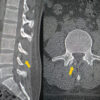
The patient was planned for staged revision spine surgery with extension of the fusion. A pre-operative computed tomography (CT) scan of the lumbar spine without contrast was obtained in July 2025, and it demonstrated post-surgical changes consistent with a laminectomy and intervertebral cage at L5–S1. The position of the cage remained unchanged and demonstrated increased endplate sclerosis with partial ankylosis as expected. CT also demonstrated interval resorption of the bone graft within the laminectomy bed, with associated soft-tissue density extending into the intraspinal canal, initially suspected to represent post-surgical scarring. Notably, a 2.14 × 2.69 cm posterior soft-tissue mass was identified superficial to the deep spinal fascia (Fig. 1). A subsequent magnetic resonance imaging (MRI) of the lumbar spine without contrast was obtained in August 2025, and revealed an indeterminate T2 hypointense soft-tissue mass in the posterior subcutaneous tissue at S1 (limited by motion artifact), severe spinal canal stenosis at L4–L5 due to disc herniation and ligamentum flavum hypertrophy with central cauda equina crowding, and anterolisthesis of L5 on S1 with caudally migrated disc extrusion causing severe right and mild-to-moderate left lateral recess stenosis with bilateral severe neural foraminal narrowing (Fig. 2).
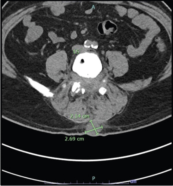
Axial lumbar spine CT without contrast demonstrating interval resorption of the osseous fragment/bony graft at the laminectomy bed with soft-tissue density extending to the intraspinal canal, inseparable and difficult to delineate from the thecal sac within the limitation of lack of contrast. A posterior soft-tissue mass measuring 2.14 by 2.69 cm and superficial to the deep spinal fascia is also seen.
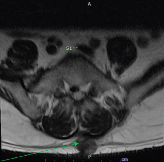
Given the severity of these findings and the patient’s correlating symptoms, he was scheduled for a staged surgical approach. Stage I included removal of the L4–L5 cage and anterior lumbar interbody fusion (ALIF) at L4–S1 with vascular surgery-assisted exposure, followed by L3–pelvis revision decompression and posterior instrumentation with fusion (Stage II).
In August 2025, the patient underwent Stage I surgery consisting of cage removal at L4–L5 and ALIF at L4–L5 and L5–S1. During cage placement, atypical anatomy was encountered, and the iliac vein was partially lacerated with associated tearing, but it was identified, and hemostasis was achieved. On the following day, the patient underwent the second stage, which consisted of L3–pelvis revision, decompression, and fusion. Intraoperatively, a palpable 2.5 × 5.1 cm mass was identified at the inferior aspect of the incision, superficial to the deep spinal fascia. The mass was dissected, and an en bloc excision was performed, with the specimen sent for frozen-section analysis. No abnormal tissue was noted deep to the fascia. Histopathology demonstrated metastatic adenocarcinoma with no definitive immunophenotype. According to the report, possible primary sites included gastric, biliary, and lung origin, with acknowledgment that approximately 20% of lung adenocarcinomas may be thyroid transcription factor-1 negative.
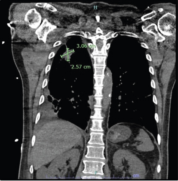
Postoperatively, the patient underwent further investigation to determine the primary site of the metastatic lesion. A CT chest without contrast was obtained in August 2025, which revealed a 3.0 by 2.6 cm spiculated cavitary mass in the right upper lobe abutting the posterior pleura and a right lower paratracheal lung node measuring 2.5 x 2.1 cm (Fig. 3 and 4). The patient underwent staging of the disease with CT cervical spine, CT thoracic spine, and MRI spine and brain, which excluded any evidence of additional metastasis. The patient was then discharged home on the seventh post-operative day and was given follow-up appointments with the orthopedic clinic and oncology for further management of his primary disease.
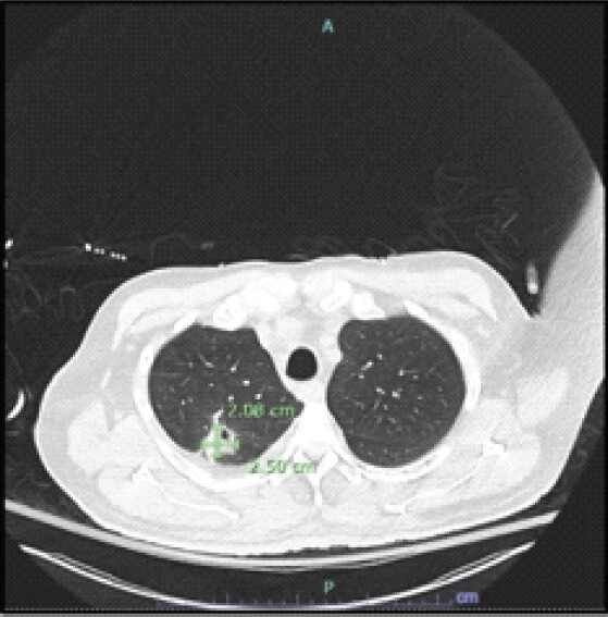
Dominant index right lower paratracheal lymph node measuring 2.5 x 2.1 cm.
On his first orthopedic clinic visit at approximately 3 weeks postoperatively, he reported significant improvement in his preoperative symptoms with only mild residual localized lumbar pain. The patient was evaluated by radiation oncology approximately 6 weeks postoperatively for staging and treatment planning given the diagnosis of metastatic lung adenocarcinoma involving the lumbar region and concern for liver lesions. Molecular testing of the tissue sample taken intraoperatively was positive for a KRAS G12C mutation and low programmed death-ligand 1 expression (1%). A positron emission tomography scan demonstrated a hypermetabolic cavitary right upper lobe, intrathoracic and intra-abdominal metastatic lymphadenopathy, and widespread metastatic involvement including bilateral lung nodules, left adrenal gland, soft-tissue lesions, and bone, specifically a hypermetabolic lytic/sclerotic destructive mass at the left ninth costovertebral junction measuring 4.4 x 2.3 cm, additional multiple foci of hypermetabolism along the lower lumbar spine, and hypermetabolic soft-tissue tumor deposits within the subcutaneous fat along the posterior lower lumbar spine and within the muscles at the right proximal thigh.
The patient was subsequently scheduled to initiate stereotactic body radiation therapy to the lower spine. He received one session of palliative external-beam radiation therapy to the lumbosacral spine (single 8 Gy fraction) for symptomatic bone metastasis from the primary right upper-lobe lung cancer and was started on pemetrexed, carboplatin, and pembrolizumab. Unfortunately, the patient died only two months following initiation of chemotherapy and radiotherapy, without an explicit cause of death.
Discussion
This case demonstrates a rare presentation of metastatic lung adenocarcinoma discovered incidentally as a superficial soft-tissue mass during staged revision lumbar spine surgery. Between 0.75 and 9% of patients with lung cancer have been shown to have soft-tissue metastases [9], with adenocarcinomas most frequently metastasizing to the cutaneous and subcutaneous tissues [10,11,12]. The vast majority of these metastases occur in the dermal and subcutaneous tissue [13], with deep non-vascular subfascial locations being rare. The presence of tumor cells at surgical sites has been hypothesized to relate to impaired lymphatic restoration or fibrin deposition, which may establish a relatively immunologically permissive microenvironment that promotes tumor cell attachment and prevents immune-mediated tumor cell removal [8,14,15,16].
The primary clinical importance is the diagnostic challenge, as atypical presentations are often misdiagnosed as benign post-operative changes such as scar formation, seromas, or local infection [2]. This is particularly relevant for patients undergoing multiple spine surgeries, where surgical changes are to be expected in the post-operative period. In this case, intraoperative frozen-section analysis was invaluable for rapid tissue characterization. Studies report frozen-section diagnostic accuracy rates of 95.1–99.0%, which help to aid the intraoperative decision [17,18,19,20]. Here, the mass’s superficial location without deeper fascial involvement represents an uncommon compartment for lung adenocarcinoma metastasis to occur, as compared to the limited-site metastases cited in the literature.
This case highlights the need for surgeons to maintain a broad differential diagnosis and consider malignancy when a soft-tissue mass is encountered within the surgical approach. Metastatic spread to surgical scars is uncommon and may be influenced by inflammation or changes to the local anatomy in the post-operative period. Special consideration should be given in revision surgery, where there has been repetitive insult to the soft tissues and multiple inflammatory events [8,21].
Intraoperative frozen-section analysis should be performed conscientiously when atypical tissue is encountered during revision surgery to enable immediate diagnosis and appropriate surgical planning, potentially avoiding redundant diagnostic procedures. Advanced imaging for soft-tissue masses alone is non-specific and may resemble benign processes, thereby necessitating tissue confirmation, especially when a primary tumor has not been identified [22,23].
A limitation of this case is that metastasis to a non-vascular superficial compartment following surgery has not previously been described and therefore may not be widely generalizable. In addition, the development of widespread metastatic disease in this patient suggests that the cancer was not confined to the surgical site but was a systemic disease process. Nevertheless, this case emphasizes that any suspicious soft-tissue finding in patients undergoing revision spine surgery warrants histopathological evaluation, as accurate and timely diagnosis is essential for appropriate oncologic staging, treatment planning, and prognostic counseling.
Conclusion
Spine surgeons should maintain a broad differential diagnosis, including malignancy, when encountering unexpected soft-tissue findings in a patient with a prior history of spine surgery and scarring. Intraoperative frozen-section analysis is a valuable tool for rapid tissue characterization and should be utilized when atypical tissue is encountered, enabling timely oncologic diagnosis and appropriate treatment planning. This represents, to the best of our knowledge, an original report of interest to spine surgeons, orthopedic and neurosurgical oncologists, and pathologists, and it advances understanding of the atypical compartments in which lung adenocarcinoma may present.
Clinical Message
Atypical soft-tissue encountered during revision spine surgery should not just be assumed to be benign post-operative scar. Even when imaging is non-specific, intraoperative frozen section enables rapid diagnosis of unexpected malignancy, guiding appropriate oncologic staging and treatment without delay.
Conflict of Interest:
Nil
Source of Support:
Nil
Consent:
The authors confirm that informed consent was obtained from the patient for publication of this article
How to Cite this Article
Sayegh J, Michael B, Carlyon T, Brito E, Moghamis IS, Alhammoud A. Metastatic Lung Cancer to Non-vascular Soft Tissue and Lumbar Spine Mimicking Postoperative Scar at L4–5 Non-union: A Case Report. J Orthop Case Rep 2026 November;16(11): 10-14. doi: 10.13107/jocr.2026.v16.i11.8370.
References
- Surov A, Köhler J, Wienke A, Gufler H, Bach AG, Schramm D . Muscle metastases: Comparison of features in different primary tumours. Cancer Imaging 2014;14:21. [Google Scholar] | [PubMed]
- Plaza JA, Perez-Montiel D, Mayerson J, Morrison C, Suster S. Metastases to soft tissue: A review of 118 cases over a 30-year period. Cancer 2008;112:193-203. [Google Scholar] | [PubMed]
- Hidaka T, Ishii Y, Kitamura S. Clinical features of skin metastasis from lung cancer. Intern Med 1996;35:459-62. [Google Scholar] | [PubMed]
- Yglesias B, Brooker M, DeVito R, Swiger A. Metastatic adenocarcinoma of the lung presenting as a soft tissue mass. Case Rep Surg 2021;2021:8856503. [Google Scholar] | [PubMed]
- Wang J, Wu R, Liu F, Yang L, Hu F, Wu Z . Case report: Lung adenocarcinoma initially presenting with cutaneous and subcutaneous metastases. Front Oncol 2022;12:925382. [Google Scholar] | [PubMed]
- Bae J, Jun D, Kim J, Choi H, Shin D, Park JH . Ulcerative skin metastasis lesion in non-small cell lung cancer. J Wound Manag Res 2020;16:106-12. [Google Scholar] | [PubMed]
- Deckey DG, Reid DB, Mallozzi S, Daniels AH. Cutaneous metastasis via surgical planes after lumbar spinal reconstruction for spinal metastatic disease. World Neurosurg 2019;123:49-53. [Google Scholar] | [PubMed]
- Otsuka I. Cutaneous metastasis after surgery, injury, lymphadenopathy, and peritonitis: Possible mechanisms. Int J Mol Sci 2019;20:3286. [Google Scholar] | [PubMed]
- Perisano C, Spinelli MS, Graci C, Scaramuzzo L, Marzetti E, Barone C . Soft tissue metastases in lung cancer: A review of the literature. Eur Rev Med Pharmacol Sci 2012;16:1908-14. [Google Scholar] | [PubMed]
- Hu SC, Chen GS, Wu CS, Chai CY, Chen WT, Lan CC. Rates of cutaneous metastases from different internal malignancies: Experience from a Taiwanese medical center. J Am Acad Dermatol 2009;60:379-87. [Google Scholar] | [PubMed]
- Dhambri S, Zendah I, Ayadi-Kaddour A, Adouni O, El Mezni F. Cutaneous metastasis of lung carcinoma: A retrospective study of 12 cases. J Eur Acad Dermatol Venereol 2011;25:722-6. [Google Scholar] | [PubMed]
- Kamble R, Kumar L, Kochupillai V, Sharma A, Sandhoo MS, Mohanti BK. Cutaneous metastases of lung cancer. Postgrad Med J 1995;71:741-3. [Google Scholar] | [PubMed]
- Fernandez-Flores A. Cutaneous metastases: A study of 78 biopsies from 69 patients. Am J Dermatopathol 2010;32:222-39. [Google Scholar] | [PubMed]
- Pendergrast WJ Jr, Futrell JW. Biologic determinants of tumor growth in healing wounds. Ann Surg 1979;189:181-8. [Google Scholar] | [PubMed]
- Al Dybiat I, Mirshahi S, Belalou M, Abdelhamid D, Shah S, Ullah M . Injured tissues favor cancer cell implantation via fibrin deposits on scar zones. Neoplasia 2020;22:809-19. [Google Scholar] | [PubMed]
- Ceelen WP, Bracke ME. Peritoneal minimal residual disease in colorectal cancer: Mechanisms, prevention, and treatment. Lancet Oncol 2009;10:72-9. [Google Scholar] | [PubMed]
- Winther C, Graem N. Accuracy of frozen section diagnosis: A retrospective analysis of 4785 cases. APMIS 2011;119:259-62. [Google Scholar] | [PubMed]
- Laokulrath N, Nasir NDM, Gudi M, Tan PH. Diagnostic accuracy and challenges of intraoperative frozen section evaluation for axillary sentinel lymph node biopsy and breast margins. Histopathology 2025;86:1121-36. [Google Scholar] | [PubMed]
- Mohamed A, Hassan MM, Zhong W, Kousar A, Takeda K, Donthi D . A quantitative and qualitative assessment of frozen section diagnosis accuracy and deferral rate across organ systems. Am J Clin Pathol 2022;158:692-701. [Google Scholar] | [PubMed]
- Gephardt GN, Zarbo RJ. Interinstitutional comparison of frozen section consultations. A college of American pathologists Q-probes study of 90,538 cases in 461 institutions. Arch Pathol Lab Med 1996;120:804-9. [Google Scholar] | [PubMed]
- Cheng X, Zhang H, Hamad A, Huang H, Tsung A. Surgery-mediated tumor-promoting effects on the immune microenvironment. Semin Cancer Biol 2022;86 Part 3:408-19. [Google Scholar] | [PubMed]
- Nishimura H, Zhang Y, Ohkuma K, Uchida M, Hayabuchi N, Sun S. MR imaging of soft-tissue masses of the extraperitoneal spaces. Radiographics 2001;21:1141-54. [Google Scholar] | [PubMed]
- Riley GM, Kwong S, Steffner R, Boutin RD. MR imaging of benign soft tissue tumors: Highlights for the practicing radiologist. Radiol Clin North Am 2022;60:263-81. [Google Scholar] | [PubMed]
© 2026 Journal of Orthopaedic Case Reports - Published by Indian Orthopaedic Research Group
About the Authors
-

Jonathan Sayegh
-

Bryce Michael
-

Thadeus Carlyon
-

Emmanuel Brito
-

Isam Sami Moghamis
-

Abduljabbar Alhammoud
Related Articles in Journal of Orthopaedic Case Reports
 November 1, 2026 Management of Retained Epidural Catheter Fragment in a Polytrauma Patient: A Case Report
 September 1, 2026 Triple Procedure for Direct Pars Repair: An Innovative, Non-fusion Approach for Managing Spondylolysis in Late Adolescence or Early Adulthood
 August 1, 2026 The Forgotten Joint and Forgotten Spine: Recalibrating Success around the Patient’s Experience
 August 1, 2026 O-arm Navigation-assisted Excision of Cervical Osteoid Osteoma Adjacent to the Vertebral Artery: Technical Note and Case Report