

O-arm-based intraoperative navigation enables precise localization and complete excision of cervical pedicular osteoid osteoma situated adjacent to the vertebral artery, minimizing neurovascular risk, preserving spinal stability, and reducing the likelihood of residual nidus and recurrence.
Dr. Sandesh Subhash Agrawal, Department of Orthopedics and Spine Surgery, Shree Narayana Hospital, Raipur - 492 001, Chhattisgarh, India. E-mail: preciouss333@gmail.com
Abstract
Introduction: Osteoid osteoma is a benign osteoblastic tumor that rarely involves the cervical spine. Lesions arising from the cervical pedicle adjacent to the vertebral artery present significant diagnostic and surgical challenges due to the complex regional anatomy and risk of neurovascular injury. Although radiofrequency ablation is an established treatment modality, surgical excision remains an important option when lesions are located close to critical neurovascular structures.
Case Report: A 20-year-old male presented with persistent nocturnal right-sided lower cervical pain for 8 months, which showed temporary relief with non-steroidal anti-inflammatory drugs. The patient failed prolonged conservative management. Computed tomography (CT) demonstrated a well-defined nidus with surrounding reactive sclerosis involving the right C7 pedicle, while magnetic resonance imaging revealed associated marrow edema and inflammatory soft-tissue changes. Due to the lesion’s proximity to the vertebral artery, the patient underwent navigation-guided excision using an intraoperative three-dimensional imaging system (O-arm® Intraoperative Imaging System, Medtronic, Minneapolis, MN, USA) through a posterior cervical approach. Intraoperative navigation enabled precise localization and complete excision of the nidus while preserving adjacent stabilizing structures and avoiding injury to surrounding neurovascular elements. Post-operative CT confirmed complete tumor removal, and histopathological examination established the diagnosis of osteoid osteoma. The patient experienced immediate pain relief and remained symptom-free without recurrence or neurological deficit during follow-up.
Conclusion: O-arm-based intraoperative navigation facilitates safe and accurate excision of cervical pedicular osteoid osteoma located adjacent to the vertebral artery. The technique allows complete nidus removal while minimizing unnecessary bone resection, preserving cervical stability, and reducing the risk of neurovascular complications. Navigation-assisted excision represents a valuable treatment option for osteoid osteomas situated in anatomically challenging regions of the cervical spine
Keywords: Osteoid osteoma, cervical spine, vertebral artery, O-arm navigation, pedicle lesion, spine surgery.

Osteoid osteoma is a benign osteoblastic skeletal tumor first described by Jaffe in 1935 [1]. It accounts for approximately 2–3% of all primary bone tumors and 10–12% of benign bone tumors [2]. Spinal involvement is reported in nearly 10–25% of cases, with the lumbar spine being the most commonly affected region, whereas cervical spine involvement is relatively rare and constitutes approximately 4% of all osteoid osteomas [2,3]. Lesions in the cervical spine are most commonly located in the posterior elements and pose unique surgical challenges due to their proximity to the spinal cord, nerve roots, and vertebral artery [3,4]. Clinically, cervical osteoid osteoma typically presents with persistent localized neck pain that worsens at night and shows partial relief with non-steroidal anti-inflammatory drugs (NSAIDs) [4]. Depending on the site of involvement, patients may also develop neck stiffness, radiculopathy, torticollis, or painful deformity [3,4]. Due to its rarity and non-specific presentation, diagnosis is frequently delayed and may mimic cervical disk disease, inflammatory disorders, or infection. Computed tomography (CT) is considered the imaging modality of choice for identifying the characteristic nidus and surrounding reactive sclerosis, whereas magnetic resonance imaging (MRI) is useful for evaluating adjacent neural and soft-tissue involvement [3,5]. Complete excision of the nidus remains the definitive treatment and is associated with excellent symptomatic relief and low recurrence rates [3]. However, lesions adjacent to the vertebral artery are technically demanding because incomplete excision may result in recurrence, and aggressive exposure may risk neurovascular injury [3,5]. O-arm–based intraoperative navigation provides real-time three-dimensional localization, facilitating precise tumor excision while minimizing injury to adjacent critical structures and preserving spinal stability. We report a rare case of cervical osteoid osteoma adjacent to the vertebral artery managed successfully using O-arm navigation-assisted surgical excision.
A 20-year-old male presented with complaints of persistent right-sided lower cervical pain for 8 months, with progressive worsening in intensity over time. The pain was predominantly nocturnal, significantly interfered with sleep, and demonstrated transient partial relief with NSAIDs. Despite prolonged conservative management, including analgesics, physiotherapy, and activity modification, the patient continued to experience persistent symptoms with significant impairment of daily activities. There was no history of trauma, fever, weight loss, constitutional symptoms, or prior cervical spine surgery. Clinical examination revealed localized tenderness over the lower cervical spine with painful restriction of terminal neck movements, particularly extension and right lateral rotation. No cervical deformity or torticollis was noted. Neurological examination was unremarkable, with preserved motor strength, intact sensation, normal deep tendon reflexes, and no signs of cervical myelopathy or radiculopathy. Plain radiographs of the cervical spine were inconclusive and did not demonstrate any obvious osseous abnormality. A CT scan of the cervical spine revealed a well-defined lucent nidus surrounded by reactive sclerosis involving the right pedicle of the C7 vertebra, consistent with osteoid osteoma. MRI diffuse signal abnormality involving the right half of the vertebral body and right posterior neural arch of the C7 vertebrae, showing mild hyperintensities on short tau inversion recovery T2-weighted image and diffuse enhancement with the presence of a small lytic lesion with internal hyperdense nidus involving the right C7 pedicle, suggestive of osteoid osteoma. Based on the characteristic clinical presentation and radiological findings, a diagnosis of osteoid osteoma involving the right C7 pedicle was established. Given the persistence of symptoms despite failed conservative treatment and the close proximity of the lesion to the vertebral artery and adjacent neural structures, surgical excision was planned. The patient underwent navigation-assisted excision of the lesion under general anesthesia using intraoperative O-arm-based three-dimensional navigation. Following posterior cervical exposure, a reference frame was secured, and intraoperative O-arm imaging was obtained to facilitate real-time CT-based navigation. Accurate localization of the nidus within the right C7 pedicle with contiguous extension into the vertebral body and ipsilateral lamina was achieved using navigation guidance. Using a high-speed burr and curettes, en bloc excision of the nidus along with the surrounding sclerotic margins was performed under continuous navigation guidance. Particular attention was paid to preserving the integrity of the adjacent vertebral artery, exiting nerve root, and surrounding stabilizing structures. Adequate preservation of the facet joint and posterior tension band obviated the need for instrumented stabilization or fusion. Repeat intraoperative O-arm imaging confirmed complete excision of the lesion without residual nidus. The post-operative course was uneventful, with marked improvement in axial neck pain immediately following surgery. Post-operative CT imaging demonstrated complete excision of the nidus with preservation of surrounding cervical anatomy (Fig. 1).
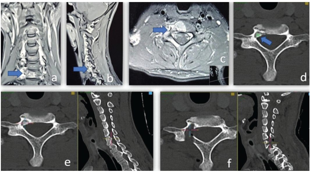
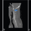
Figure 1: Pre-operative magnetic resonance imaging (MRI), computed tomography (CT), and post-operative CT imaging of right C7 pedicle osteoid osteoma. (a) Coronal MRI showing inflammatory marrow edema and soft-tissue reaction involving the right C7 posterior element (blue arrow). (b) Sagittal MRI demonstrating a focal lesion involving the right C7 pedicle with surrounding inflammatory changes (blue arrow). (c) Axial MRI revealing edema and inflammatory soft-tissue changes adjacent to the right C7 pedicle in close proximity to the vertebral artery (blue arrow). (d) Axial CT image demonstrating a well-defined radiolucent nidus measuring approximately 0.49 × 0.43 cm with surrounding reactive sclerosis involving the right C7 pedicle (blue arrow). (e) Axial and sagittal pre-operative CT-based images demonstrating precise localization of the nidus within the right C7 pedicle. (f) Axial and sagittal postoperative CT images confirming complete excision of the nidus with preservation of the surrounding cervical osseous anatomy.
Histopathological examination revealed interlacing trabeculae of osteoid and woven bone lined by prominent osteoblasts within a fibrovascular stroma, confirming the diagnosis of osteoid osteoma (Fig. 2). At latest follow-up, the patient remained asymptomatic with no evidence of recurrence, cervical instability, or neurological deficit.
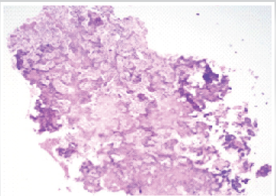
Figure 2: Photomicrograph showing tissue fragments composed of thin interconnecting trabeculae of immature woven bone lined by osteoblasts. No significant pleomorphism, mitotic activity, or necrosis is identified. Histomorphological features are suggestive of osteoid osteoma (Hematoxylin and Eosin stain, ×100).
Cervical osteoid osteoma is an infrequently encountered benign osteogenic neoplasm and represents a relatively small proportion of spinal osteoid osteomas, with cervical involvement reported in approximately 4% of all cases [3,5]. Despite its benign biological behavior, cervical osteoid osteoma remains clinically significant due to its intimate relationship with critical neurovascular structures, including the spinal cord, cervical nerve roots, and vertebral artery [5]. The diagnosis is frequently delayed due to the lesion’s small size, variable inflammatory response, and non-specific symptomatology, often resulting in misdiagnosis as cervical disk disease, inflammatory spondylopathy, or infective pathology [4,6]. The pathophysiology of pain in osteoid osteoma is attributed to markedly elevated prostaglandin concentrations within the nidus and the presence of abundant unmyelinated neural fibers within the vascular osteoid matrix [7]. This biochemical activity explains the characteristic nocturnal exacerbation of pain and transient responsiveness to NSAIDs. In spinal lesions, reactive muscle spasm and localized inflammatory response may additionally produce painful restriction of cervical motion, secondary deformity, or radicular symptoms depending on lesion location [3,4]. In the present case, persistent nocturnal axial neck pain with temporary NSAID responsiveness and failure of prolonged conservative management strongly correlated with the classical clinical profile described in previous literature. The posterior elements are the predominant site of involvement in spinal osteoid osteoma due to increased biomechanical stress concentration and relatively rich vascularity [3]. Pedicular lesions within the cervical spine present a unique surgical challenge due to the limited osseous corridor and proximity to the vertebral artery and exiting nerve roots. Lesions involving the C7 pedicle are particularly demanding because excessive resection may compromise facet integrity and posterior column biomechanics, whereas incomplete nidation excision is associated with persistent symptoms and recurrence [5,8]. Previous reports have consistently demonstrated that recurrence in osteoid osteoma is predominantly related to residual nidus tissue rather than aggressive tumor biology [5]. Consequently, precise intraoperative localization and complete excision of the nidus remain the fundamental principles of surgical management. Radiologically, conventional radiographs are frequently non-diagnostic, particularly in cervical lesions, due to the small nidus size and obscuration by overlapping osseous anatomy [5]. CT remains the diagnostic gold standard due to its superior spatial resolution and ability to delineate the nidus, internal mineralization, cortical involvement, and surrounding reactive sclerosis [3,5]. In the present case, CT imaging precisely localized the nidus within the right C7 pedicle and facilitated detailed preoperative surgical planning. MRI, although sensitive for identifying marrow edema, adjacent inflammatory changes, and neural compression, may occasionally obscure the nidus due to extensive reactive inflammatory response and therefore may mimic infective or neoplastic lesions [2,9]. This emphasizes the importance of CT-MRI correlation in the evaluation of persistent unexplained cervical pain in young patients. Percutaneous radiofrequency ablation (RFA) has demonstrated high success rates in appendicular osteoid osteoma and is increasingly considered a minimally invasive therapeutic option [10]. However, its application in cervical spinal lesions remains limited due to the potential risk of thermal injury to adjacent neural and vascular structures [5,10]. Lesions located within the pedicle or adjacent to the vertebral artery are particularly unsuitable for ablative techniques due to the inability to reliably control heat dispersion within the confined cervical anatomy. In addition, lack of tissue diagnosis and the possibility of incomplete nidus ablation remain important limitations of RFA in spinal lesions [5]. Therefore, surgical excision continues to represent the preferred treatment strategy in cervical osteoid osteoma located adjacent to critical neurovascular structures. Recent advances in intraoperative imaging and spinal navigation have substantially enhanced the accuracy and safety of complex cervical spinal procedures [11,12]. O-arm-based navigation provides real-time intraoperative three-dimensional imaging and allows precise localization of the nidus with high spatial accuracy. In the present case, CT-based navigation was particularly advantageous because the lesion was situated adjacent to the vertebral artery within a narrow operative corridor. Navigation facilitated accurate trajectory planning, controlled pedicular drilling, and targeted excision of the nidus while minimizing unnecessary bone removal and preserving adjacent stabilizing structures. Preservation of the facet complex and posterior tension band avoided the need for instrumented stabilization or fusion. Furthermore, repeat intraoperative O-arm imaging enabled immediate confirmation of complete excision, thereby minimizing the likelihood of residual lesions and recurrence. Histopathological examination remains essential for definitive diagnosis and characteristically demonstrates interlacing trabeculae of woven bone and osteoid rimmed by osteoblasts within a highly vascular fibroconnective stroma [1,7]. The patient in the present report demonstrated immediate post-operative resolution of nocturnal pain and sustained symptomatic improvement without recurrence or cervical instability on follow-up evaluation. The present case highlights the clinical utility of O-arm-based navigation in achieving safe, anatomically precise, and complete excision of cervical pedicular osteoid osteoma adjacent to the vertebral artery while preserving spinal stability and minimizing neurovascular risk.
Cervical osteoid osteoma involving the pedicle is a rare and diagnostically challenging lesion that should be considered in young patients presenting with persistent nocturnal neck pain responsive to NSAIDs. Lesions adjacent to the vertebral artery require meticulous surgical planning because incomplete excision may result in recurrence, while excessive resection may compromise spinal stability. O-arm-based intraoperative navigation enables precise nidus localization, facilitates safe complete excision, and minimizes neurovascular injury and unnecessary bone removal. Navigation-guided excision represents an effective and reliable treatment strategy for complex cervical osteoid osteoma.
Cervical osteoid osteoma should be considered in young patients presenting with persistent nocturnal neck pain that responds to NSAIDs. In lesions located adjacent to critical neurovascular structures such as the vertebral artery, O-arm-based navigation provides a safe and effective method for complete nidus excision while minimizing unnecessary bone removal and preserving cervical stability.
References
- 1. Jaffe HL. Osteoid-osteoma: A benign osteoblastic tumor composed of osteoid and atypical bone. Arch Surg 1935;31:709-28. [Google Scholar] [PubMed]
- 2. Achar TS, Raj AA, Krishnan KA, Hegde SK. Osteoid osteoma of the body of cervical vertebra-a case report of an unusual presentation. Indian Spine J 2025;8:75-80. [Google Scholar] [PubMed]
- 3. Gadiya A, Shah K, Patel P, Nene A. Osteoid osteoma of cervical spine: A case report and review of literature. J Orthop Case Rep 2019;9:78-81. [Google Scholar] [PubMed]
- 4. Kaner T, Sasani M, Oktenoglu T, Aydin S, Ozer AF. Osteoid osteoma and osteoblastoma of the cervical spine: The cause of unusual persistent neck pain. Pain Physician 2010;13:549-54. [Google Scholar] [PubMed]
- 5. Laus M, Albisinni U, Alfonso C, Zappoli FA. Osteoid osteoma of the cervical spine: Surgical treatment or percutaneous radiofrequency coagulation? Eur Spine J 2007;16:2078-82. [Google Scholar] [PubMed]
- 6. Pettine KA, Klassen RA. Osteoid osteoma and osteoblastoma of the spine. J Bone Joint Surg Am 1986;68:354-61. [Google Scholar] [PubMed]
- 7. O’Connell JX, Nanthakumar SS, Nielsen GP, Rosenberg AE. Osteoid osteoma: The uniquely innervated bone tumor. Mod Pathol 1998;11:175-80. [Google Scholar] [PubMed]
- 8. Faraj A, Byrne P, Mehdian H. Osteoid osteoma of the lateral mass of C5. Should excision be combined with fusion? Eur Spine J 1998;7:242-5. [Google Scholar] [PubMed]
- 9. Barbiera F, Murmura E, Murmura B, Accardi M, Botta A, Aiello A, et al. Osteoid Osteoma: Typical and Atypical Radiological Findings. C-2922 European Congress of Radiology (ECR 2018); 2018. [Google Scholar] [PubMed]
- 10. Hadjipavlou AG, Lander PH, Marchesi D, Katonis PG, Gaitanis IN. Minimally invasive surgery for ablation of osteoid osteoma of the spine. Spine (Phila Pa 1976) 2003;28:E472-7. [Google Scholar] [PubMed]
- 11. Rajasekaran S, Kanna RM, Shetty AP. Intraoperative navigation in spine surgery: Evolution and current applications. Asian Spine J 2010;4:1-10. [Google Scholar] [PubMed]
- 12. Costa F, Cardia A, Ortolina A, Fabio G, Zerbi A, Fornari M. Spinal navigation: Standard pre-operative versus intraoperative computed tomography data set acquisition for computer-guidance system: Radiological and clinical study in 100 consecutive patients. Spine 2011;36:2094-8. [Google Scholar] [PubMed]






Related Articles in Journal of Orthopaedic Case Reports
 September 1, 2026 Pediatric Cervical Spine Aneurysmal Bone Cyst with Recurrent Atlantoaxial Rotatory Subluxation Requiring C1–C2 Fusion: A 7-Year Follow-Up Case Report
 September 1, 2026 Triple Procedure for Direct Pars Repair: An Innovative, Non-fusion Approach for Managing Spondylolysis in Late Adolescence or Early Adulthood
 September 1, 2026 Combined Ultrasound- and Fluoroscopy-guided Percutaneous Cementoplasty and Ablation for a C2 Pathologic Fracture: A Case Report
 August 1, 2026 The Forgotten Joint and Forgotten Spine: Recalibrating Success around the Patient’s Experience