


Introduction
Pelvic-ring injuries are uncommon in children, and sacroiliac (SI) joint disruption is a particularly rare pattern. The growing pelvis differs from the adult pelvis in its greater elasticity and cartilaginous growth regions; in young children, SI-joint and pubic-symphysis injuries are reported among characteristic pelvic-ring injury patterns. Pediatric pelvic injuries are generally associated with high-energy trauma, and occult injury to the posterior ring may be difficult to appreciate on plain radiographs, making cross-sectional imaging useful when clinical suspicion persists [1].
The acetabulum is involved in 4–20% of these fractures and is often associated with a pelvic ring fracture. It is vital to note that these fractures frequently occur in the setting of high-energy trauma and polytrauma. The vast majority of them are motor vehicle-related accidents: A car-pedestrian collision in 40–80% of instances, or a passenger in a light vehicle in 10–30% of cases [2]. Given the circumstances, practitioners must be aware of the heightened risk of related and perhaps fatal injuries. This is especially true in young children, where the triradiate cartilage is still open and protected by the flexibility of the pubic symphysis and SI joints [3].
In pediatric pelvic injuries, management should aim to restore pelvic stability while preserving the growth potential of the acetabulum and surrounding physes. In children with open growth plates, anatomical reduction of significantly displaced acetabular cartilage is essential to minimize long-term hip dysfunction. Surgical intervention may be considered after a short delay when substantial displacement persists and considerable acetabular growth potential remains. Temporary fixation using Kirschner wires that spare the growth plates for 4–6 weeks may be employed to maintain reduction while minimizing growth disturbance [4]
Long-term follow-up is important because residual pelvic asymmetry, SI-joint changes, back pain, and leg-length discrepancy have been reported after childhood SI-joint disruption. In a retrospective series of 18 children, follow-up averaged 14 years, and radiographic SI-joint changes did not consistently correspond to symptoms [5]. We report a rare case of neglected traumatic SI-joint dislocation with anterior pelvic-ring disruption in a young child, successfully treated by delayed open reduction and Kirschner-wire fixation. The case highlights the importance of appropriate management and continued clinical and radiographic surveillance during growth.
Case Report
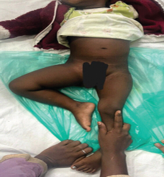
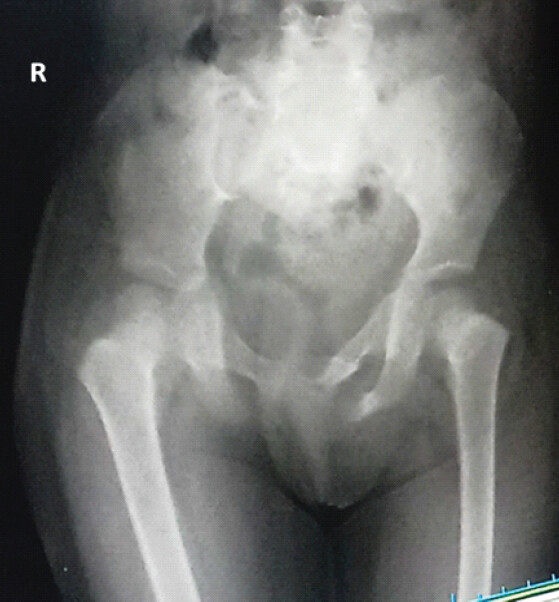
An 18-month-old girl, who had been walking independently before the injury, sustained trauma following a fall from a tractor. She was initially managed conservatively at another hospital and presented 3 months after the injury with an inability to walk and pain localized to the right lower back. The right lower limb was held in flexion, abduction, and external rotation (Fig. 1). Clinical examination revealed restricted movements of the right hip and tenderness over the right SI joint.
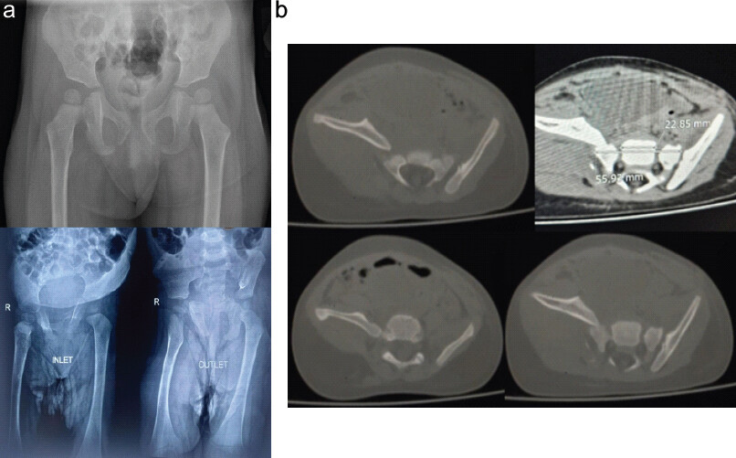
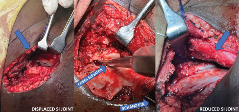
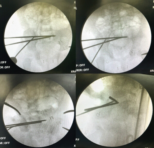
Radiographs and computed tomography demonstrated separation of the right SI joint, anterior displacement of the right hemipelvis, and disruption of the pubic symphysis (Fig. 2a and b). Open reduction of the right SI joint was performed using a Cobb elevator and a Schanz pin placed in the iliac crest to facilitate reduction (Fig. 3). The reduced joint was stabilized using 3-mm Kirschner wires (Fig. 4).
Postoperatively, the child was permitted to perform bed exercises while maintaining non-weight-bearing status for 6 weeks, after which mobilization was initiated. The Kirschner wires were removed 3 months after surgery, and the child regained independent walking at 6 months postoperatively. At the latest follow-up, the 3-year-old child was able to walk independently and sit cross-legged comfortably. Radiographs obtained after 1-year post-surgery demonstrated a well-reduced right SI joint without secondary displacement or loss of reduction. Continued clinical and radiological follow-up was maintained to monitor functional recovery and pelvic development (Figs. 5 and 6).
Discussion
Traumatic SI-joint disruption in a young child is rare and may be overlooked because the immature pelvic ring is flexible and the injury may coexist with anterior-ring disruption. The clinical findings in this child – persistent lower-back pain, inability to walk, abnormal limb posture, and focal SI tenderness – were therefore important indicators of a clinically significant posterior-ring injury despite the delayed presentation. Cross-sectional imaging confirmed separation of the right SI joint, anterior displacement of the hemipelvis, and pubic-symphysis disruption. Comparable pediatric reports describe both isolated and combined SI injuries, emphasizing that the injury pattern and the child’s clinical status must be assessed together rather than inferred from plain radiographs alone [6].
The decision to operate was guided by persistent displacement, associated anterior-ring disruption, pain, and major functional disability rather than by the time elapsed since injury alone. In a 4-year-old child with complete anterior SI dislocation, Elnahal et al. achieved stabilization using K-wires and an external fixator [7], while Abdelgawad et al. reported that ilio-sacral screw fixation can be performed in children when the fixation corridor is carefully planned and imaging guidance is used [8]. Brown et al. likewise described reduction aids and bilateral SI screws for an adolescent with bilateral dislocation. Together, these reports support an individualized strategy based on age, pelvic size, displacement pattern, associated injuries, and the available safe fixation corridors [6]. In the present child, a Cobb elevator and an iliac Schanz pin permitted controlled mobilization of the displaced hemipelvis, and 3-mm Kirschner wires provided temporary stabilization in a very small pelvis while avoiding more extensive implants. The broader pediatric series by Wang et al. also supports tailoring the fixation method to fracture morphology and associated pelvic injuries [9].
The post-operative course was favorable: Protected mobilization began after 6 weeks of non-weight-bearing, independent walking returned by 6 months, and comfortable cross-legged sitting was subsequently achieved. At 1-year follow-up, radiographs demonstrated maintained SI-joint reduction without secondary displacement or loss of alignment. This outcome is consistent with the generally favorable early functional results reported after operative treatment of unstable pediatric pelvic injuries; in a prospective series of 45 surgically treated children, Arafa et al. reported excellent functional results in 93.3% of patients, although complications – including pin-tract infection and limb-length discrepancy – were also observed [10]. The 5-year follow-up reported by Elnahal et al. further illustrates the value of surveillance beyond the early post-operative period [7]. Nevertheless, this remains a single case with limited follow-up relative to skeletal growth. Continued clinical and radiological surveillance through maturity is therefore required to assess pelvic symmetry, SI-joint development, gait, chronic pain, and limb-length equality.
Conclusion
Neglected SI joint disruption in young children can present with significant functional disability and poses a challenging management dilemma. This case demonstrates that carefully planned delayed open reduction and temporary Kirschner-wire fixation can restore pelvic alignment and achieve satisfactory functional recovery, even 3 months after injury. However, long-term clinical and radiological surveillance is essential to monitor pelvic growth and identify potential late complications.
Clinical Message
Persistent pain, inability to walk, and abnormal limb positioning following pediatric pelvic trauma should raise suspicion of an underlying pelvic-ring injury, including SI joint disruption. Delayed presentation should not preclude surgical intervention when significant displacement and functional impairment persist. Careful reduction, preservation of growth-sensitive structures, and long-term follow-up are a key to achieving favorable outcomes.
Conflict of Interest:
Nil
Source of Support:
Nil
Consent
The authors confirm that informed consent was obtained from the patient for publication of this article
How to Cite this Article
Mahadevuni V, Srinivas PL, Ramesh L, Sale PKA. Neglected Traumatic Sacroiliac Joint Disruption in a Young Child: Delayed Open Reduction and Functional Recovery. J Orthop Case Rep. 2026 November;16(11): 309-313. doi: 10.13107/jocr.2026.v16.i11.8482.
References
- Aprato A, Giudice C, Bedino P, Audisio A, Massè A. Current concepts in pediatric pelvic ring fractures: A narrative review. Trauma Care 2022;2:359-72. [Google Scholar] | [PubMed]
- Guillaume JM, Pesenti S, Jouve JL, Launay F. Pelvic fractures in children (pelvic ring and acetabulum). Orthop Traumatol Surg Res 2020;106:S125-33. [Google Scholar] | [PubMed]
- Leonard M, Ibrahim M, Mckenna P, Boran S, McCormack D. Paediatric pelvic ring fractures and associated injuries. Injury 2011;42:1027-30. [Google Scholar] | [PubMed]
- Liporace FA, Ong B, Mohaideen A, Ong A, Koval KJ. Development and injury of the triradiate cartilage with its effects on acetabular development: review of the literature. J Trauma Acute Care Surg 2003;54:1245-9. [Google Scholar] | [PubMed]
- Heeg M, Klasen HJ. Long-term outcome of sacroiliac disruptions in children. J Pediatr Orthop 1997;17:337-41. [Google Scholar] | [PubMed]
- Brown M, Simister S, Paul AV, Helmkamp JK, McGowan S, DeBaun M. Bilateral sacroiliac joint dislocation, discussion and surgical strategies. JBJS Case Connect 2023;13. ???. [Google Scholar] | [PubMed]
- Elnahal WA, Fahmy M, Acharya MR. Open complete anterior dislocation of the sacro-iliac joint in a 4-year-old boy: A case report of a rare injury with 5-year follow-up. Strategies Trauma Limb Reconstr 2018;13:51-5. [Google Scholar] | [PubMed]
- Abdelgawad AA, Davey S, Salmon J, Gurusamy P, Kanlic EM. Ilio-sacral (IS) screw fixation for sacral and sacroiliac joint (SIJ) injuries in children. J Pediatr Orthop 2016;36:117-21. [Google Scholar] | [PubMed]
- Wang Y, Du X, Tomaszewski R, Journeau P, Mayr J. Operative management of sacroiliac joint dislocation in children with unstable pelvic fractures – A STROBE-compliant investigation. J Orthop 2024;52:6-11. [Google Scholar] | [PubMed]
- Arafa MW, Khalifa AA, Fergany A, Abdelhafez MA, Mohamedean A, Adam FF. Surgical management of paediatric pelvic fractures: A prospective case series and early experience from a level one Egyptian trauma centre. Int Orthop 2022;46:2315-28. [Google Scholar] | [PubMed]
© 2026 Journal of Orthopaedic Case Reports - Published by Indian Orthopaedic Research Group
About the Authors
-

Vishwanath Mahadevuni
-

P L Srinivas
-

L Ramesh
-

Pradeep Kumar A Sale
Related Articles in Journal of Orthopaedic Case Reports
 December 1, 2025 Recurrent Giant Cell Tumor of the Carpal Bones in an Adult Male Managed by En-bloc Excision, Extended Curettage, Bone Grafting, and all-Kirschner Wire Fixation: A Rare Case Report and Literature Review
 February 1, 2025 Stealth Navigation with iFuse-TORQ Implant for Sacroiliac Joint Fusion Technique Guide
 December 1, 2024 Percutaneous Doxycycline Ablation for Recurrent Aneurysmal Bone Cyst in the Sacroiliac Joint: A Case Report
 June 10, 2022 Management of Partial Heel Pad Avulsion using Multiple Kirschner wire Anchorage Technique – Case Report