


Introduction
Lateral condyle fractures of the humerus account for approximately 10–20% of pediatric elbow fractures and represent one of the most important physeal injuries around the elbow in children [1]. Although the majority of these fractures heal with appropriate and timely management, non-union is a well-recognized complication, with a reported incidence ranging from 1.1% to 2.2% [2]. Non-union most commonly occurs as a result of missed or unrecognized fractures, delayed presentation, or inadequate primary treatment, including insufficient reduction or stabilization [3]. The diagnosis can be challenging, particularly in younger children, because of the predominantly cartilaginous distal humerus and minimal displacement visible on initial radiographs, which increases the risk of misdiagnosis and inappropriate management [4]. Several biological and mechanical factors contribute to the development of non-union. These include impaired vascularity of the metaphyseal fragment, interposition of synovial fluid between fracture fragments, and continuous traction forces exerted by the forearm extensor muscles, which prevent fracture healing [5]. In addition, technical errors during treatment, such as inadequate fixation or premature immobilization removal, may further increase the risk [5]. If left untreated, non-union can lead to significant long-term complications, including persistent pain, elbow instability, progressive cubitus valgus deformity, and tardy ulnar nerve palsy [6]. The development of cubitus valgus is particularly concerning in children, as the lateral condyle contributes to the growth and stability of the distal humerus, and progressive deformity may worsen with skeletal growth.
The management of fractures of the lateral condyle of the humerus with delayed presentation in children remains controversial. Early surgical intervention is generally recommended to achieve union, restore anatomy, and prevent progressive deformity and neurological complications [4]. Various surgical options have been described, including open reduction and internal fixation, in situ fixation, bone grafting, corrective osteotomy, and anterior transposition of the ulnar nerve, depending on the chronicity of non-union and associated deformity [7,8,9]. However, there is no clear consensus regarding the optimal timing and method of treatment, and outcomes reported in the literature remain variable. Therefore, further evaluation of non-union of the lateral condyle humerus in children is essential to determine the most effective management strategies.
Materials and Methods
Study design and setting
This was a retrospective-prospective study conducted at a tertiary care center after obtaining approval from the Institutional Ethics Committee (Ref. No.: VIMS/IEC/2024/95). The retrospective arm included patients treated between 2022 and 2024, and the prospective arm included patients enrolled between 2024 and 2025. Written informed consent was obtained from the parents or legal guardians of all patients.
Inclusion criteria
The study included children who met the following criteria: (1) Age <16 years. (2) Displaced lateral condyle fracture of the humerus. (3) Delayed presentation, defined as presentation more than 3 weeks after injury. (4) Minimum follow-up of 3 months. (5) Symptomatic: Pain, stiffness, deformity. (6) For established non-union: Large metaphyseal fragment, displacement of <1 cm from joint surface, an open, viable lateral condylar physis (Flynn criteria [10]).
Exclusion criteria
Patients were excluded if they had (1) closed physes. (2) Undisplaced fractures. (3) Pathological fractures. (4) Previous surgical intervention for the same fracture. (5) Associated fractures around the same elbow. (6) Inadequate follow-up.
A total of 19 patients satisfying the inclusion criteria were included in the study.
Clinical and radiological evaluation
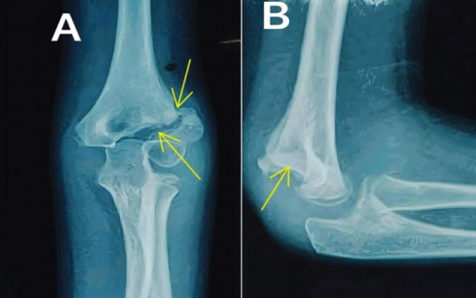
Baseline demographic data, including age, sex, side involved, mechanism of injury, and duration between injury and presentation, were recorded. Clinical evaluation included assessment of pain, elbow range of motion measured using a goniometer, presence of deformity, Mayo Elbow Performance Score (MEPS) [11], and ulnar nerve function. Radiological evaluation was performed using standard anteroposterior and lateral radiographs of the elbow (Fig. 1). Fracture displacement, presence of sclerosis, and features suggestive of delayed union or non-union were assessed.
Surgical technique
All patients were treated surgically under general anesthesia with a tourniquet. Antibiotic prophylaxis was given with 1.5 g of cefuroxime 30 min before the incision. A standard lateral approach to the elbow was used. The fracture site was exposed, fibrous tissue was removed, and the fracture ends were freshened. Reduction was achieved, and internal fixation was performed using a single cannulated cancellous screw (4 mm), two screws, or a combination of Kirschner wire (1.5–2 mm) and screw, depending on the size of the fragment and stability achieved intra-operatively. In cases of established non-union, the metaphyseal fragment of the lateral condyle and distal humeral metaphysis are exposed, without attempting to anatomically realign the articular surface. Intra-articular and posterior dissection was minimized to avoid osteonecrosis and further loss of movement. Bone grafting was not performed in any patient.
In two patients who presented 1 year after injury with established cubitus valgus deformity, corrective dome osteotomy and fixation with plate were performed in addition to anterior transposition of the ulnar nerve in one patient who had developed features of ulnar neuropathy.
Post-operative protocol
The limb was immobilized in an above-elbow plaster backslab for 4–6 weeks. Following removal of plaster, gradual active range-of-motion exercises were initiated. Patients were followed up at regular intervals with clinical and radiological evaluation until fracture union.
Outcome measures
Radiological union was assessed based on the presence of bridging trabeculae across the fracture site on follow-up radiographs. Clinical outcome assessment included pain, range of motion, carrying angle, and complications such as infection, implant failure, or ulnar nerve palsy. Functional outcome was evaluated using MEPS (a score of 90–100 is excellent, 75–89 is good, 60–74 is fair, and <60 is poor).
Statistical analysis
Data were recorded and analyzed using the Statistical Package for the Social Sciences (version 26). Descriptive statistics were used to summarize the data. Continuous variables were expressed as mean and range, and categorical variables were expressed as frequencies and percentages.
Descriptive statistics were used to summarize the data. A paired t-test was used to compare pre-operative and post-operative range of motion and MEPS. Spearman correlation was used to assess association between age and functional outcome. The Chi-square test was used to assess the association between categorical variables. P < 0.05 was considered statistically significant.
Results
A total of 19 patients with delayed presentation of lateral condyle fracture of the humerus were included in the present study.
The mean age of the patients was 8.6 ± 2.87 years (range: 5–15 years). Of the total participants, 10 patients (52.6%) were male, and 9 (47.4%) were female. The right elbow was involved in 11 patients (57.9%), whereas the left elbow was affected in eight patients (42.1%). The mean duration between injury and presentation was 13.6 ± 12.57 weeks. The most common mode of injury was fall from height, accounting for 15 patients (78.9%), whereas road traffic accidents were responsible for injury in 4 patients (21.1%) (Table 1).
Demographic characteristics of the study population (n=19)
| Variable | Value |
|---|---|
| Age (years), mean±SD | 8.6±2.87 |
| Sex (n %) | |
| Male | 10 (52.6) |
| Female | 9 (47.4) |
| Side involved (n %) | |
| Right | 11 (57.9) |
| Left | 8 (42.1) |
| Duration from injury to presentation (weeks), mean±SD | 13.6±12.57 |
| Mode of injury – Fall from height (n %) | 15 (78.9) |
| Mode of injury – Road traffic accident (n %) | 4 (21.1) |
SD: Standard deviation
The most common presenting complaint was pain associated with decreased range of motion, which was observed in 12 patients (63.2%). Pain and stiffness were reported in 3 patients (15.8%). Lateral condyle prominence was noted in 2 patients (10.5%). Valgus deformity was present in 1 patient (5.3%), whereas 1 patient (5.3%) presented with valgus deformity associated with ulnar neuropathy. The mean follow-up after surgery was 29.77 ± 7.3 weeks (range 13–55 weeks).
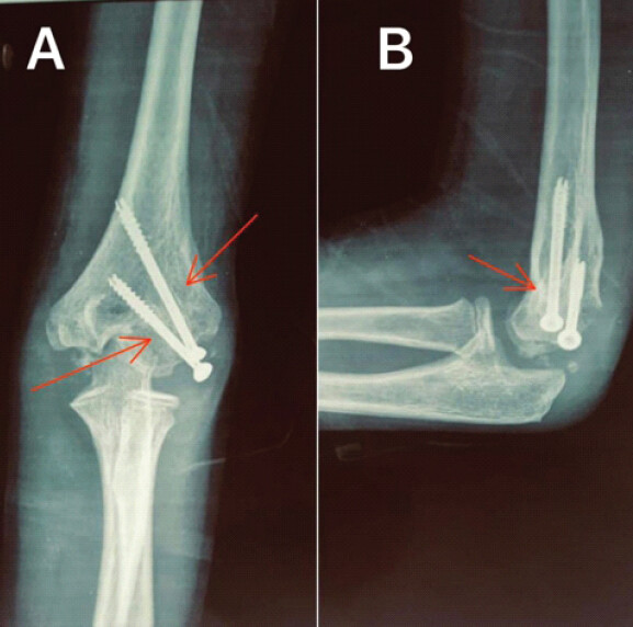
Union was achieved in all patients (Fig. 2).
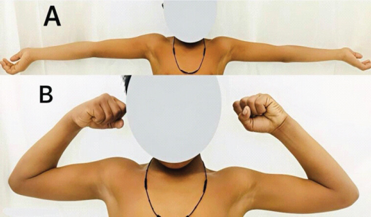
Functional outcome (MEPS) and range of motion (Table 2). The mean pre-operative MEPS was 74.6 ± 6.57, which improved to 77.5 ± 8.37 postoperatively, with a mean improvement of 2.9 points. However, statistical analysis using the paired t-test demonstrated that this improvement was not statistically significant (P = 0.18). Based on post-operative MEPS grading, 3 patients (15.8%) achieved excellent outcomes, 9 patients (47.4%) had good outcomes, and 7 patients (36.8%) had fair outcomes, whereas no patient had a poor outcome. Overall, 63.2% of patients achieved good to excellent functional outcomes following surgical intervention. The mean pre-operative elbow flexion was 116.23° ± 14.6°, which improved to 128.63° ± 10.24° postoperatively, representing a mean improvement of 12.4°. This improvement was found to be statistically highly significant (paired t-test, P < 0.001). The mean pre-operative elbow extension deficit was –10.45° ± 12.8°, which improved to 2.7° ± 4.4° postoperatively, representing a mean improvement of 13.15°. This improvement was also statistically highly significant (paired t-test, P < 0.001) (Fig. 3).
Comparison of pre-operative and post-operative outcomes (Paired t-test)
| Variable | Pre-operative Mean±SD | Post-operative Mean±SD | t -statistic | P -value |
|---|---|---|---|---|
| MEPS | 74.6±6.57 | 77.5±8.37 | t=1.19 | 0.18 |
| Elbow flexion (°) | 116.23±14.6 | 128.63±10.24 | t=3.03 | <0.001 |
| Elbow extension (°) | –10.45±12.8 | 2.7±4.4 | t=11.1 | <0.001 |
MEPS: Mayo elbow performance score, SD: Standard deviation
Association between demographic variables and functional outcome: Spearman correlation analysis demonstrated a weak positive correlation between age and post-operative MEPS (r = 0.23), which was not statistically significant (P = 0.35). The association between sex and functional outcome, as well as side of injury and functional outcome, was analyzed using Fisher’s exact test due to the small sample size. No statistically significant association was observed between sex and post-operative functional outcome (P > 0.05) or between side of injury and post-operative functional outcome (P > 0.05).
Complications were observed in 10 patients (52.6%). Superficial infection occurred in three patients and resolved with antibiotic therapy. K-wire migration was noted in two patients, which required early removal of the wires. Lateral condylar prominence was observed in two patients. Fishtail deformity and wound dehiscence were noted in one patient each, with the wound dehiscence requiring re-suturing. In addition, one patient presented 2 months after surgery with elbow stiffness, which subsequently improved and regained functional range of motion following physiotherapy and range-of-motion exercises.
Discussion
The present study demonstrates that open reduction and internal fixation for lateral condyle humerus fractures with delayed presentation leads to significant improvement in elbow range of motion, particularly flexion and extension. However, the improvement in the mean MEPS from 74.6 ± 6.57 to 77.5 ± 8.37 was not statistically significant (P = 0.18). The most common presenting complaint in our study was pain associated with decreased range of motion, followed by pain with stiffness, lateral condyle prominence, and valgus deformity. These findings highlight the functional impairment and deformity commonly associated with delayed presentation of lateral condyle fractures.
Delayed presentation of lateral condyle humerus fractures is frequently associated with pain, stiffness, cubitus valgus deformity, tardy ulnar nerve palsy, and occasionally elbow instability [2]. Management of established non-union poses several technical challenges. With progressive displacement, the lateral condyle fragment migrates proximally relative to the medial condyle, resulting in asymmetry and incongruity of the distal humeral articular surface [12]. In addition, the fragment often develops fibrous non-union with the distal humerus, making anatomical reduction difficult. Restoration of articular alignment requires careful mobilization of the fragment and may increase the risk of avascular necrosis due to the extensive soft-tissue dissection required for fragment mobilization [12]. Furthermore, long-standing valgus deformity may need to be addressed to restore normal elbow alignment and biomechanics. In the present study, dome corrective osteotomy was performed in two patients with valgus deformity, and one of them also required anterior transposition of the ulnar nerve due to symptomatic ulnar neuropathy.
The timing of surgical intervention in delayed lateral condyle humeral fractures has been widely debated. Earlier authors such as Jakob et al. [13] and Fontanetta et al. [14] cautioned against delayed open reduction due to concerns regarding vascular compromise and the risk of avascular necrosis. Conversely, other authors have suggested that because of the superior healing potential in children compared to adults, surgical intervention may still yield satisfactory results in the pediatric population even after delayed presentation [15,16]. More recent studies have further supported the role of surgical fixation even in long-standing non-union, reporting favorable functional outcomes following operative management [17,18].
In a retrospective study of 27 patients with neglected lateral humeral condyle fractures, Trisolino et al. [4] reported a mean time from injury to presentation of 27 months. The presenting symptoms were variable, with pain and/or stiffness observed in 85% of patients. 16 (59%) had a relatively preserved range of motion with an arc greater than 100°, whereas 12 patients had a flexion contracture exceeding 30°. Deformity was present in eight patients, including valgus deformity in six, three of whom also demonstrated ulnar neuropathy. The treatment modalities included nonoperative management, in situ fixation, open reduction and internal fixation, and corrective osteotomy. At a mean follow-up of 7 years, complications were reported in 59% of patients, although only 22% were considered major. Despite this, pain and functional outcomes improved in 85% of patients. The mean MEPS improved from 62 preoperatively to 98 postoperatively, and the Dhillon score improved from five to eight points.
Liu et al. [7] evaluated 16 children presenting at a mean of 7.4 weeks after injury and reported significant improvement in functional outcomes following surgical treatment. The mean Dhillon functional score improved from 3.3 to 5.6, and the overall functional score improved from 5.6 to 8.5 at final follow-up. Complications such as fishtail deformity and premature physeal closure were noted; however, no cases of avascular necrosis were reported. Importantly, they found that neither age, delay in surgery, nor pre-operative functional status significantly influenced complication rates. These findings are comparable with those of the present study, where significant improvement in elbow range of motion was observed, and complications such as fishtail deformity occurred in a small number of patients without significantly affecting functional outcomes. Furthermore, similar to Liu et al. [7], our study did not demonstrate a statistically significant correlation between age and post-operative MEPS, suggesting that satisfactory outcomes can be achieved across different pediatric age groups.
Shrestha and Hutchison [19] reported outcomes in 40 patients with delayed presentation who underwent surgical treatment. They observed fracture union in 97.5% of patients and excellent functional outcomes, with a mean MEPS of 93. They also reported a mean improvement in the arc of elbow motion exceeding 40°, indicating substantial restoration of joint mobility following surgical fixation. A multicenter retrospective study [20] involving 63 patients from eight medical centers with humeral lateral condylar non-union reported a mean patient age of 7.3 years and a mean follow-up duration of 7 years. All cases of non-union achieved successful union following treatment, with restoration of the humeroulnar angle. The reported rate of avascular necrosis was 16.7%. Overall, patients demonstrated excellent post-operative range of motion and MEPS at final follow-up. Multivariable linear regression analysis revealed that fracture displacement and duration from initial injury were statistically significant predictors of functional outcome. Similarly, Eamsobhana and Kaewpornsawan [21] evaluated 17 patients with lateral condyle non-union and reported excellent to good outcomes in all cases, with a mean MEPS of 94 and successful fracture union in nearly all patients. They also demonstrated a negative correlation between the duration of neglect and functional outcome, suggesting that prolonged delay may adversely affect elbow function.
Agarwal et al. [22], in a study of 22 children with delayed presentation, reported pain, swelling, restriction of elbow motion, lateral condylar prominence, and valgus deformity as the most common presenting symptoms. Union was achieved in 20 cases, whereas one patient developed malunion and another experienced condylar resorption following post-operative infection and avascular necrosis. Additional radiographic findings included prominent lateral condyle, fishtail deformity, and premature physeal closure. The average Liverpool Elbow Score at final follow-up was 8.12, reflecting satisfactory functional outcomes. Zhang et al. [2], in a systematic review involving 177 patients, reported post-operative union in 159 patients (89.8%). Mixed-effects logistic regression analysis demonstrated that percutaneous fixation was associated with lower union rates compared with open fixation. Age at surgery did not significantly affect union rates. However, increased age at surgery and reduction of the fracture fragment were associated with reduced post-operative range of motion, whereas female sex and corrective osteotomy were associated with improved post-operative range of motion.
The findings of the present study are consistent with previous reports demonstrating improvement in elbow motion following surgical fixation. In our cohort, mean flexion improved by 12.4°, whereas extension improved by 13.15°. However, the mean post-operative MEPS in our study was lower than that reported in some previous studies. This difference may be attributed to variations in follow-up duration, pre-operative functional status, and duration of neglect. In our study, the mean duration from injury to presentation was 13.6 ± 12.5 weeks, with the majority of patients presenting with pain and decreased elbow motion. In addition, demographic factors such as age, gender, side of injury, and mechanism of injury did not demonstrate a statistically significant correlation with functional outcome. This may be related to the relatively small sample size and narrower range of delay before presentation. An important observation in the present study was the absence of a significant correlation between patient age and functional outcome. This finding is consistent with the observations of Liu et al. [7], who also reported no association between age and complication rate. However, it differs from the findings of Eamsobhana and Kaewpornsawan [21], who reported a negative correlation between increasing age and functional outcome. These discrepancies may be explained by differences in sample size, duration of neglect, and patient demographics across studies.
The complication profile observed in our study was comparable with that reported in previous studies. These complications were generally minor and were managed successfully without adversely affecting the final functional outcome. Similar complication patterns have been reported by Liu et al. [7], Shrestha and Hutchison [19], and Eamsobhana and Kaewpornsawan [21], suggesting that surgical fixation remains a relatively safe and reliable treatment when performed with careful surgical technique.
The present study has several limitations. The small sample size of 19 patients limits statistical power, subgroup analysis, and generalizability. The retrospective-prospective, single-center design may have introduced selection bias and heterogeneity in data collection, whereas the absence of a control group prevents comparison with alternative treatment strategies. The wide variation in delay to presentation and inclusion of both delayed fractures and established non-unions further increased clinical heterogeneity. There was also heterogeneity in surgical management, including different fixation constructs and corrective osteotomy in two patients, limiting meaningful comparison of surgical techniques. As bone grafting was not performed, its potential role in selected non-unions could not be assessed. Although elbow flexion and extension improved significantly, the improvement in MEPS was not statistically significant, and its subjective components may introduce measurement bias. Potential confounding factors such as fracture displacement, duration of neglect, deformity, and pre-operative functional status could not be adequately controlled because of the small sample size. Radiological assessment was based primarily on plain radiographs without standardized advanced imaging for assessment of articular congruity, avascular necrosis, or physeal status. The mean follow-up duration was 29.77 ± 7.3 weeks (range, 13–55 weeks). Although the minimum follow-up of 3 months was defined as an inclusion criterion, the relatively short and variable follow-up limits assessment of long-term functional outcomes and late complications, including growth disturbance, premature physeal closure, progressive cubitus valgus, tardy ulnar neuropathy, and avascular necrosis. Larger prospective multicenter studies with longer follow-up and standardized treatment and outcome assessment are required to better define the long-term outcomes of delayed lateral condyle fractures and non-unions.
Conclusion
Open reduction and internal fixation without bone grafting were associated with fracture union and significant improvement in elbow range of motion in children presenting late with lateral condyle fractures of the humerus. Although the improvement in MEPS was not statistically significant, most patients achieved satisfactory short-term functional outcomes. However, the small sample size, absence of a comparison group, variation in fracture chronicity and surgical techniques, and limited follow-up restrict the strength and generalizability of these findings. Surgical treatment may provide satisfactory short-term clinical and radiological outcomes in appropriately selected patients; however, larger prospective multicenter studies with standardized treatment protocols, comparative analyses, and longer follow-up are required to better define the optimal management strategy and long-term outcomes of delayed lateral condyle fractures and established non-unions in children.
Clinical Message
Delayed symptomatic lateral condyle fractures in children can achieve union and satisfactory elbow function with surgical fixation without bone grafting in selective patients. Careful assessment, individualized planning, and long-term follow-up are essential.
Conflict of Interest:
Nil
Source of Support:
Nil
Consent
The authors confirm that informed consent was obtained from the patient for publication of this article
How to Cite this Article
Kumar A, Kumar S, Gupta D, Hussain A, Ul Islam MS. Outcomes of Open Reduction and Internal Fixation of Pediatric Lateral Condyle Fractures of Humerus with Delayed Presentation. J Orthop Case Rep. 2026 November;16(11): 314-321. doi: 10.13107/jocr.2026.v16.i11.8484.
References
- Tan SH, Dartnell J, Lim AK, Hui JH. Paediatric lateral condyle fractures: A systematic review. Arch Orthop Trauma Surg 2018;138:809-17. [Google Scholar] | [PubMed]
- Zhang S, Tan SH, Lim AK, Hui JH. Surgical outcomes in paediatric lateral condyle non-union: A systematic review and meta-analysis. Orthop Traumatol Surg Res 2022;108:102933. [Google Scholar] | [PubMed]
- Li XT, Shen XT, Wu X, Chen XL. A novel transverse ultrasonography technique for minimally displaced lateral humeral condyle fractures in children. Orthop Traumatol Surg Res 2019;105:557-62. [Google Scholar] | [PubMed]
- Trisolino G, Antonioli D, Gallone G, Stallone S, Zarantonello P, Tanzi P. Neglected fractures of the lateral humeral condyle in children; Which treatment for which condition?. Children (Basel) 2021;8:56. [Google Scholar] | [PubMed]
- Salgueiro L, Roocroft JH, Bastrom TP, Edmonds EW, Pennock AT, Upasani VV. Rate and risk factors for delayed healing following surgical treatment of lateral condyle humerus fractures in children. J Pediatr Orthop 2017;37:1-6. [Google Scholar] | [PubMed]
- Toh S, Tsubo K, Nishikawa S, Inoue S, Nakamura R, Harata S. Long-standing nonunion of fractures of the lateral humeral condyle. J Bone Joint Surg Am 2002;84:593-8. [Google Scholar] | [PubMed]
- Liu TJ, Wang EB, Dai Q, Zhang LJ, Li QW, Zhao Q. Open reduction and internal fixation for the treatment of fractures of the lateral humeral condyle with an early delayed presentation in children: A radiological and clinical prospective study. Bone Joint J 2016;98 -B:244-8. [Google Scholar] | [PubMed]
- Saraf SK, Khare GN. Late presentation of fractures of the lateral condyle of the humerus in children. Indian J Orthop 2011;45:39-44. [Google Scholar] | [PubMed]
- Abed Y, Nour K, Kandil YR, El-Negery A. Triple management of cubitus valgus deformity complicating neglected nonunion of fractures of lateral humeral condyle in children: A case series. Int Orthop 2018;42:375-84. [Google Scholar] | [PubMed]
- Flynn JC, Richards JF Jr., Saltzman RI. Prevention and treatment of non-union of slightly displaced fractures of the lateral humeral condyle in children. An end-result study. J Bone Joint Surg Am 1975;57:1087-92. [Google Scholar] | [PubMed]
- Morrey BF. Functional evaluation of the elbow Morrey BF, Sanchez-Sotelo J, Morrey ME. Morrey's The Elbow and Its Disorders.. 5th ed. Philadelphia, PA. Elsevier 2018:66-74. [Google Scholar] | [PubMed]
- Su F, Dang Y, Li M, Qu J, Wang B, Wang X. A standardized rehabilitation protocol following two-stage surgery for nonunion of lateral humeral condyle fracture with cubitus valgus in children: Functional and radiographic outcomes. BMC Musculoskelet Disord 2025;26:1005. [Google Scholar] | [PubMed]
- Jakob R, Fowles JV, Rang M, Kassab MT. Observations concerning fractures of the lateral humeral condyle in children. J Bone Joint Surg Br 1975;57:430-6. [Google Scholar] | [PubMed]
- Fontanetta P, Mackenzie DA, Rosman M. Missed, maluniting, and malunited fractures of the lateral humeral condyle in children. J Trauma 1978;18:329-35. [Google Scholar] | [PubMed]
- Masada K, Kawai H, Kawabata H, Masatomi T, Tsuyuguchi Y, Yamamoto K. Osteosynthesis for old, established non-union of the lateral condyle of the humerus. J Bone Joint Surg Am 1990;72:32-40. [Google Scholar] | [PubMed]
- Shimada K, Masada K, Tada K, Yamamoto T. Osteosynthesis for the treatment of non-union of the lateral humeral condyle in children. J Bone Joint Surg Am 1997;79:234-40. [Google Scholar] | [PubMed]
- Gong MQ, Huang XW, Wang C, Zha YJ, Li Y, Zhang LD. Management of chronic neglected lateral condyle elbow non-union in adults: Functional results of a cohort study and a proposed treatment algorithm. Int Orthop 2017;41:157-64. [Google Scholar] | [PubMed]
- Ibrahim MA, Ismail MS. Corrective osteotomy and in situ fusion for late-presenting nonunion of lateral condyle fractures of the humerus in adults. J Shoulder Elbow Surg 2019;28:520-4. [Google Scholar] | [PubMed]
- Shrestha S, Hutchison RL. Outcomes for late presenting lateral condyle fractures of the humerus in children: A case series. J Clin Orthop Trauma 2020;11:251-8. [Google Scholar] | [PubMed]
- Adulkasem N, Lewsirirat S, Adulyanukosol V, Sirirungruangsarn Y, Phongprapapan P, Unprasert P. Long-term outcome of nonunion of the lateral humeral condyle fracture in children: A multicentre retrospective study. Int Orthop 2024;48:1785-91. [Google Scholar] | [PubMed]
- Eamsobhana P, Kaewpornsawan K. Should we repair nonunion of the lateral humeral condyle in children?. Int Orthop 2015;39:1579-85. [Google Scholar] | [PubMed]
- Agarwal A, Qureshi NA, Gupta N, Verma I, Pandey DK. Management of neglected lateral condyle fractures of humerus in children: A retrospective study. Indian J Orthop 2012;46:698-704. [Google Scholar] | [PubMed]
© 2026 Journal of Orthopaedic Case Reports - Published by Indian Orthopaedic Research Group
About the Authors
-

Ashok Kumar
-

Sandeep Kumar
-

Dhruv Gupta
-

Altaf Hussain
-

Mir Shahid Ul Islam
Related Articles in Journal of Orthopaedic Case Reports
 May 1, 2026 Outcomes of Internal Fixation in Proximal Humerus Head Split Fractures in Young Adults – How Often Is It Missed and Can We Really Afford to Do So
 April 1, 2026 Novel Technique for Fracture Non-Union in Children – A Case Report
 March 1, 2026 Clavicle Fracture Non-Union in a 3-Year-Old Child: A Case Report and Literature Review
 January 1, 2026 Successful Management of a 40-Year-Old Non-union of the Humeral Shaft with Pseudoarthrosis with Plate Fixation and Bone Grafting: A Case Report