


Introduction
Distraction osteogenesis, popularized by Ilizarov, has become an established technique for limb lengthening, deformity correction, management of bone defects, and treatment of complex non-unions [1,2,3]. The procedure is based on the principle of gradual distraction across a biologically active corticotomy site, stimulating new bone formation through the tension-stress effect while simultaneously promoting adaptation of surrounding soft tissues.[1,2,4].
Successful distraction osteogenesis depends upon multiple biological and mechanical factors, such as preservation of vascularity using low-energy corticotomy, stability of fixation, latency period, distraction rate and rhythm, and rehabilitation. Of these variables, the corticotomy technique plays a pivotal role because it directly influences the biological environment responsible for regenerate formation [1,2,4,5,6,7].
Multiple drill-hole corticotomy, as described by De Bastiani et al. [5], and the Gigli saw corticotomy popularized by Paley [6] are among the most commonly used low-energy osteotomy techniques. Although both the methods aim to minimize soft tissue injury, the relative superiority of either technique remains controversial with existing literature [8,9,10,11].
The present study was undertaken to compare regenerate quality, post-operative pain, functional outcomes, operative duration, and complications following lower limb corticotomy performed using multiple Drill-Hole and Gigli Saw techniques.
Materials and Methods
Twenty patients who underwent lower limb distraction osteogenesis between October 2019 and April 2022 at our center were included in our study. All patients were older than 18 years of age. Patients with pre-existing metabolic diseases and who underwent corticotomy for deformity correction were not included in the study. These patients were observed in two groups of ten patients each based on the type of corticotomy performed. The choice of corticotomy technique was made independently by the treating surgeon as part of routine clinical practice. Monolateral fixator limb reconstruction system (LRS) is used following corticotomy involving the femur, whereas Ilizarov ring fixator is used in all cases involving the tibia. Patients were subsequently categorized according to the technique utilized. Group A included patients who underwent corticotomy using multiple Drill-Hole technique and Group B with Gigli Saw technique.
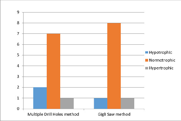
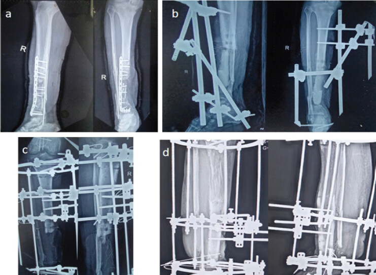
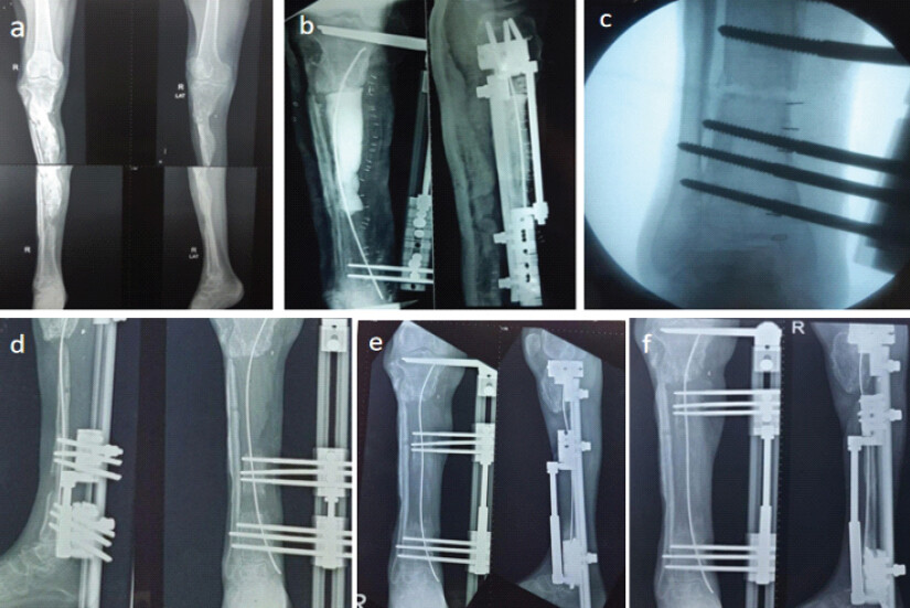
In both groups, a latency period of 7 days was observed before the start of distraction. Distraction was done at a rate of 1 mm in 24 h, divided as 0.25 mm every 6 h in both groups. The patients were monitored clinically and radiologically by weekly X-rays during the distraction phase and monthly in the consolidation phase. Radiological regenerate quality was assessed after six completed weeks of distraction and categorized as hypotrophic regenerate, normotrophic regenerate, hypertrophic regenerate as described by Catagni [12] (Fig. 1, 2, 3). Post-operative pain was assessed using Numeric Pain Rating Scale (NPRS) on post-operative day (POD) 1,7 and 28.
Statistical analysis
Continuous variables were analyzed using the Mann–Whitney U test, while categorical variables were compared using Fisher’s Exact test.
Results
A total of 20 patients were included in the study, with ten patients in each group. The mean age was 31.8 years in Group A and 31.7 years in Group B. The corticotomy was done in the tibia for 8 patients of Group A, with the remaining 2 being femur, whereas in Group B, 9 cases involved tibia and 1 was femur. All cases of femoral lengthening were done using a monolateral fixator LRS, whereas all cases of tibia underwent lengthening using Ilizarov ring fixator. All cases of femoral lengthening were done using proximal diaphyseal corticotomy. However, proximal metaphyseal corticotomy was utilised in all cases involving tibia except 2 cases in each group, where a distal metaphyseal corticotomy was done. Except 1 case of post-traumatic bone loss in each group, all other patients were a case of infected non-union.
The average time taken exclusively for the corticotomy part during the surgery was 10.7 min (95% confidence interval [CI]: 9.48–11.92) in Group A in comparison to 19.9 min (95% CI: 17.96–21.84) in Group B (Mann-Whitney U = 0, P < 0.0001), showing a statistically significant difference between the two groups.
The quality of regenerate was compared radiographically at 6 completed weeks of distraction in all the participants. There were 7 cases of normotrophic, 2 hypotrophic, and 1 hypertrophic regenerate in Group A whereas 8 normotrophic, 1 hypotrophic and 1 hypertrophic regenerate in Group B; with Fisher’s Exact test, P = 1.000. The result showed no statistically significant difference between two groups (Table 1).
Radiological quality of regenerate
| Regenerate type | Group A Multiple drill holes method n (%) | Group B Gigli saw method n (%) |
|---|---|---|
| Hypotrophic | 2 (20) | 1 (10) |
| Normotrophic | 7 (70) | 8 (80) |
| Hypertrophic | 1 (10) | 1 (10) |
The NPRS score was recorded on PODs 1, 7, and 28 (Table 2). The difference in the two groups was not significant, with Mann–Whitney U test values of 45.0,47.5,44.0 and P = 0.63,0.51 and 0.48, respectively. The mean duration of distraction was 58.3 days in Group A and 67.5 days in Group B.
Post-operative pain scores
| NPRS | POD 1 | POD 7 | POD 28 |
|---|---|---|---|
| Group A | 7.1 | 5.4 | 3.6 |
| (95% CI : 6.52–7.68) | (95% CI : 5.05–5.75) | (95% CI : 3.24–3.96) | |
| Group B | 6.9 | 5.35 | 3.3 |
| (95% CI : 6.34–7.46) | (95% CI : 4.98–5.72) | (95% CI : 2.96–3.64) | |
| Mann-Whitney U test | 45 | 47.5 | 44 |
| P-value | 0.63 | 0.51 | 0.48 |
NPRS: Numeric pain rating scale, CI: Confidence interval, POD: Post-operative day
Complications
Complication rates were comparable between groups. Pin tract infection occurred in two patients in each group, which was managed with local wound care and oral antibiotics. One patient in Group A developed posterior tibial artery pseudoaneurysm, which was managed conservatively. One patient in Group A developed chronic infection requiring surgical debridement. Pre-mature regenerate consolidation requiring repeat corticotomy occurred in one patient in Group A. One patient in each group developed equinus deformity requiring tendo-Achilles lengthening. One patient in Group B developed soft tissue impingement at the compression site for which he underwent surgical release. However, no cases of osteomyelitis, perioperative fracture, incomplete corticotomy, valgus deformity, or recurvatum deformity were observed.
Discussion
Distraction osteogenesis remains one of the most powerful biological methods for limb reconstruction and is founded on the tension-stress principles described by Ilizarov. Successful regenerate formation depends upon multiple factors, such as preservation of vascularity, mechanical stability, and adherence to appropriate distraction protocols [1,2,4]. Among these variables, the corticotomy technique plays a pivotal role because it directly influences the biological environment responsible for regenerate formation.
The principal finding of the present study was that both multiple Drill-Hole and Gigli Saw corticotomy techniques produced comparable regenerate quality during distraction osteogenesis. Normotrophic regenerate was the predominant regenerate pattern in both groups, and no statistically significant difference was observed in regenerate morphology. Similarly, post-operative pain scores, functional outcomes, and complication rates were comparable between the two techniques.
The theoretical basis for comparing these techniques lies in their differing effects on periosteal and endosteal blood supply. The Gigli Saw technique has traditionally been regarded as biologically advantageous because the corticotomy is performed through a subperiosteal passage of the saw, minimizing soft tissue disruption and preserving periosteal vascularity. In contrast, the multiple Drill-Hole technique requires repeated cortical perforations and completion with an osteotome, theoretically increasing local tissue trauma. Despite these theoretical differences, both techniques are low-energy corticotomies designed to preserve osteogenic potential.
The present findings demonstrated no statistically significant difference in regenerate morphology, post-operative pain, functional outcomes, or complication rates between the Multiple Drill-Hole and Gigli Saw corticotomy techniques. These observations are consistent with the findings of Peek et al., who compared two proximal tibial osteotomy techniques and reported comparable healing characteristics despite technical differences between procedures [11].
In contrast, Eralp et al. reported significantly superior healing indices following Gigli saw corticotomy and attributed this advantage to improved preservation of periosteal vascularity and reduced soft-tissue trauma [8]. Similarly, Makhdoom et al. observed significantly lower modified healing indices in patients undergoing Gigli saw osteotomy and concluded that preservation of periosteal blood supply may enhance regenerate consolidation [9].
More recently, Rana et al. reported superior healing indices in the multiple drill-hole group and suggested that meticulous preservation of the biological environment, rather than the specific osteotomy technique itself, may be the determining factor for successful regenerate formation [10].
The regenerate morphology observed in the present study may be interpreted according to the regenerate patterns described by Catagni [12]. Normotrophic regenerate, which predominated in both groups, reflects an optimal biological response characterized by adequate vascularity, balanced osteogenesis, and appropriate mechanical stability. Hypotrophic regenerate is generally associated with impaired biological activity or inadequate vascularity, whereas hypertrophic regenerate may represent abundant osteogenic activity occurring under conditions of relative instability. The predominance of normotrophic regenerate in both groups suggests that both corticotomy techniques successfully preserved the biological pre-requisites necessary for distraction osteogenesis.
The conflicting findings reported by Eralp et al., Makhdoom et al., Peek et al. [11], and Rana et al. suggest that the biological influence of corticotomy technique is likely smaller than the influence of fixation stability, distraction protocol, and surgical execution. The present findings support this concept, as theoretical biological advantages did not translate into significant differences in regenerate quality or functional outcomes.
Furthermore, regenerate assessment was based on conventional radiographic morphology. Recent literature suggests that quantitative assessment methods, such as pixel value ratio analysis, bone densitometry, and computed tomography-based evaluation may provide more objective measurements of regenerate maturation and healing potential [13].
An important finding of the present study was the significantly shorter operative duration associated with the multiple Drill-Hole technique, which is in concurrence with a recent study by Rana et al. [10]. Although regenerate quality and clinical outcomes were comparable, the reduced operative time may offer practical advantages, particularly in lengthy reconstructive procedures where surgical efficiency is desirable.
The strengths of the present study lie in its prospective observational design standardized follow-up protocol, and direct comparison of two commonly employed low-energy corticotomy techniques within the same clinical setting. Furthermore, the simultaneous assessment of radiological regenerate quality, post-operative pain, functional outcomes, operative duration, and complications provides a comprehensive evaluation of treatment effectiveness. The study also contributes to the limited body of comparative literature examining the influence of corticotomy technique on outcomes during distraction osteogenesis.
However, this study has several limitations. The sample size was relatively small, limiting statistical power. The study was conducted at a single center and included predominantly young patients with minimal comorbidities, potentially limiting generalizability. Surgical outcomes in distraction osteogenesis are influenced by surgeon experience, rehabilitation protocols, and patient compliance, factors that may not be completely controlled in an observational study design. Finally, regenerate assessment was based on conventional radiographic morphology rather than quantitative techniques, such as pixel value ratio analysis or computed tomography-based assessment.
Conclusion
Both multiple Drill-Hole and Gigli Saw corticotomy techniques produced satisfactory regenerate formation and comparable clinical outcomes during distraction osteogenesis, with no statistically significant differences observed in regenerate quality, post-operative pain, functional outcomes, or complication rates. The multiple Drill-Hole technique was associated with a significantly shorter operative duration. These findings suggest that meticulous preservation of the biological environment and adherence to the principles of distraction osteogenesis may be more important determinants of successful bone regeneration than the specific corticotomy technique employed. Both methods therefore remain effective options for lower limb reconstruction.
Ethics Committee Approval
This study was approved by the Ethics Committee of Armed Forces Medical College vide no.IEC/2019/72 on October 21, 2019.
Clinical Message
Both Multiple Drill-Hole and Gigli Saw corticotomy techniques are reliable low-energy corticotomy methods for distraction osteogenesis. When meticulous surgical technique and standard distraction protocols are followed, regenerate quality and functional outcomes are comparable irrespective of the corticotomy technique employed.
Conflict of Interest:
Nil
Source of Support:
Nil
Consent
The authors confirm that informed consent was obtained from the patient for publication of this article
How to Cite this Article
Venunathan G, Prakash S, Shekhar R, Prasad M, Garg R. Outcomes Following Lower Limb Corticotomy Using Multiple Drill-Hole and Gigli Saw Techniques During Distraction Osteogenesis: A Prospective Observational Comparative Study. J Orthop Case Rep. 2026 November;16(11):498-503. doi: 10.13107/jocr.2026.v16.i11.8536.
References
- Ilizarov GA. The tension-stress effect on the genesis and growth of tissues. Part I. The influence of stability of fixation and soft-tissue preservation. Clin Orthop Relat Res 1989;238:249-81. [Google Scholar] | [PubMed]
- Ilizarov GA. The tension-stress effect on the genesis and growth of tissues: Part II. The influence of the rate and frequency of distraction. Clin Orthop Relat Res 1989;239:263-85. [Google Scholar] | [PubMed]
- Cattaneo R, Catagni MA, Johnson EE. The treatment of infected nonunions and segmental defects of the tibia by the methods of Ilizarov. Clin Orthop Relat Res 1992;280:143-52. [Google Scholar] | [PubMed]
- Gubin AV, Borzunov DY, Malkova TA. The ilizarov paradigm: Current concerns and future research. Int Orthop 2013;37:1533-9. [Google Scholar] | [PubMed]
- De Bastiani G, Aldegheri R, Renzi-Brivio L, Trivella G. Limb lengthening by callus distraction (callotasis). J Pediatr Orthop 1987;7:129-34. [Google Scholar] | [PubMed]
- Paley D. Current techniques of limb lengthening. J Pediatr Orthop 1988;8:73-92. [Google Scholar] | [PubMed]
- Aronson J. Experimental and clinical experience with distraction osteogenesis. Cleft Palate Craniofac J 1994;31:473-81. discussion 481-2. [Google Scholar] | [PubMed]
- Eralp L, Kocaoglu M, Ozkan K, Türker M. A comparison of two osteotomy techniques for tibial lengthening. Arch Orthop Trauma Surg 2004;124:298-300. [Google Scholar] | [PubMed]
- Makhdoom A, Kumar J, Siddiqui AA. Ilizarov external fixation: Percutaneous gigli saw versus multiple drill-hole osteotomy techniques for distraction osteogenesis. Cureus 2019;11:e4973. [Google Scholar] | [PubMed]
- Rana R, Behera HB, Behera S, Prabhat V, Raj M, Panda S. Comparative analysis of distraction osteogenesis with Ilizarov external fixation using percutaneous Gigli saw osteotomy versus multiple drill-hole osteotomy techniques: A prospective observational study. Ann Afr Med 2026 2026. Aril07. https://pesquisa.bvsalud.org/portal/resource/en/mdl-41943438. [Google Scholar] | [PubMed]
- Peek AC, Timms A, Chin KF, Calder PR, Goodier WD. Patterns of healing: A comparison of two proximal tibial osteotomy techniques. Strategies Trauma Limb Reconstr 2016;11:59-62. [Google Scholar] | [PubMed]
- Catagni MA. The radiographic classification of bone regenerate during distraction Bianchi-Maiocchi A, Aronson J. Operative Principles of Ilizarov. Philadelphia, PA. Williams and Wilkins 1991:53-7. [Google Scholar] | [PubMed]
- Cardoso GS, Amorim R. Bone regenerate evaluation methods. Rev Bras Ortop (Sao Paulo) 2024;59:e1-9. [Google Scholar] | [PubMed]
© 2026 Journal of Orthopaedic Case Reports - Published by Indian Orthopaedic Research Group
About the Authors
-

Gopinath Venunathan
-

Syam Prakash
-

Raj Shekhar
-

Manish Prasad
-

Rahul Garg
Related Articles in Journal of Orthopaedic Case Reports
 June 1, 2025 Ilizarov-Assisted Reduction of Femoral Condyle Locked in Tibial Fracture: A Case Report
 November 1, 2026 Immediate Limb Lengthening Following Vascular Repair of Iatrogenic Arterial Lesions – A Report of Two Cases
 November 1, 2026 Compression of the Diseased Segment and Regeneration through Healthy Bone for Salvage of Infected Recurrent Congenital Pseudarthrosis of the Tibia in an Adult: A Case Report
 October 1, 2026 Soft-Tissue Closure through Acute Deformation and Gradual Correction Using Taylor Spatial Frame in a Gustilo-Anderson Type IIIB Open Tibial Pilon Fracture: A Case Report