


Introduction
Pes calcaneus is a sagittal-plane hindfoot deformity characterized by persistent ankle dorsiflexion, commonly seen in neurological conditions such as cerebral palsy, myelodysplasia, and spina bifida [1,2,3]. While the distal consequences of calcaneus deformity are well described, its long-term influence on proximal joint biomechanics remains insufficiently characterized [4]. In a fixed dorsiflexed hindfoot, premature anterior displacement of the ground reaction force (GRF) vector occurs, shifting the sagittal mechanical axis anterior to the center of knee rotation, generating an external hyperextension moment [5].
Chronic exposure to an anteriorly directed sagittal load may lead to progressive attenuation of the posterior capsule and structural adaptation of periarticular soft tissues [5,6]. Over time, this can culminate in severe genu recurvatum and secondary tibiofemoral degeneration. Although coronal-plane interactions between foot deformity and knee osteoarthritis have been investigated, particularly in planovalgus alignment [4], the specific relationship between pes calcaneus deformity and progressive sagittal knee instability has received limited attention.
Genu recurvatum in the setting of neuromuscular disease poses unique reconstructive challenges [7]. Unconstrained or semi-constrained knee designs may inadequately control sagittal instability when posterior capsular structures are incompetent, and quadriceps strength is compromised [7,8]. Recurrence of hyperextension has been reported with less constrained implants, particularly in neurologically impaired patients [8,9,10]. Rotating hinge prostheses, by providing intrinsic sagittal stability independent of soft-tissue integrity, therefore represent a logical reconstructive solution in advanced cases [11,12].
The present report describes two cases of long-standing, untreated pes calcaneus deformity secondary to spina bifida that developed progressive genu recurvatum and end-stage knee osteoarthritis. These cases illustrate a potential distal-to-proximal sagittal failure cascade linking hindfoot dorsiflexion to chronic knee hyperextension and degenerative joint collapse. Recognition of this biomechanical relationship may have implications not only for arthroplasty strategy but also for early deformity correction to prevent secondary proximal joint degeneration.
Case Report
Case 1
A 53-year-old housewife presented with pain in her right knee and difficulty in walking for the past 5 years. She has had bilateral foot deformity since her childhood secondary to a spina bifida at the lower lumbar level. She had not undergone any corrective surgeries in her spine or on her foot in the past. She had undergone an intramedullary nailing for a closed traumatic fracture of the right (ipsilateral) tibia about 8 years ago. She reports progressively increasing hyperextension deformity in her right knee (Fig. 1) for a decade, which was normal in her childhood as far as she remembers.
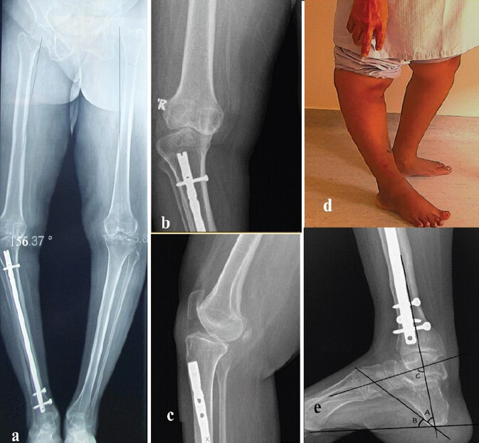
Clinical examination of the right knee revealed varus alignment of about 10° and hyperextension deformity of 25°, and full flexion. The left knee showed 10° varus and approximately 5° of hyperextension. The quadriceps and hip abductors were normal bilaterally. Examination of the feet revealed callosities on the heel pad, calcaneus-valgus alignment of the hind foot, a high longitudinal arch, and a supinated forefoot. These findings were more pronounced on the right side.
The radiographs showed increased calcaneal pitch (44°), decreased tibiocalcaneal angle (36°), and increased talocalcaneal angle (61°), suggestive of severe pes calcaneus deformity (Fig. 1e). Pre-operative limb alignment was 17° of varus at the knee.
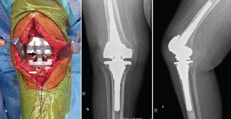
The previous tibial intramedullary nail was buried at the entry point and was inaccessible to the nail-extraction device. A 2-cm long cortical window was created medial to the tibial tuberosity to access the nail (Fig. 2a). She underwent right total knee arthroplasty (TKA) using S-ROM NOILES rotating hinge knee (RHK) System (DePuy, USA, Fig. 2b and 2c). She recovered uneventfully in the post-operative period. She returned with pain in her contralateral left knee 4 years after the right TKA. As this knee had no significant deformities, a posterior-stabilized, primary knee prosthesis was used.
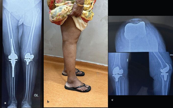
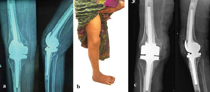
At 5 years of follow-up (Fig. 3), she reported no pain, was ambulant without any walking aids, and had a mild limp likely due to her foot deformities. Post-operative limb alignment was 3° of varus at the knee. She declined further corrective surgery for her foot deformities. Her Oxford knee score improved from 5 to 44.
Case 2
A 73-year-old woman presented with right knee pain and difficulty in walking for about 5 years; her quality of life was significantly affected, and she was dependent on a walker frame to walk (Fig. 4a). She had bilateral foot deformities since childhood secondary to spina bifida at the L5 level. She had not undergone any surgical procedures on her spine and feet in the past.
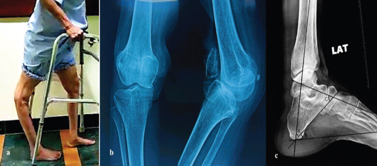
On examination, the right knee exhibited a 10° varus and 30° hyperextension deformity compared to the left knee, which showed a 5° varus and 20° hyperextension deformity. Bilateral feet showed hindfoot calcaneo-valgus deformity and high-arched midfoot. Quadriceps power on the right knee was 3/5, and bilateral plantar flexors were 3/5; all other muscles were 5/5, with no sensory deficits.
Radiographs showed advanced osteoarthritis in the right knee with varus and hyperextension deformity (Fig. 4b). Foot and ankle radiographs demonstrated increased calcaneal pitch (38°), decreased tibiocalcaneal angle (42°), and increased talocalcaneal angle (65°), suggestive of pes calcaneus deformity (Fig. 4c). Preoperatively, limb alignment was 8° of varus at the knee.
She underwent a rotating-hinge TKA using an Endo-Modell rotating-hinge prosthesis (Waldemar LINK GmbH and Co., Hamburg, Germany). She recovered well postoperatively without any surgical complications. Her rehabilitation process was prolonged due to a weak quadriceps mechanism. She had no pain, no recurrence of deformity, and no loosening of the prosthesis at 4-year follow-up (Fig. 5). Postoperatively, limb alignment was 2° of varus at the knee. At the last follow-up, she could ambulate with a walking stick, partially due to contralateral lower-limb involvement. Oxford knee score improved from 2 to 27 at the latest follow-up.
Discussion
The present cases illustrate a potential distal-to-proximal biomechanical cascade in which untreated chronic pes calcaneus deformity may have contributed to progressive sagittal instability of the knee and secondary osteoarthritis. Both patients required complex knee reconstruction with a RHK prosthesis. While the local effects of pes calcaneus deformity in neuromuscular conditions are well documented, its long-term influence on knee sagittal alignment has received comparatively limited attention. Compensatory mechanisms between joints, especially in response to coronal-plane deformities, have been documented in a few studies [1,2]. However, there is limited literature on secondary sagittal-plane deformities of the knee and their surgical management after the development of degenerative arthritis. Unlike primary osteoarthritis associated with coronal malalignment, this pattern represents a predominantly sagittal-plane failure mechanism.
Pes calcaneus deformity is characterized by fixed ankle dorsiflexion resulting from paralysis or weakness of the gastrocnemius–soleus complex, with relative preservation of dorsiflexors and evertors [2,13]. In normal gait, the plantar flexors modulate forward tibial progression during mid-stance and help maintain the GRF vector within a controlled relationship to the knee joint center [13,14]. Loss of plantarflexion power, combined with fixed dorsiflexion, causes premature anterior displacement of the GRF relative to the tibiofemoral axis [6]. This anterior shift creates an external hyperextension moment at the knee [6]. In patients with quadriceps weakness, the knee may adopt hyperextension as an adaptive stabilizing strategy to achieve passive stability during stance [1,6,10].
Chronic exposure to repetitive hyperextension loading may progressively attenuate the posterior capsule and ligamentous stabilizers, leading to fixed sagittal instability [9,10,11]. Altered tibiofemoral mechanics and abnormal contact loading may further accelerate degenerative changes within the knee joint [11,15]. Previous literature has emphasized coronal-plane coupling between foot deformity and knee osteoarthritis, particularly in pes planovalgus alignment and medial compartment loading [4]. However, reports specifically linking fixed calcaneus deformity to progressive genu recurvatum and secondary sagittal degeneration remain sparse [6,16]. The present cases extend this understanding by suggesting persistent hindfoot dorsiflexion may act as a distal contributor to progressive sagittal knee instability.
From a reconstructive perspective, TKA in the presence of fixed recurvatum presents substantial challenges. Conventional cruciate-retaining or posterior-stabilized implants depend on intact soft tissue tension and extensor mechanism function to maintain sagittal stability [8]. In neuromuscular patients with posterior capsular incompetence and quadriceps weakness, recurrence of hyperextension has been reported with less constrained designs [8,17]. RHK prostheses offer a promising solution by stabilizing the knee without relying on the already compromised bony and soft-tissue structures [17]. The extension-stopper design in rotating-hinge prostheses helps prevent recurrence and correct instability (Fig. 2a). RHK has been successfully used in neuromuscular conditions, such as poliomyelitis, with excellent pain relief, improved function, and enhanced activities of daily living [8,17]. However, concerns regarding the recurrence of recurvatum and the need for revision in approximately 7% of cases, as early as 6.2 years, have also been reported [12].
Although some literature exists regarding the influence of pes planovalgus deformity on TKA outcomes [4], studies on the pes calcaneus are lacking. The present cases underscore the potential long-term consequences of untreated pes calcaneus deformity. While early correction of hindfoot malalignment in childhood is primarily advocated to prevent ulceration and functional gait dysfunction [1,2], its role in mitigating proximal joint degeneration warrants further study. Early correction of the pes calcaneus deformity in childhood can provide a plantigrade foot, potentially preventing secondary changes in the proximal joints [1,18]. Methods of surgical treatment of pes calcaneus deformity can be divided into two categories. In young and adolescent patients, the standard treatment is surgical correction of soft-tissue deformities and tendon transfer from functionally preserved muscles [1,18]. At an older age, triple arthrodesis, corrective osteotomy of the calcaneus, and wedge-shaped foot resections are performed [1,18].
Conclusion
These cases suggest untreated pes calcaneus may represent an underrecognized distal contributor to progressive sagittal knee failure in selected neuromuscular populations. However, other factors, including intrinsic quadriceps weakness and age-related degeneration, likely contributed to the development of osteoarthritis. Nonetheless, the consistent pattern of a long-standing pes calcaneus deformity, progressive recurvatum, and advanced sagittal instability suggests a plausible mechanical association warranting further investigation.
Clinical Message
Long-term effects of uncorrected pes calcaneus deformity on the proximal joints and axial skeleton are less studied. In this report, a distal-to-proximal deformity cascade leading to severe genu recurvatum needing a rotating hinge knee arthroplasty is described. Long-term studies are needed to evaluate the causative association and the role of early surgical correction of foot deformities in preventing this complex condition.
Conflict of Interest:
Nil
Source of Support:
Nil
Consent
The authors confirm that informed consent was obtained from the patient for publication of this article
How to Cite this Article
Hulse N, Reddy SC, Charankumar GG. Pes Calcaneus Leading to Genu Recurvatum: A Distal to Proximal Deformity Cascade Treated by Rotating Hinge Arthroplasty: Two Case Reports. J Orthop Case Rep. 2026 November;16(11):231-235. doi: 10.13107/jocr.2026.v16.i11.8452.
References
- Rodrigues RC, Dias LS. Calcaneus deformity in spina bifida: Results of anterolateral release. J Pediatr Orthop 1992;12:461-4. [Google Scholar] | [PubMed]
- Swaroop VT, Dias L. Orthopaedic management of spina bifida-part II: Foot and ankle deformities. J Child Orthop 2011;5:403-14. [Google Scholar] | [PubMed]
- Williams JJ, Graham GP, Dunne KB, Menelaus MB. Late knee problems in myelomeningocele. J Pediatr Orthop 1993;13:701-3. [Google Scholar] | [PubMed]
- Almeheyawi RN, Bricca A, Riskowski JL, Barn R, Steultjens M. Foot characteristics and mechanics in individuals with knee osteoarthritis: Systematic review and meta-analysis. J Foot Ankle Res 2021;14:24. [Google Scholar] | [PubMed]
- Kerrigan DC, Todd MK, Della Croce U. Knee recurvatum in gait: Associated biomechanical factors and implications. Gait Posture 1996;4:235-46. [Google Scholar] | [PubMed]
- Yngve DA. Recurvatum of the knee in cerebral palsy: A review. Cureus 2021;13:e14408. [Google Scholar] | [PubMed]
- Da Silva AG, Giglio PN, Fernandes DA, De Oliveira DT, Da Palma IM, Araújo HR. Primary total knee arthroplasty in patients who have neuromuscular disorders and genu recurvatum using a rotating-hinge implant: A case series with a mean 4-year follow-up. J Arthroplasty 2026;41:1446-52. [Google Scholar] | [PubMed]
- Kouk S, Rathod PA, Maheshwari AV, Deshmukh AJ. Rotating hinge prosthesis for complex revision total knee arthroplasty: A review of the literature. J Clin Orthop Trauma 2018;9:29-33. [Google Scholar] | [PubMed]
- Mortazavi SMJ, Razzaghof M, Noori A, Okati A. Late-onset de novo genu recurvatum after primary total knee arthroplasty: A case series and review. J Knee Surg 2020;33:282-9. [Google Scholar] | [PubMed]
- Gaitán-Lee H, Correa WP, Herrera WP, Villegas JC, Guzmán Nalus F, Gaitán-Duarte H. Genu recurvatum in total knee arthroplasty: A review. J Arthrosc Jt Surg 2024;11(2)92-98. [Google Scholar] | [PubMed]
- Kowalczewski J, Marczak D, Synder M, Sibiński M. Primary rotating-hinge total knee arthroplasty: Good outcomes at mid-term follow-up. J Arthroplasty 2014;29:1202-6. [Google Scholar] | [PubMed]
- Rodríguez-Merchán EC. Total knee arthroplasty using hinge joints: Indications and results. EFORT Open Rev 2019 Apr 25 4(4)121-132. doi: 10.1302/2058-5241.4.180056. PMID: 31057949; PMCID: PMC6492056 [Google Scholar] | [PubMed] | [CrossRef]
- Sobel E, Glockenberg A. Calcaneal gait. Etiology and clinical presentation. J Am Podiatr Med Assoc 1999;89:39-49. [Google Scholar] | [PubMed]
- Ong CF, Geijtenbeek T, Hicks JL, Delp SL. Predicting gait adaptations due to ankle plantarflexor muscle weakness and contracture using physics-based musculoskeletal simulations. PLoS Comput Biol 2019;15:e1006993. [Google Scholar] | [PubMed]
- Dejour D, Bonin N, Locatelli E. Tibial antirecurvatum osteotomies. Oper Tech Sports Med 2000;8:67-70. [Google Scholar] | [PubMed]
- Heijink A, Gomoll AH, Madry H, Drobnič M, Filardo G, Espregueira-Mendes J. Biomechanical considerations in the pathogenesis of osteoarthritis of the knee. Knee Surg Sports Traumatol Arthrosc 2012;20:423-35. [Google Scholar] | [PubMed]
- Moran MC. Functional loss after total knee arthroplasty for poliomyelitis. Clin Orthop Relat Res 1996;323:243-6. [Google Scholar] | [PubMed]
- Kozhevnikov OV, Kralina SE, Gribova IV, Ivanov AV. Pes calcaneus deformity in children and methods of surgical correction. Turner 2019;13:11073. [Google Scholar] | [PubMed]
© 2026 Journal of Orthopaedic Case Reports - Published by Indian Orthopaedic Research Group
About the Authors
-

Narayan Hulse
-

Santhosh C Reddy1
-

G G Charankumar
Related Articles in Journal of Orthopaedic Case Reports
 February 1, 2026 Single-Stage Correction of Genu Recurvatum with Dual-Pillar Plating – A Case Report
 November 1, 2025 Restoring Sagittal Alignment: Anterior Open-Wedge Flexion Valgus Osteotomy for Post-Traumatic Genu Recurvatum with Reversed Posterior Tibial Slope – A Rare Case Report
 October 29, 2014 Malignant Pigmented Villonodular Synovitis-A Rare Entity
 June 15, 2021 Severe Kyphotic Deformity Involving Thoracolumbar Region – A Management Dilemma