


Introduction
Intramuscular myxoma is an uncommon benign mesenchymal tumor arising within skeletal muscle and typically involving large muscle groups, particularly the thigh and gluteal region [1]. The entity was classically described by Stout [2]. Histologically, it is characterized by hypocellularity, bland spindle or stellate cells, abundant myxoid stroma, scant collagen, and low proliferative activity [1,2]. Complete surgical excision is usually curative, and recurrence is uncommon [3].
Magnetic resonance imaging (MRI) may strongly suggest the diagnosis, but its findings are not specific enough to exclude other deep myxoid soft-tissue tumors, including malignant lesions [4]. Intramuscular myxoma may also occur in association with fibrous dysplasia as Mazabraud syndrome [5]. The present case is noteworthy because the lesion became clinically apparent in late pregnancy, enlarged rapidly to giant size, and caused severe sitting intolerance with sciatic-type symptoms, thereby mimicking a more aggressive process.
Case Report
A 34-year-old woman presented to the orthopedic outpatient clinic with a left gluteal mass that caused discomfort and pain while sitting. She reported no relevant prior symptoms in this region. Physical examination revealed a firm, mobile, non-tender mass located to the left of the intergluteal cleft and extending toward the posterior proximal thigh. The externally estimated size was approximately 15 × 15 cm, and no overlying skin change was noted.
The patient had delivered a healthy infant by cesarean section 3 months earlier. She reported that she had first noticed a small mass approximately 3 months before delivery, during the third trimester of her first pregnancy, and that it had enlarged rapidly thereafter. At presentation, she was breastfeeding and stated that she could feed her infant only while lying down because sitting was intolerable. She also reported right shoulder pain, which she attributed to prolonged positioning while nursing. During the preceding 10 days, she had developed burning pain radiating from the buttock along the entire lower extremity in the sciatic nerve distribution.
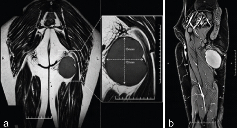
MRI performed at another institution demonstrated an approximately 154 × 155 × 156 mm well-defined intramuscular mass in the left gluteal region adjacent to the ischium, with diffuse subcutaneous fluid accumulation and no osseous destruction. The lesion was reported to lie adjacent to and compress the sciatic nerve. Although the imaging appearance suggested a benign soft-tissue tumor because no contrast enhancement was observed, the radiology report recommended exclusion of a malignant mesenchymal neoplasm because of focal degenerative areas (Fig. 1).
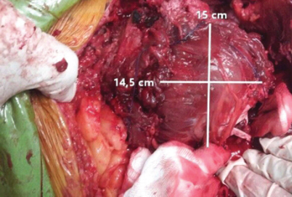
A preoperative core needle biopsy was performed; histopathologic examination showed fibrous and myxoid tissue, with no atypia or malignant cells identified in the sampled material. Because of the severity of the patient’s symptoms, the radiologic impression of a well-circumscribed, resectable lesion, and the preoperative biopsy findings, complete surgical excision was planned. The patient was placed in the prone position, and the procedure was performed under spinal anesthesia. A longitudinal incision was made, and the subcutaneous tissues and fascia were divided in layers. The sciatic nerve was identified, carefully protected, and gently retracted throughout the procedure. A giant mass adherent to the surrounding musculature was excised en bloc with grossly negative margins (Fig. 2).
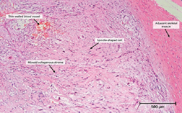
Gross pathologic examination revealed a solid brown-gray tumor measuring 155 × 150 × 145 mm in maximal dimensions. The closest distance to the surgical margin was 1 cm. Microscopic examination demonstrated a hypocellular neoplasm composed of bland spindle cells with elongated nuclei embedded in abundant myxoid stroma, with minimal collagen deposition. Immunohistochemical analysis demonstrated focal CD34 positivity, while smooth muscle actin, desmin, S-100, and CD1a were negative. The Ki-67 proliferation index was approximately 1%, indicating low proliferative activity. These findings were consistent with intramuscular myxoma (Fig. 3).
The postoperative course was uneventful. The patient’s sitting difficulty and sciatic-type pain resolved, and she was able to continue breastfeeding. She was followed clinically in the orthopedic outpatient clinic for 24 months with interval history taking and local physical examination. No postoperative complications or clinically evident local recurrence were observed during this follow-up period.
Discussion
Intramuscular myxoma is typically a slowly growing benign tumor of large skeletal muscles. Published series report a predominance in middle-aged women, with mean ages ranging from the early 50s to early 60s and lesion sizes generally smaller than in the present case [1,3,4,6]. In contrast, our patient was 34 years old; the lesion became clinically apparent during late pregnancy and demonstrated unusually rapid growth over approximately 6 months, ultimately reaching giant size and resulting in marked functional limitation with sciatic-type pain.
The gluteal location of intramuscular myxoma is common. Previous reports have described gluteal lesions presenting with sciatica-like symptoms or mimicking other deep soft-tissue tumors [7,8,9]. The distinctive feature of the present case is therefore not merely its location, but the combination of late-pregnancy onset, unusually rapid enlargement to giant size, postpartum functional impairment, and symptomatic sciatic compression in a young patient.
From a diagnostic perspective, this case highlights the limitations of imaging. MRI is the most informative preoperative modality for intramuscular myxoma and typically demonstrates a well-circumscribed lesion with low signal intensity on T1-weighted images and high signal intensity on fluid-sensitive sequences, sometimes accompanied by a surrounding fatty rind or adjacent muscle edema [4]. Nevertheless, these findings are not pathognomonic, and the differential diagnosis of a large deep myxoid mass includes myxoid liposarcoma, low-grade myxofibrosarcoma, low-grade fibromyxoid sarcoma, and peripheral nerve sheath tumors [4,6]. In the present case, the rapid enlargement of the lesion and the radiologic recommendation to exclude mesenchymal sarcoma contributed to substantial preoperative diagnostic uncertainty, particularly in distinguishing the lesion from malignant myxoid neoplasms.
The preoperative biopsy showed fibrous and myxoid tissue, with no malignant cells or atypia identified in the sampled material; however, the definitive diagnosis was established on the resection specimen. Histologically, intramuscular myxoma is typically a hypocellular and hypovascular neoplasm composed of bland spindle cells within abundant myxoid stroma, with scant collagen and low mitotic activity [1,10]. The low proliferative index and supportive immunophenotype in our case were compatible with this diagnosis. Recent studies suggest that GNAS alteration analysis may be helpful in diagnostically difficult cases, particularly when tissue samples are limited; however, it was not required here because the morphology and immunophenotype were characteristic [11]. Intramuscular myxoma may also occur in association with fibrous dysplasia as part of Mazabraud syndrome [5]. However, no fibrous dysplasia or other osseous abnormality was identified in this patient, arguing against that association.
Complete excision remains the treatment of choice, and recurrence is uncommon after adequate resection [3,6]. A recent diagnostic and therapeutic pathway analysis suggested that standardized radiologic follow-up is not routinely required after resection of a histopathologically confirmed intramuscular myxoma [6]. In the present case, because the lesion was completely excised and the diagnosis was confirmed histopathologically, postoperative surveillance was performed clinically. No recurrent symptoms or clinically detectable mass were observed during 24 months of follow-up. Although routine postoperative MRI was not performed and recurrence not detectable by clinical examination alone cannot be excluded with certainty, the clinical course was reassuring.
The temporal association with late pregnancy is clinically noteworthy. Pregnancy-related hand and wrist disorders, particularly carpal tunnel syndrome and de Quervain’s tenosynovitis, are well recognized during pregnancy and lactation [12,13,14]. In addition, a histopathologic study of postpartum de Quervain’s disease demonstrated myxoid degeneration [15]. Although a direct hormonal or mechanistic relationship cannot be established from a single case, the temporal association raises the hypothesis that pregnancy-related hormonal or extracellular-matrix changes may have contributed to the unusually rapid enlargement observed in this patient. Further clinicopathologic studies are needed to determine whether such changes can influence the growth kinetics of intramuscular myxoma.
Conclusion
This case demonstrates that intramuscular myxoma, although histologically benign, can rarely behave in a clinically alarming manner, presenting in a younger woman as a rapidly enlarging giant gluteal mass during late pregnancy and mimicking a malignant myxoid soft-tissue neoplasm. When MRI demonstrates a well-circumscribed myxoid lesion but the clinical course is unusually aggressive, histopathologic evaluation remains essential for definitive diagnosis. Complete excision can relieve symptoms and provide durable local control.
Clinical Message
Intramuscular myxoma may rarely present in a young woman as a rapidly enlarging giant gluteal mass during late pregnancy. MRI may strongly suggest the diagnosis, but its findings are not specific enough to exclude other deep myxoid soft-tissue tumors, including malignant lesions. Histopathologic evaluation remains essential for definitive diagnosis, and complete excision can relieve symptoms and provide durable local control.
Conflict of Interest:
Nil
Source of Support:
None
Consent:
The authors confirm that informed consent was obtained from the patient for publication of this article
How to Cite this Article
Güneri C, Köker Y, Benli IT. Rapidly Enlarging Giant Intramuscular Myxoma of the Distal Gluteus Maximus during Late Pregnancy. J Orthop Case Rep. 2026 November;16(11):147-151. doi: 10.13107/jocr.2026.v16.i11.8420.
References
- Hashimoto H, Tsuneyoshi M, Daimaru Y, Enjoji M, Shinohara N.. Intramuscular myxoma. A clinicopathologic, immunohistochemical, and electron microscopic study. Cancer 1986;58:740-7. [Google Scholar] | [PubMed]
- Stout AP.. Myxoma, the tumor of primitive mesenchyme. Ann Surg 1948;127:706-19. [Google Scholar] | [PubMed]
- Baltu Y, Arikan SM, Dolen UC, Uzun H, Alkan BI, Aydin O.. Intramuscular myxoma: Clinical and surgical observation notes on eleven cases. Int Orthop 2017;41:837-43. [Google Scholar] | [PubMed]
- Bancroft LW, Kransdorf MJ, Menke DM, O'Connor MI, Foster WC.. Intramuscular myxoma: Characteristic MR imaging features. AJR Am J Roentgenol 2002;178:1255-9. [Google Scholar] | [PubMed]
- Kabukcuoglu F, Kabukcuoglu Y, Yilmaz B, Erdem Y, Evren I.. Mazabraud's syndrome: Intramuscular myxoma associated with fibrous dysplasia. Pathol Oncol Res 2004;10:121-3. [Google Scholar] | [PubMed]
- Reiter A, Trumm K, Ballhause TM, Weiss S, Frosch KH, Korthaus A. Diagnostic and therapeutic pathways of intramuscular myxoma. Diagnostics (Basel) 2022;12:1573. [Google Scholar] | [PubMed]
- Islam S, Ramsingh C, Bheem V, Harnarayan P, Naraynsingh V.. Intramuscular myxoma of the gluteal region presenting as sciatica-like symptoms. Int J Curr Res 2017;9:52591-4. [Google Scholar] | [PubMed]
- Yamashita H, Endo K, Takeda C, Teshima R, Osaki M, Yoshida H.. Intramuscular myxoma of the buttock mimicking low-grade fibromyxoid sarcoma: Diagnostic usefulness of MUC4 expression. Skeletal Radiol 2013;42:1475-9. [Google Scholar] | [PubMed]
- Maslehaty H, Barth H, Mahvash M, Hugo HH, Mehdorn HM.. Rapid growing intramuscular myxoma mimicking a peripheral nerve sheath tumor. J Neurol Res 2011;1:69-70. [Google Scholar] | [PubMed]
- Nielsen GP, O'Connell JX, Rosenberg AE.. Intramuscular myxoma: A clinicopathologic study of 51 cases with emphasis on hypercellular and hypervascular variants. Am J Surg Pathol 1998;22:1222-7. [Google Scholar] | [PubMed]
- Libbrecht L, Vanden Bempt I, Schubert T, Sciot R, Galant C.. Next generation sequencing for GNAS uncovers CD34 as a sensitive marker for intramuscular myxoma. Ann Diagn Pathol 2019;43:151409. [Google Scholar] | [PubMed]
- Ablove RH, Ablove TS.. Prevalence of carpal tunnel syndrome in pregnant women. WMJ 2009;108:194-6. [Google Scholar] | [PubMed]
- Afshar A, Tabrizi A.. Pregnancy-related hand and wrist problems. Arch Bone Jt Surg 2021;9:345-9. [Google Scholar] | [PubMed]
- Bae KJ, Baek GH, Lee Y, Lee J, Jo YG.. Incidence and risk factors for pregnancy-related de Quervain's tenosynovitis in South Korea: A population-based epidemiologic study. Clin Orthop Surg 2023;15:145-52. [Google Scholar] | [PubMed]
- Read HS, Hooper G, Davie R.. Histological appearances in post-partum de Quervain's disease. J Hand Surg Br 2000;25:70-2. [Google Scholar] | [PubMed]
© 2026 Journal of Orthopaedic Case Reports - Published by Indian Orthopaedic Research Group
About the Authors
-

Cem Güneri
-

Yasin Köker
-

İsmet Teoman Benli
Related Articles in Journal of Orthopaedic Case Reports
 May 1, 2026 Spinal Aneurysmal Bone Cyst in a Pregnant Patient Presenting with Neurological Symptoms: A Case Report
 May 1, 2026 Surgical Management of Pott’s Paraplegia in a 27-Week Pregnant Woman with Continuation of Pregnancy: A Case Report
 October 1, 2025 A Rare Clinical Dilemma: Humeral Shaft Fracture in a Full-Term Pregnant Patient – A Case Report with Review of Literature
 October 1, 2025 Glomus Tumour of Sciatic Nerve – A Rare Presentation Mimicking Sciatica