


Introduction
Chronic elbow dislocations are unreduced dislocations persisting beyond 3 weeks, often associated with soft tissue contractures, fibrosis, and ligament insufficiency, which lead to instability and stiffness. Traditional immobilization frequently results in poor functional recovery. Dynamic external fixation allows joint stability while permitting early controlled mobilization. This study evaluates the outcomes of this technique in six patients with chronic unstable elbow dislocations.
The elbow requires a close balance between osseous congruity, ligamentous stability, and a functional arc of motion. Once a dislocation remains unreduced for several weeks, this balance becomes increasingly difficult to restore because contracted periarticular tissues and fibrosis may mechanically prevent reduction, while injured ligaments may be insufficient to maintain stability after reduction. For this reason, treatment of a chronic dislocation differs from treatment of an acute simple dislocation and frequently requires open release and reconstruction rather than repeated forceful closed manipulation [1,2,3].
A central problem after reconstruction is the competing risk of stiffness and instability. Immobilization may protect a tenuous reduction, but the elbow is particularly vulnerable to loss of motion after prolonged immobilization. Conversely, unrestricted early movement may threaten stability when ligamentous and capsular structures have only recently been repaired. Dynamic or motion-capable external fixation was developed to bridge this therapeutic conflict by maintaining alignment while permitting a controlled rehabilitation program [4,5,6,7].
Published reports have described hinged external fixation, dynamic external fixation, and other motion-preserving stabilization methods for unstable or unreduced elbow injuries [3,4,5,6,7,8,9]. However, chronic unstable dislocations remain uncommon, and the practical details of staged postoperative mobilization are clinically important. The present study therefore evaluated a six-patient series managed with open reduction, removal of obstructing fibrous tissue and bony fragments, ligament repair, and an AO external fixator configured for controlled uniplanar mobilization. The primary clinical focus was restoration of a stable elbow with progressive improvement in range of motion (ROM) and Mayo elbow performance score (MEPS) while documenting treatment-related complications.
Materials and Methods
Study design and participants
Prospective case series conducted at a tertiary care hospital between January 2023 and June 2025.
Inclusion criteria
-
Chronic elbow dislocation >3 weeks
-
Clinical and radiological instability
-
Age >18 years.
Exclusion criteria
-
Open injuries
-
Associated fractures requiring internal fixation
-
Neurovascular injury.
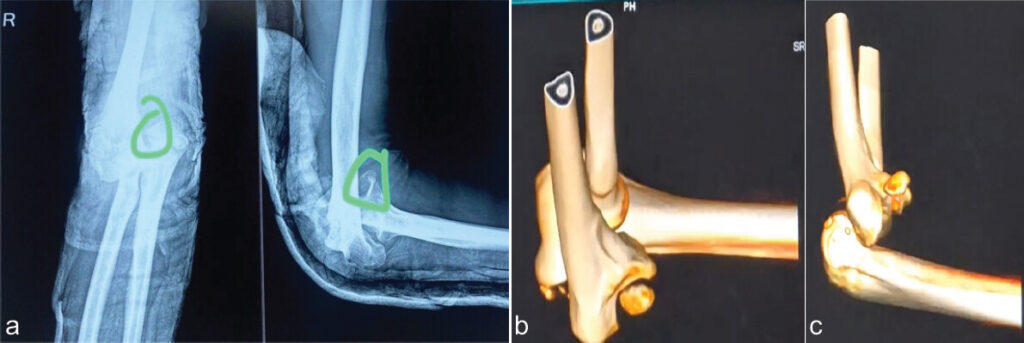
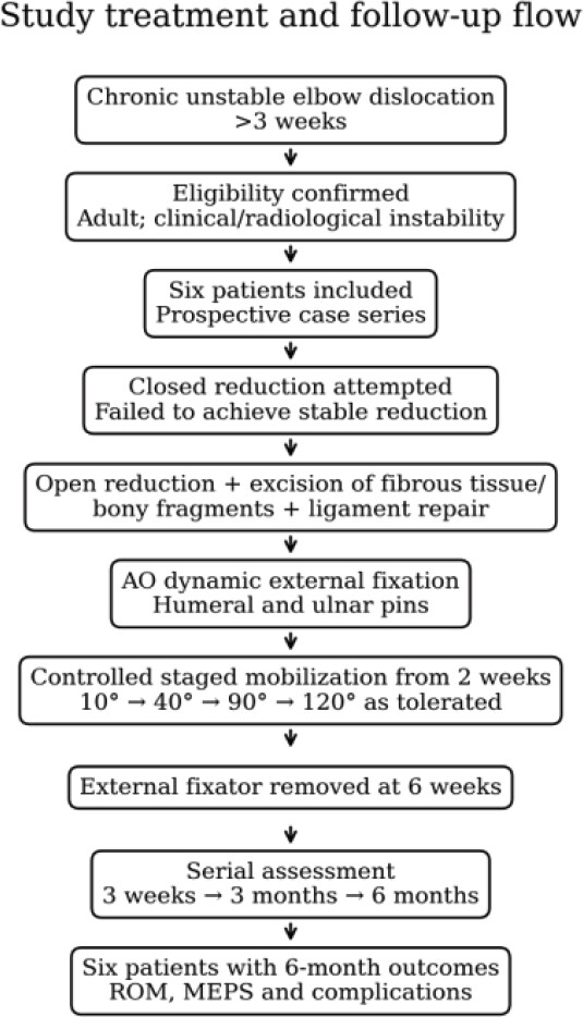
Preoperative assessment included plain radiographs, with computed tomography (CT) imaging used to demonstrate the chronic dislocation and bony malalignment illustrated in Fig. 1. A closed reduction was attempted in each patient but could not provide a satisfactory stable reduction. The subsequent operative strategy was therefore directed at obtaining open reduction, clearing tissue or bony obstacles to congruity, repairing torn ligamentous structures, and adding external fixation to protect the reconstructed elbow during the early rehabilitation period.
Surgical technique
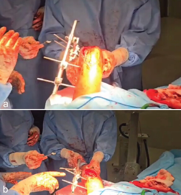
All patients initially underwent attempted closed reduction, which failed due to persistent instability. Open reduction was performed through a posterior approach, with excision of fibrous tissue and bony fragments. Torn ligaments were repaired, and an AO external fixator was applied using two pins in the ulna and two in the humerus, connected with rods and clamps to allow controlled uniplanar mobilization.
The ligamentous and capsular structures were assessed intraoperatively after open reduction and removal of interposed fibrous tissue and loose bony fragments. The injuries were predominantly partial tears and button-holing/attenuation of the anteromedial soft-tissue and ligamentous structures rather than complete ligament disruption requiring formal reconstruction. Where a discrete tear was identified, end-to-end repair was performed with non-absorbable sutures; partially torn or attenuated structures were reinforced with the adjacent capsuloligamentous tissue as required. Formal ligament reconstruction was not performed. Because the injury pattern and residual tissue quality varied between patients, repair was directed to the structures found to be insufficient at surgery rather than to a separate reconstruction protocol. Stability and concentric reduction were reassessed clinically and under fluoroscopy through flexion-extension and varus-valgus stress before application of the external fixation construct.
The external fixation construct was also standardized. Two Schanz pins were placed in the lateral column of the distal humerus and two in the subcutaneous border of the proximal ulna, away from the radial and ulnar nerves, and were connected by rods and clamps as a single lateral uniplanar frame. The clamp nearest the joint was positioned so that the effective axis of the construct approximated the center of the capitellum on the lateral fluoroscopic view, allowing flexion and extension when that clamp was released and rigid protection when it was locked. The frame was not a purpose-built hinged device, so motion was permitted in a controlled, intermittent, and supervised manner rather than continuously.
Rehabilitation protocol
Mobilization started at 2 weeks postoperatively using a staged protocol: 10° lock for a few hours, followed by 40°, 90°, and 120° locks, each for a few hours per day, as tolerated. External fixator was removed at 6 weeks.
Outcome measures
-
ROM
-
MEPS
-
Complications.
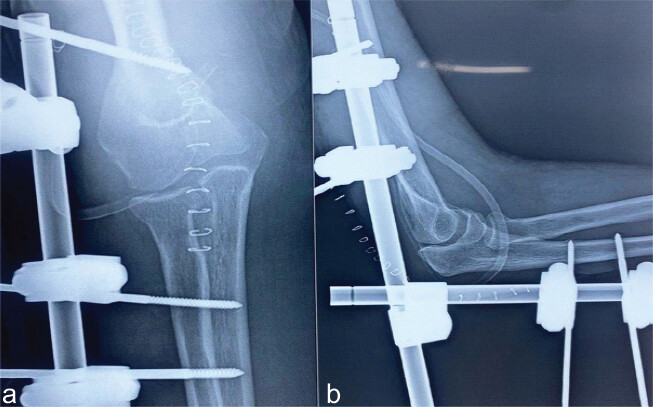
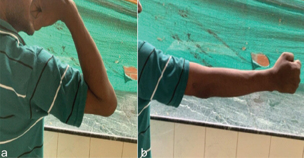
Clinical follow-up focused on three predefined outcome domains: Flexion-extension ROM, MEPS, and complications. ROM and MEPS were documented preoperatively and during post-operative follow-up at 3 weeks, 3 months, and 6 months. Complications were recorded during follow-up, including pin-tract infection, stiffness, and recurrent instability. Radiographic and clinical progression was also documented, with immediate post-operative radiographs used to demonstrate alignment and final follow-up imaging and clinical photographs used to document maintained reduction and functional motion.
Outcome assessment
ROM was measured at every visit with a universal long-arm goniometer, with the patient seated and the shoulder in neutral, using the lateral humeral epicondyle as the fulcrum, the line toward the acromion as the proximal arm and the line toward the ulnar styloid as the distal arm. Maximum active extension and active flexion were recorded and the flexion-extension arc was taken as the difference between them; three consecutive measurements were made and the mean, rounded to the nearest degree, was recorded. The MEPS was administered at the same visits and scored across its pain, motion, stability, and daily-function components. As this was a single-center series without dedicated research staff, both ROM and MEPS were recorded by a member of the treating orthopedic team; assessments were therefore not blinded, and no independent assessor was available. No additional validated patient-reported outcome measure was administered.
Complication surveillance
Complications were sought actively rather than recorded only when volunteered. Pin sites were inspected at each visit and graded using the Checketts-Otterburn classification. Clinical examination at every follow-up included varus and valgus stress testing and assessment for posterolateral rotatory instability to detect residual or recurrent instability. Radiographs taken immediately postoperatively, at fixator removal, at 3 months, and at 6 months were reviewed for maintenance of a concentric reduction, for heterotopic ossification graded by the Hastings and Graham classification, and for early degenerative change. Ulnar nerve symptoms, wound problems, and any requirement for a secondary procedure were also recorded.
Statistical analysis
Because the cohort comprised only six patients, the analysis was descriptive. Continuous variables are reported as means with ranges and categorical variables as counts. No inferential statistical testing was performed: a sample of this size has insufficient power for hypothesis testing and any p-value derived from it would be uninterpretable, so no claim of statistical significance is made anywhere in this report. Individual patient values are presented in full in Table 1 so that the consistency of change can be judged directly.
Serial functional outcomes and complications
| Patient | Pre-op ROM (°) | 3-week ROM (°) | 3-month ROM (°) | 6-month ROM (°) | Pre-op MEPS | 3-week MEPS | 3-month MEPS | 6-month MEPS | Complication |
|---|---|---|---|---|---|---|---|---|---|
| 1 | 20° | 50° | 90° | 110° | 45 | 60 | 85 | 90 | Nil |
| 2 | 30° | 55° | 85° | 100° | 40 | 55 | 80 | 85 | Pin infection |
| 3 | 25° | 60° | 95° | 115° | 50 | 65 | 90 | 95 | Nil |
| 4 | 35° | 50° | 90° | 105° | 40 | 55 | 78 | 80 | Stiffness |
| 5 | 28° | 55° | 90° | 110° | 45 | 60 | 85 | 88 | Nil |
| 6 | 30° | 50° | 88° | 105° | 42 | 58 | 82 | 85 | Pin infection |
ROM: Range of motion, MEPS: Mayo elbow performance score. All patients achieved stable reduction with progressive improvement in ROM and MEPS
Ethics
This study was approved by the Institutional Ethics Committee (IEC No. 86/SVMCH/IEC-CERT/MAY23; approval date: May 16, 2023). Written informed consent was obtained from all patients before enrolment. The study was conducted in accordance with the principles of the Declaration of Helsinki.
Results
A total of six patients with chronic unstable elbow dislocations were treated with open reduction, ligament repair, and dynamic external fixation. Patient demographics are summarized in Table 2. The mean duration of dislocation was 5.8 weeks (range 4–8 weeks). Mechanisms of injury included falls in three patients and road traffic accidents in three patients.
Patient demographic and injury characteristics
| Patient | Age (years) | Sex | Duration of dislocation (weeks) | Side | Mechanism of injury |
|---|---|---|---|---|---|
| 1 | 35 | M | 4 | Right | Fall |
| 2 | 42 | F | 5 | Left | RTA |
| 3 | 29 | M | 6 | Right | Fall |
| 4 | 50 | M | 7 | Left | RTA |
| 5 | 38 | F | 5 | Right | Fall |
| 6 | 46 | M | 8 | Left | RTA |
RTA: Road traffic accident
Functional outcomes
The mean pre-operative flexion-extension arc was 28° (range 20–35°), which improved to a mean of 108° (range 100–115°) at 6 months postoperatively. The mean MEPS improved from 44 (range 40–50) preoperatively to 87 (range 80–95) at 6 months. All patients achieved stable reduction. Two patients developed superficial pin tract infections that resolved with oral antibiotics, and one patient experienced mild stiffness, which improved with physiotherapy (Table 1).
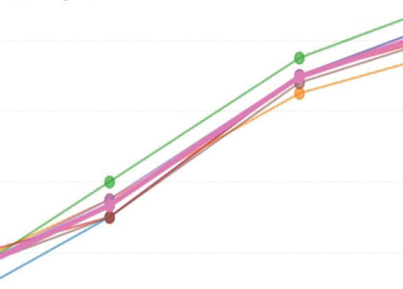
Serial assessment demonstrated progressive recovery of elbow motion. Individual pre-operative flexion-extension arcs ranged from 20° to 35°. At 3 weeks, ROM ranged from 50° to 60°; by 3 months, it ranged from 85° to 95°; and at 6 months, it ranged from 100° to 115°. The mean flexion-extension arc increased from 28° preoperatively to 108° at 6 months. Thus, the observed improvement was not confined to a single patient but was seen across the entire six-patient series.
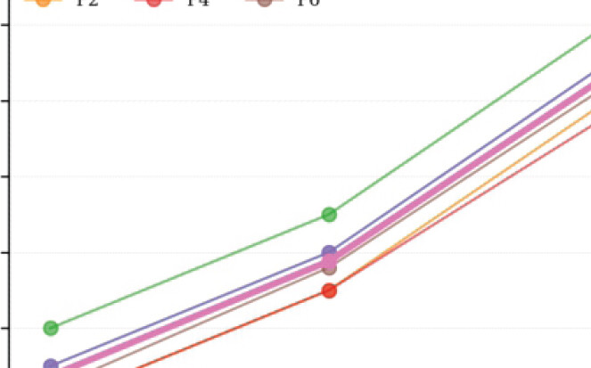
Functional scores followed the same pattern. Pre-operative MEPS values ranged from 40 to 50. At 3 weeks, scores ranged from 55 to 65; at 3 months from 78 to 90; and at 6 months from 80 to 95. The mean MEPS improved from 44 preoperatively to 87 at final follow-up. All patients maintained a stable reduction through the 6-month assessment.
Because the interval between injury and surgery ranged from 4 to 8 weeks, outcomes were examined against this duration. The three elbows treated at 4-5 weeks reached a mean 6-month arc of 107° (range 100–110°) with a mean MEPS of 88 (range 85–90), while the three treated at 6–8 weeks reached a mean arc of 108° (range 105–115°) with a mean MEPS of 87 (range 80–95). The patient with the longest delay of 8 weeks achieved a 6-month arc of 105° and a MEPS of 85. No consistent relationship between longer delay and poorer outcome was apparent, although with three elbows in each group this comparison is descriptive only and supports no statistical inference.
Active surveillance for late complications was negative at the 6-month assessment. The two pin-tract infections were Checketts-Otterburn grade 2 and settled with oral antibiotics and pin-site care, without pin removal or debridement. No heterotopic ossification was identified on the 3-month or 6-month radiographs of any patient, and no early degenerative change was visible. Varus, valgus, and posterolateral rotatory stress testing were negative in all six elbows at final review, and no patient required a secondary procedure such as capsular release, revision stabilization, or implant removal beyond routine fixator removal at 6 weeks. There were no ulnar nerve symptoms, deep infections, or pin-site fractures.
Imaging and clinical progression
Preoperative imaging revealed anteromedial dislocation in all cases, confirmed with X-rays and CT scans (Fig. 1a, Fig. 1b, c). AO external fixators were applied to the humerus and ulna to allow controlled uniplanar mobilization (Fig. 2a). Staged flexion-extension was achieved as per the rehabilitation protocol (Fig. 2b and c). Immediate post-operative X-rays confirmed well-aligned elbows (Fig. 3a and b). At 6-month follow-up, clinical photographs showed functional flexion and extension (Fig. 4c and d).
Discussion
Chronic elbow dislocation represents a difficult reconstructive problem because the pathology is no longer limited to simple loss of congruity. With increasing duration after injury, periarticular fibrosis, capsular contracture, shortening or insufficiency of the collateral ligament complex, and adaptive soft-tissue changes can coexist with articular malalignment. These changes explain why delayed closed reduction may be unsuccessful and why prolonged immobilization after reduction can further compromise motion. The therapeutic goal is therefore twofold: Restoration of a stable concentric reduction and preservation of sufficient motion to avoid a functionally stiff elbow [1,2].
The principal finding of the present prospective case series was that open reduction and ligament/capsular repair, supplemented by dynamic external fixation and a staged mobilization protocol, maintained stable alignment in all six chronic unstable dislocations. The mean flexion-extension arc improved from 28° before surgery to 108° at 6 months, while mean MEPS increased from 44 to 87. Patient-level data also demonstrated progressive improvement between the early, 3-month, and 6-month assessments. These observations support further evaluation of protected controlled motion after reconstruction of chronic unstable elbow dislocation, but the contribution of the external fixator cannot be separated from the other components of treatment in this uncontrolled series.
A major rationale for dynamic or hinged external fixation is that the construct can protect the repaired or healing soft tissues while permitting controlled movement. Biomechanical work has demonstrated the stabilizing role of a lateral unilateral dynamic external fixator after ligament injury [3]. Clinically, operative treatment of chronic elbow dislocation with strategies that allow early ROM has also been described with favorable functional recovery [4]. The current series applies the same broad principle to a simple AO external fixator configuration used to permit controlled uniplanar mobilization after open reduction and ligament repair.
The timing and progression of rehabilitation are particularly important in a chronic dislocation. In this series, mobilization was not initiated immediately after surgery; instead, controlled movement began at 2 weeks and progressed through staged locks of 10°, 40°, 90°, and 120° for several hours according to tolerance. The fixator was removed at 6 weeks. This protocol was intended to provide an initial period of protection while avoiding prolonged immobilization. The progressive improvement in ROM from the 3-week assessment through the 6-month follow-up(Fig 4a and 4b), (Fig 5, 6) suggests that useful motion can continue to develop after fixator-assisted rehabilitation.
Variability in the interval between injury and surgery is intrinsic to any chronic dislocation series and is a potential source of heterogeneity here. Capsular contracture, periarticular fibrosis, and secondary articular change all progress with time, so an elbow treated at 8 weeks is not biologically equivalent to one treated at 4 weeks and might be expected to need more extensive release and to regain less motion. In the present patients, the descriptive comparison between shorter and longer delays showed no such gradient, but six elbows are far too few to exclude one, and any true effect of delay could easily be masked by differences in the original injury pattern, patient age, and rehabilitation compliance. Stratification by duration in a larger series would be required to answer this question.
The magnitude of recovery observed here is consistent with the broader literature supporting hinged or dynamic fixation in difficult elbow instability. Jupiter and Ring reported the use of hinged external fixation for unreduced elbow dislocations [5], and Iordens et al. described good functional recovery in complex elbow dislocations managed with hinged external fixation in a prospective multicenter study [6]. Motion-capable external fixation has also been described in fracture-dislocations of the elbow [7]. Although these populations and constructs are not identical to the present six chronic dislocations, collectively they support the principle that a stable mechanical environment can facilitate rehabilitation without relying on prolonged rigid immobilization.
An important clinical observation in the present series was the absence of recurrent dislocation during the reported follow-up. Recurrent instability is a major concern when mobilization is introduced after reconstruction of a chronically dislocated elbow. The combination of open reduction, removal of obstructing fibrous tissue and bony fragments, ligament repair, and external stabilization was used to address both the mechanical block to reduction and the soft-tissue insufficiency. Previous work on elbow subluxation and dislocation has emphasized the importance of stability and appropriate management of the injured stabilizing structures [10]. In the current series, stable reduction was maintained despite progressive mobilization.
Complications were relatively minor. Two patients developed superficial pin-tract infections that resolved with oral antibiotics, and one patient had mild stiffness that improved with physiotherapy. No major complication was reported. Pin-related problems are an inherent consideration with external fixation, and careful pin-site care and surveillance remain necessary. At the same time, the low number of serious adverse events in this small series suggests that the staged protocol was feasible when accompanied by clinical and radiographic monitoring.
The choice of outcome instruments also deserves comment. The MEPS(Fig 7) is widely used in elbow trauma and is convenient, but it is a clinician-administered composite in which motion and stability are graded by the observer, and its weighting can yield a good score despite residual patient dissatisfaction. It is therefore observer-dependent, and in this series, it was scored by a member of the treating team rather than by an independent or blinded assessor, which may bias values upwards. No validated patient-reported outcome measure, such as the Disabilities of the Arm, Shoulder and Hand score, the Oxford Elbow Score, or the Patient-Rated Elbow Evaluation, was administered, so the patients’ own perception of pain, confidence in the elbow, and satisfaction was not captured. Future evaluation of this technique should pair a blinded objective ROM measurement with at least one patient-reported instrument.
The external fixation strategy used here may be particularly relevant where access to dedicated hinged devices is limited. More complex hinged constructs are designed to reproduce elbow motion around the flexion-extension axis, but they may require specific equipment and technical familiarity. Alternative methods of maintaining stability while permitting motion include internal fixation devices; Schultz et al. described treatment of a chronic elbow dislocation using an internal fixator [8]. Static and dynamic external fixation has also been compared in unstable elbow fracture-dislocations, with both approaches reported as effective [9]. These reports illustrate that several stabilization strategies are available and that selection should be individualized to the injury pattern, soft-tissue condition, surgeon experience, and available resources.
Several limitations should be considered when interpreting the results. First, only six patients were included, which limits precision and prevents meaningful subgroup or inferential statistical analysis. Second, this was a single-center case series without randomization or a comparator group, so improvements cannot be attributed to the external fixator independently of open reduction, debridement, ligament repair, physiotherapy, or natural recovery. Third, follow-up was limited to 6 months; longer observation is required to determine whether motion, stability, and MEPS are maintained and to evaluate late problems such as recurrent instability, degenerative change, or persistent stiffness. Finally, the staged locking protocol was advanced according to patient tolerance, which introduces a degree of clinical variability.
Further limitations relate to selection, measurement, and external validity. The series was not randomized and patients were enrolled consecutively at a single tertiary center under one surgical team, so selection and treatment bias are both possible and the findings may not transfer to other institutions, levels of surgical experience, or implant availability. The exclusion criteria restrict generalizability further: Open injuries, fractures requiring internal fixation, and neurovascular injury were all excluded, yet these are precisely the features that define the most complex chronic dislocations, so the results should not be extrapolated to that group. Outcome assessment was neither blinded nor supplemented by a patient-reported measure, and no dedicated hinged external fixator arm was available for comparison, so no claim of equivalence or superiority over a purpose-built hinged device can be made. Complication ascertainment was also limited by the 6-month horizon, since heterotopic ossification, post-traumatic arthritis, late ligamentous attenuation, and pin-site sequelae may all declare themselves later. Recovery should accordingly be presented to patients as progressive rather than immediate.
Despite these limitations, the study provides a clinically useful description of a reproducible staged approach to a relatively uncommon and challenging condition. The consistent maintenance of reduction, improvement in ROM and MEPS across all six patients, and manageable complication profile support further evaluation of controlled early mobilization after reconstruction of chronic unstable elbow dislocation. Larger prospective studies with longer follow-up and direct comparison between fixation strategies would be valuable to define patient selection, optimal timing of mobilization, duration of fixation, and long-term functional outcomes.
Conclusion
In this six-patient prospective case series, open reduction and ligament/capsular repair supplemented by dynamic external fixation and controlled staged mobilization maintained stable reduction and were associated with progressive improvement in elbow motion and MEPS at 6 months. Mean flexion-extension arc improved from 28° to 108° and mean MEPS from 44 to 87. The technique provided a practical means of protecting stability while allowing rehabilitation, with only minor pin-tract infection and stiffness observed. Larger comparative studies with longer follow-up are required before definitive conclusions regarding superiority or long-term outcomes can be made.
Clinical Message
Following open reduction and soft-tissue repair for chronic unstable elbow dislocation, staged controlled mobilization using dynamic external fixation can provide protected motion while maintaining reduction during early rehabilitation.
Conflict of Interest:
Nil
Source of Support:
Nil
Consent
The authors confirm that informed consent was obtained from the patient for publication of this article
How to Cite this Article
Velayudham K, Suganya S, Iyyanarappan P, Anand K. Restoring Stability and Motion: Controlled Early Uniplanar Mobilization using Dynamic External Fixation in Chronic Unstable Elbow Dislocations – A Case Series. J Orthop Case Rep. 2026 November;16(11):388-396. doi: 10.13107/jocr.2026.v16.i11.8506.
References
- Morrey BF. Current concepts in the management of complex elbow trauma. Surgeon 2009;7:151-61. [Google Scholar] | [PubMed]
- Duckworth A, Ring D, Kulijdian A, McKee MD. Unstable elbow dislocations. J Shoulder Elbow Surg 2008;17:281-6. [Google Scholar] | [PubMed]
- Anderson DR, Haller JM, Anderson LA, Hailu S, Chala A, O'Driscoll SW. Surgical treatment of chronic elbow dislocation allowing for early range of motion: Operative technique and clinical results. J Orthop Trauma 2018;32:196-203. [Google Scholar] | [PubMed]
- Kamineni S, Hirahara H, Neale P, O'Driscoll SW, An KN, Morrey BF. Effectiveness of the lateral unilateral dynamic external fixator after elbow ligament injury. J Bone Joint Surg Am 2007;89:1802-9. [Google Scholar] | [PubMed]
- Jupiter JB, Ring D. Treatment of unreduced elbow dislocations with hinged external fixation. J Bone Joint Surg Am 2002;84:1630-5. [Google Scholar] | [PubMed]
- Iordens GI, Den Hartog D, Van Lieshout EM, Tuinebreijer WE, De Haan J, Patka P. Good functional recovery of complex elbow dislocations treated with hinged external fixation: A multicenter prospective study. Clin Orthop Relat Res 2015;473:1451-61. [Google Scholar] | [PubMed]
- Pennig D, Gausepohl T, Mader K. Transarticular fixation with motion capacity in fracture dislocations of the elbow. Injury 2000;31 Suppl 1:35. [Google Scholar] | [PubMed]
- Schultz BJ, Lowe DT, Pean CA, Egol KA. Treatment of a chronic elbow dislocation with an internal fixator. J Orthop Trauma 2021;35 Suppl 2:S13-4. [Google Scholar] | [PubMed]
- AlQahtani S, Aibinder WR, Parry JA, Seltser A, Athwal GS, King GJ. Static and dynamic external fixation are equally effective for unstable elbow fracture-dislocations. J Orthop Trauma 2021;35:e82-8. [Google Scholar] | [PubMed]
- O'Driscoll SW, Morales CM, Korinek S, An KN, Morrey BF. Elbow subluxation and dislocation. A spectrum of instability. Clin Orthop Relat Res 1992;280:186-97. [Google Scholar] | [PubMed]
© 2026 Journal of Orthopaedic Case Reports - Published by Indian Orthopaedic Research Group
About the Authors
-

Karthik Velayudham
-

Sree Suganya
-

Premkumar Iyyanarappan
-

Karthik Anand
Related Articles in Journal of Orthopaedic Case Reports
 November 1, 2026 Outcomes of Open Reduction and Internal Fixation of Pediatric Lateral Condyle Fractures of Humerus with Delayed Presentation
 October 1, 2026 A Functional Outcome of Closed Intra-articular Fracture of Proximal Interphalangeal Joint of Hand Managed By Suzuki Frame – A Longitudinal Study
 September 1, 2026 Posterior Cruciate Ligament Avulsion Fracture with Fibular Avulsion of Lateral Collateral Ligament-biceps Femoris Complex Avulsion Injury in Ipsilateral Diaphyseal Tibia Fracture – A Rare Complex Injury
 May 1, 2026 A Novel Failure Interface in Constrained Total Hip Arthroplasty: Dissociation at the Constrained Liner Insert-Bipolar Head Junction Following Periprosthetic Joint Infection