


Introduction
Displaced olecranon fractures frequently require open reduction and internal fixation through a posterior midline approach. Traditional positioning for posterior elbow exposure involves prone or lateral decubitus placement to allow gravity-assisted elbow flexion and direct posterior access [1]. Positioning a patient in the lateral decubitus can be cumbersome and time intensive, and an assistant is often needed to maintain elbow flexion. Polytrauma can further complicate placing patients into the lateral decubitus position, and airway access may be complicated by this positioning [2].
Supine positioning has the benefit of being generally easier to set up with improved airway access for anesthesia control. The primary limitation of supine positioning for posterior elbow surgery is maintaining stable elbow flexion and posterior exposure without an assistant or bulky positioning equipment. Wijeratna et al. [3], Metikala et al. [4], Patel et al. [5], Sandrowski [6], and others have described different variations of supine positioning for posterior elbow access.
We describe a simple, low-cost suspension technique – the Montague–Iliff Maneuver – that allows reproducible posterior access in the supine position using sterile Kerlix and IV poles.
Surgical Technique
Supplies needed
-
IV poles x2
-
Non-sterile weights, ~ 20 lbs. (i.e., bean bags or traction weights)
-
Sterile Kerlix x2
-
Sterile Coban
-
Hemostat x2
Patient positioning
The patient is placed supine on a standard operating table. A bump may be placed under the ipsilateral scapula to facilitate shoulder positioning if needed.
Approximately 10 lbs of weights are placed at the base of each IV pole. These counterweights prevent IV pole instability during the procedure. One IV pole is positioned toward the head of the contralateral side of the patient while the other IV pole is positioned toward the patient’s feet still on the contralateral side.
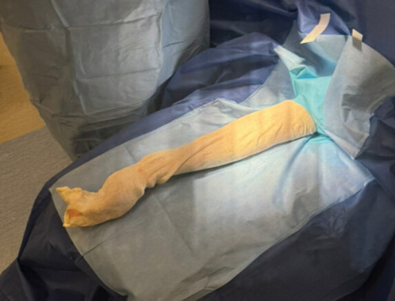
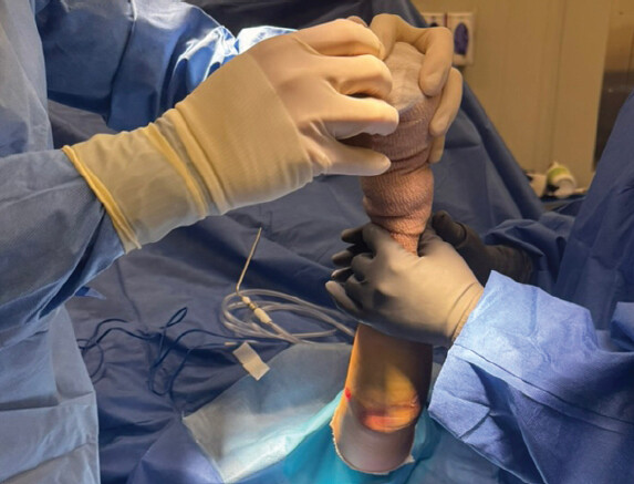
The operative extremity is prepared and draped in standard sterile fashion with a stockinette over the operative arm and either held by an assistant or placed on an arm board at the side of the bed (Fig. 1). After exsanguination and inflation of the tourniquet, the stockinette is cut circumferentially just below the drape and rolled down to the distal 1/3rd of the forearm, securely wrapping the hand and wrist with Coban dressing (Fig. 2). The shoulder is then positioned in 90° forward flexion and approximately 90° internal rotation. The elbow is flexed to 90°, bringing the olecranon superiorly and exposing the posterior proximal ulna. The extremity is held in this position until arm suspension is achieved.
Arm suspension
A 4” sterile Kerlix is fastened with a hemostat around the patient’s wrist over the Coban-wrapped stockinette. The opposite end of the Kerlix is then passed off the operative field toward the contralateral side. The now unsterile free end of the Kerlix is placed over a wrung on an IV pole which is positioned toward the head of the patient opposite to the operative side. The anesthesiologist or other assistant applies gentle tension and secures the non-sterile end of the Kerlix to the bed rail to maintain suspension of the operative arm.
A second sterile Kerlix is fastened in similar fashion to the first on the patient’s operative wrist and the free end of this second Kerlix is thrown over a second IV pole positioned toward the foot of the patient opposite to the operative extremity and tied down again by anesthesia or an assistant (Figs. 3 and 4).
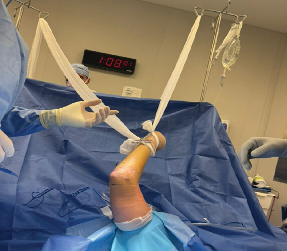
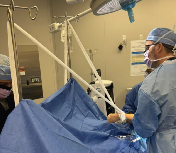
The triangulation of the two Kerlix to suspend the arm creates stable positioning of the arm with maintained elbow flexion without requiring a surgical assistant to hold the limb for the duration of the case. The amount of tension can be adjusted intraoperatively with the hemostats to optimize exposure while avoiding excessive traction. The arm board may be removed after the arm has been suspended.
Discussion
Several techniques have been described in an effort to improve posterior elbow access in the supine position for displaced olecranon fracture repairs. Wijeratna et al. (2013) first reported a supine arm-over-chest method for open elbow surgery: The arm is supported on the chest by a Lloyd-Davies leg support (or padded L-bar) attached to the table, with a rolled towel under the ipsilateral shoulder to elevate it [3]. Metikala et al. described a supine forearm-flexed technique specifically for olecranon fractures [4]. In their method, the patient lies supine with the arm on an arm table; a saline bag and radiolucent triangle support the humerus while the elbow is flexed over a sterile bolster on the chest. They secure the arm with bolsters to prevent sliding. Patel et al. (Ann R Coll Surg 2019) also described a supine variant for tension-band wiring: the arm lies over an arm board, shoulder abducted, and externally rotated 90°, with the elbow resting on a kidney dish [5]. Sandrowski describes yet another technique: The arm is placed supine hanging slightly over the table edge, supported by two cylindrical rollers forming a “V” cradle under the arm, with Esmarch wraps securing it [6].
The Montague-Iliff Maneuver addresses several limitations of traditional posterior positioning techniques [7]. It has the advantage of being simple and reproducible and uses almost universally available materials. It is cost effective and the patient can be positioned very quickly. Potential limitations of the maneuver include the fact that the anesthesiologist or other assistant must help in fastening the far end of the Kerlix. The surgeon may also need to familiarize himself with the orientation of the elbow in this position [8, 9]. Care must be taken to avoid excessive traction to avoid displacement of the IV pole and subsequent sterile field contamination. Care must also be taken to ensure that there is adequate padding beneath the wrist where the Coban is tied to it. Reduced maneuverability of the patient’s shoulder may also present challenges to using this positioning technique [10].
In our experience, the technique provides stable exposure without traction-related complications when properly tensioned, and excellent access to the posterior elbow which can be attained in 3 min or less after sterile draping has been applied.
Conclusion
The Montague-Iliff Maneuver is a safe, cost-effective, and reproducible supine positioning technique for posterior olecranon fracture fixation. It provides stable elbow flexion and posterior exposure without the need for prone or lateral positioning, specialized equipment, or dedicated assistants. Further evaluation in larger clinical series may help validate its reproducibility and outcomes.
Clinical Message
For selected patients undergoing posterior olecranon fixation, the Montague-Iliff Maneuver offers a practical supine option for maintaining elbow flexion and posterior exposure, provided the wrist is padded and sling tension is carefully controlled.
Conflict of Interest:
Nil
Source of Support:
Nil
Consent
The authors confirm that informed consent was obtained from the patient for publication of this article
How to Cite this Article
Jones H, Montague M, Iliff A. The Montague-Iliff Maneuver: A Simple Supine Suspension Positioning Technique for Olecranon Fracture Open Reduction Internal Fixation. J Orthop Case Rep. 2026 November;16(11): 322-325. doi: 10.13107/jocr.2026.v16.i11.8486.
References
- Thorne T, Mau M, Dong W, Lisitano L, DaSilva Z, Rothberg DL. Intramedullary screw fixation for simple olecranon fractures. JBJS Essent Surg Tech 2025;15:e23.00077. [Google Scholar] | [PubMed]
- Camp CL, Degen RM, Dines JS, Altchek DW, Sanchez-Sotelo J. Basics of elbow arthroscopy part III: Positioning and diagnostic arthroscopy in the lateral decubitus position. Arthrosc Tech 2016;5:e1351-5. [Google Scholar] | [PubMed]
- Wijeratna MD, Thomas S, Van Rensburg L. The supine position for elbow surgery. Ann R Coll Surg Engl 2012;94:446. [Google Scholar] | [PubMed]
- Metikala S, Anibas JJ, Price R, Cheppalli N. A novel positioning technique for olecranon fracture fixation. Cureus 2023;15:e38338. [Google Scholar] | [PubMed]
- Patel A, Shepherd K, Karthik K. Patient positioning for tension band wiring of olecranon fractures. Ann R Coll Surg Engl 2019;101:228-9. [Google Scholar] | [PubMed]
- Sandrowski K. AAHS - a New Technique for Patient Positioning During Olecranon Fixation. American Association for Hand Surgery Annual Meeting 2018. Available from: https://meeting.handsurgery.org/abstracts/2018/ep147.cgi#:~:text=technique%3a%20the%20patientis%20placed%20supine,figure%201. [Last accessed on 15 Dec 2018]. [Google Scholar] | [PubMed]
- DeBernardis DA, Sandrowski K, Padegimas EM, Rivlin M. A simplified technique for patient positioning during olecranon fracture fixation. Tech Shoulder Elbow Surg 2020;21:107-11. [Google Scholar] | [PubMed]
- Aggarwal S, Paknikar K, Sinha J, Compson J, Reichert I. Comprehensive review of surgical approaches to the elbow. J Clin Orthop Trauma 2021;20:101482. [Google Scholar] | [PubMed]
- Camp CL, Degen RM, Dines JS, Sanchez-Sotelo J, Altchek DW. Basics of elbow arthroscopy part II: Positioning and diagnostic arthroscopy in the supine position. Arthrosc Tech 2016;5:e1345-9. [Google Scholar] | [PubMed]
- Kwee MM, Ho YH, Rozen WM. The prone position during surgery and its complications: A systematic review and evidence-based guidelines. Int Surg 2015;100:292-303. [Google Scholar] | [PubMed]
© 2026 Journal of Orthopaedic Case Reports - Published by Indian Orthopaedic Research Group
About the Authors
-

Hansan Jones
-

Michael Montague
-

Anthony Iliff
Related Articles in Journal of Orthopaedic Case Reports
 September 1, 2025 Modern Perspectives on Unicompartmental Knee Arthroplasty: An Editorial Review
 September 1, 2025 Intertrochanteric Femur Fracture in a High-Above-Knee Amputee: Technical Tips for Intraoperative Planning and Implant Selection with Literature Review
 March 1, 2025 Biological Stabilization of Proximal Tibiofibular Joint Recurrent Instability with Semitendinosus Graft – Surgical Technique and Case Report
 January 1, 2025 Fragment-specific Plate Fixation in a Case of Mayo Type IIB Olecranon Fracture: A Case Report