[box type=”bio”] Learning Point for the Article: [/box]
Vascular injury should actively be ruled out in all cases of glenohumeral dislocation, regardless of presentation.
Case Report | Volume 8 | Issue 4 | JOCR July – August 2018 | Page 53-56| Steven Magister, Andrew Bridgforth, Seth Yarboro. DOI: 10.13107/jocr.2250-0685.1158
Authors: Steven Magister[1], Andrew Bridgforth[1], Seth Yarboro[1]
[1]Department of Orthopaedics, University of Virginia, Charlottesville, Virginia, USA.
Address of Correspondence:
Dr. Seth Yarboro,
PO Box 800159 HSC, Dept Orthopaedic Surgery, University of Virginia, Charlottesville, VA 22903.
E-mail: sry2j@hscmail.mcc.virginia.edu
Abstract
Introduction: Axillary artery injury is a rare and potentially devastating sequelae of glenohumeral dislocation. While neurovascular exam is critical in all presentations, the presence of “soft” and/or “hard” signs should prompt a more thorough examination and possible employment of advanced imaging techniques.
Case Report: We present a case of a 51-year-old male with an axillary artery injury associated with an anterior glenohumeral dislocation. The patient was initially evaluated at an outside hospital where the vascular injury was not immediately identified, and then was subsequently transferred to our institution where he underwent bypass grafting without significant sequela. Additional prophylactic fasciotomies were also performed due to concern for reperfusion compartment syndrome.
Conclusion: Although rare, clinicians should actively rule out vascular injuries when evaluating shoulder dislocations, especially in the elderly patient with a known history of atherosclerotic disease, those with evidence of chronic joint instability, and in the setting of high energy injury mechanisms. Hard signs of vascular injury including diminished distal pulses are the hallmark of this complication, and should always prompt vascular surgery consultation.
Keywords: Axillary artery, shoulder dislocation, bypass repair.
Introduction
Given the shallow geometry of the glenoid fossa and large range of motion permitted, glenohumeral joint dislocation is a relatively common injury with an incidence of 24 per 100,000 persons per year [1]. Frequent complications of shoulder dislocation include deltoid weakness, rotator cuff tear, nerve injury, bony deformities, persistent joint laxity, and in rare instances axillary artery damage [2].Vascular injury is identified by “hard signs” including: active pulsatile hemorrhage, expanding hematoma, palpable/audible bruit, overt limb ischemia, or diminished pulses. Further, “soft signs” of vascular injury include: hypotension or shock, neurologic deficits, stable non-pulsatile hematoma, or proximity of injury to major vascular structures. When a vascular injury is suspected urgent evaluation and treatment should be initiated in order to prevent limb ischemia and possible irreversible damage. Here we present and discuss a unique case of an anterior shoulder dislocation with associated axillary artery injury treated with reduction and prosthetic bypass grafting.
Case Report
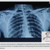
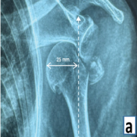
A 51-year-oldintoxicated, homeless man with a history of alcohol abuse initially presented to an outside hospital after being found unconscious at a gas station. Initial survey revealed a right shoulder deformity, and X-ray demonstrated an anterior glenohumeral dislocation (Fig. 1). There was no evidence of bony deformities seen on X-ray. Following successful reduction under sedation with etomidate, the patient became increasingly apneic and desaturated. He was subsequently intubated in order to protect his airway given his level of intoxication coupled with the sedative used for reduction. At this time, the decision was made to transfer the patient urgently via helicopter to a tertiary care center for definitive care. Upon presentation to our level 1 trauma center as a mid-level trauma, the man remained intubated and sedated. Initial assessment was significant for diminished, but still present, distal pulses in the right upper extremity. Additionally, the capillary refill was delayed to 5 s and the extremity was cooler compared to the left. Due to concerns for vascular involvement, a computerized tomography (CT) angiogram of the right upper extremity was performed and revealed an occlusion of the axillary artery just distal to the takeoff of the subscapular artery (Fig. 2). Reconstitution of the brachial artery was found 11.5 cm proximal to the antecubital fossa. Presence of enhanced extraluminal contrast at the point of occlusion on delayed images was consistent with active extravasation. In addition, an expanding hematoma, which measured 7.3 cm× 7.7 cm× 12 cm was noted in the proximal arm. Incidentally, CT imaging also showed evidence of multiple corticated and incompletely corticated osseous fragments along the superolateral humeral head, and proximal humeral shaft. Additionally, a Hill Sachs deformity was noted in the humeral head. At this time, approximately 7 h after initial presentation to the outside hospital, vascular surgery was notified, and it was decided that immediate surgery was necessary in order to establish hemostasis based on the active extravasation seen on vascular imaging. Given that distal pulses were still present, albeit diminished, and that there was reconstitution distally on imaging, it was thought tissue ischemia would be limited. Still, the risk of reperfusion injury was high given the anticipated relative change in vascular flow following bypass, so orthopaedic surgery was consulted in order to perform prophylactic fasciotomies. Vascular surgery performed a right common carotid artery to mid brachial artery bypass with a 40 cmvascular allograft (CryoArtery, CryoLife-Kennesaw, GA). The decision to use a Cryo Arteryallograft rather than saphenous vein autograft was made intraoperatively due to the small size of the patient’s saphenous vein after exposure. Given the extensive amount of hemarthrosis in the shoulder the vascular surgery team chose to tunnel the bypass superficially. For similar reasons, the common carotid was used as an inflow source, as opposed to a more distal segment closer to the apparent vascular injury. Following successful bypass, arm and forearm fasciotomies as well as carpal tunnel release were performed by the orthopaedic surgery team. Upon inspection, the forearm muscles and surrounding tissue appeared viable and not necrotic. Fasciotomy was extended to include carpal tunnel release due to swelling that was noted more distally in the hand. The patient was then brought to the intensive care unit for monitoring post-operatively. The patient returned to the operating room on post-operativedays 2, 4, and 5 from the index procedure for irrigation, debridement, and delayed primary closure of all wounds. Throughout his hospital course, vascular examination was performed routinely, and showed no evidence of stenosis, thrombosis, or bypass failure. The right upper extremity remained well perfused with palpable distal pulses and brisk capillary refill. In addition, distal neurologic and musculoskeletal function such as wrist flexion and extension and grip strength were intact and equal to that of the contralateral limb. Attempts were made to transition the patient to a skilled nursing facility in order to properly care for his wounds, but the patient discharged against medical advice on day 6 of his hospital stay. At 2 week follow up, his operative extremity remained well perfused and neurovascular exam was normal. Forearm and wrist flexors, extensors as well as hand intrinsic strength remained strong, however the patient did express mild stiffness at the wrist during active range of motion. The patient plans to follow up at 3 months with vascular surgery for a graft ultrasound to ensure adequate patency and flow.
Discussion
Shoulder dislocations are a relatively common injury, and are often associated with a variety of transient complications such as shoulder weakness, axillary paresthesia, and joint instability. Although much more rare, a major complication of shoulder dislocation is axillary artery transection, and is associated with a high morbidity if not properly diagnosed. The axillary artery begins as a direct continuation of the subclavian artery as it passes lateral to the first rib. It is further subdivided into three parts, proximal, posterior, and distal, which are defined by the location relative to the pectoralis minor muscle [3]. Major branches of the third portion include the subscapular and circumflex humeral arteries. The distal portion is most commonly injured given its fixed position, which subjects the artery to significant shear forces during traumatic events such as glenohumeral dislocation or subsequent reduction [4,5]. Clinically, axillary artery injury after an anterior shoulder dislocation is rare [6]. Most commonly, this injury is seen in elderly populations over the age of 50 and in the setting of atherosclerotic disease [7]. Atherosclerotic disease leads to decreased arterial elasticity and laxity, which reduces the threshold of shear force required for injury to occur during dislocation or even during dislocation reduction [4]. Axillary artery injury has also been reported in the setting of recurrent shoulder dislocations [8]. Specifically in the setting of relocation, one study showed that close to 70% of patients who underwent closed reduction for a chronically dislocated shoulder experienced a vascular injury [8].Mechanistically, chronic subluxation and spontaneous relocations lead to increased fibrous tissue deposition over time. As the surrounding soft tissue becomes increasingly stiff so too do the surrounding vascular structures. Via a similar mechanism to that of atherosclerosis, this lowers the shear force threshold needed for vascular injury. CT findings of a Hill Sachs deformity and osseous fragments in this case suggest this patient was subject to chronic joint pathology lending this presentation to previously reported cases. While not known at the time of initial presentation and reduction, care should be taken in the select populations where chronic joint pathology or atherosclerosis is a concern. This could potentially include using more aggressive anesthesia to allow for greater muscle relaxation and easier reduction. Closed reduction under general anesthesia could also be employed especially in patients with an already tenuous cardiopulmonary status as in the case here. Lastly, vascular injury should also be of greater concern in the setting of shoulder dislocations secondary to high energy mechanisms, such as motor vehicle accidents. One recent report documented an axillary artery injury secondary to a fracture dislocation of the shoulder as a result of a high-speed motor vehicle versus tree mechanism [9]. With higher energy injuries, greater force across the joint can lead to fractures, and as a result bony fragments could act to transect the axillary artery. Every patient with a shoulder dislocation requires thorough neurovascular examination, since failure to recognize an underlying arterial injury could lead to limb loss and possibly death [10]. Prompt diagnosis and revascularization is critical, ideally within 2–3 h [11].Clinically, the triad of expanding hematoma, anterior dislocation, and diminished distal pulses has been reported [7]. However, given the extensive collateral circulation of the upper extremity, axillary injury has been reported without evidence of decreased pulses [12].Collateral circulation could propose a mechanism whereby irreversible tissue ischemia can be prevented during extended periods of ischemia. As in this case, CT angiogram showed revascularization distal to the arterial injury consistent with an extensive collateral circulation. This presumably played a significant role in the prevention of tissue necrosis and overall return of limb function post operatively. If clinical suspicion is high for vascular injury, advanced angiographic imaging should be employed to better quantify the injury. If an injury is confirmed, vascular surgery should be consulted urgently. Most commonly, axillary artery injuries leading to near or complete occlusion are treated surgically by vein or prosthetic bypass grafting [13].In the case here, surgery was primarily performed in order to establish hemostasis and prevent further active extravasation. In addition, prophylactic forearm fasciotomies should be considered to prevent reperfusion injury associated compartment syndrome [14].One retrospective study of 62 patients with a variety of upper and lower extremity injuries found the incidence of compartment syndrome to be 60% (37 patients) following revascularization [15]. The majority (31/37) of these patients had a prophylactic fasciotomy at the time of revascularization. Of the remaining patients (6/37), a second surgery was required after revascularization. In these six patients rhabdomyolysis was found in five patients. Three patients required amputation. Additionally, in a recent report of 378 patients with varying vascular injuries of the extremities, six patients required amputation following revascularization as a result of compartment syndrome and subsequent tissue necrosis [16].These studies highlight the importance and potentially devastating sequelae of reperfusion injury, specifically in the setting of delayed revascularization. Regardless of the warm ischemic time or the presence of collateral circulation, fasciotomies should always be performed if there is concern for reperfusion injury as failure to do so could lead to potentially irreversible limb necrosis.
Conclusion
While uncommon, axillary artery injury is a potentially devastating sequelae of glenohumeral dislocation. Regardless of the clinical context all patients with a glenohumeral dislocation should have distal pulses palpated both pre and post reduction. As in the case presented here, vascular injury was properly identified upon arrival at our institution and subsequent treatment resulted in recovery without permanent sequelae.
Clinical Message
Vascular injury following glenohumeral dislocation is rare, but failure to recognize such an injury can prove devastating. Therefore, all patients with a glenohumeral dislocations should be thoroughly evaluated for vascular compromise, both pre and post reduction. While lack of distal pulses is the hallmark of this sequelae it may not be present, and thus clinical judgement should guide patient care. Advanced imaging, and orthopaedic and vascular surgery consultations should always be employed when evidence of vascular injury becomes apparent.
References
1. Zacchilli MA, Owens BD, Epidemiology of shoulder dislocations presenting to emergency departments in the United States. J Bone Joint Surg Am 2010;92:542-9.
2. Beeson MS. Complications of shoulder dislocation. Am J Emerg Med 1999;17:288-95.
3. Agur A, Dalley A. Grant’s Atlas of Anatomy: Upper Limb. In: Lippincott Williams and Wilkins, Philadelphia USA; 2009. p. 504-5.
4. Kelley SP, Hinsche AF, Hossain JF. Axillary artery transection following anterior shoulder dislocation: Classical presentation and current concepts. Injury 2004;35:1128-32.
5. Milton GW. The circumflex nerve and dislocation of the shoulder. Br J Phys Med 1954;17:136-8.
6. Julia J, Lozano P, Gomez F, Corominas C. Traumatic pseudoaneurysm of the axillary artery following anterior dislocation of the shoulder. Case report. J Cardiovasc Surg (Torino) 1998;39:167-9.
7. Allie B, Kilroy DA, Riding G, Summers C. Rupture of axillary artery and neuropraxis as complications of recurrent traumatic shoulder dislocation: Case report. Eur J Emerg Med 2005;12:121-3.
8. Calvet E, Leroy J, Lecroix F. Luxations de l’epaule et lesions vasculaires. J Chir (Paris) 1941;58:337.
9. Chehata A, Morgan FH, Bonato L. Axillary artery injury after an anterior shoulder fracture dislocation and “periosteal sleeve avulsion of the rotator cuff” (SARC). Case report and review of the literature. Trauma Case Reports 2017;8:5-10.
10. Thorsness R, English C, Gross J, Tyler W, Voloshin I, Gorczyca J, et al. Proximal humerus fractures with associated axillary artery injury. J Orthop Trauma 2014;28:659-63.
11. Feliciano DV. Management of peripheral arterial injury. CurrOpinCrit Care 2010;16:602-8.
12. Adovasio R, Visintin E, Sgarbi G. Arterial injury of the axilla: an unusual case after blunt trauma of the shoulder. J Trauma 1996;41:754-6.
13. Byrd RG, Byrd RP, Roy TM. Axillary artery injuries after proximal fracture of the humerus. Am J Emerg Med 1998;16:154-6.
14. Percival TJ, White JM, Ricci MA. Compartment syndrome in the setting of vascular injury. PerspecVasc Surg EndovascTher2011;23:119-24.
15. Rozycki GS, Tremblay LN, Feliciano DV, McClelland WB. Blunt vascular trauma in the extremity: Diagnosis, management, and outcome. J Trauma 2003;55:814-24.
16. Li Z, Zhao L, Wang K, Cheng J, Zhao Y, Ren W. Characteristics and treatment of vascular injuries: A review of 387 cases at a Chinese center. Int J Clin Exp Med 2014;7:4710-9.
 |
 |
 |
| Dr. Steven Magister | Dr. Andrew Bridgforth | Dr. Seth Yarboro |
| How to Cite This Article: Magister S, Bridgforth A, Yarboro S. Axillary Artery Injury Following Closed Reduction of an Age-Indeterminate Anterior Glenohumeral Dislocation. Journal of Orthopaedic Case Reports 2018 Jul-Aug; 8(4): 53-56. |
[Full Text HTML] [Full Text PDF] [XML]
[rate_this_page]
Dear Reader, We are very excited about New Features in JOCR. Please do let us know what you think by Clicking on the Sliding “Feedback Form” button on the <<< left of the page or sending a mail to us at editor.jocr@gmail.com


