
Prioritization of 2nd MTP joint reduction during open reduction of combined dislocation of lesser MTP joints.
Dr. Vipin Mundakattu, Department of Orthopedics, Government Medical College, Manjeri, Kerala, India. E-mail: vipmaldia@gmail.com
Abstract
Introduction: Combined dislocation of lesser metatarsophalangeal (MTP) joints is a rare occurrence. This case report details the successful open reduction of combined lesser MTP dislocation.
Case Report: A 54-year-old male presented with trauma to his right foot following a fall from height. The patient had dorsolateral dislocation of the second, third, and fifth MTP joints, accompanied by a displaced fracture of the fourth metatarsal neck with an intact fourth MTP joint. The initial closed reduction attempt proved unsuccessful, leading to a planned open reduction and K-wire fixation. Unexpected challenges were encountered during the procedure, with the unreducible nature of the fourth metatarsal fracture prompting a strategic shift in the approach.
Conclusion: This case highlights the significance of understanding the anatomy of MTP joints and the need for a systematic reduction strategy, with a focus on the second MTP joint, to achieve stability.
Keywords: 2nd metatarsophalangeal, combined dislocation, plantar plate, deep transverse metatarsal ligaments.
Closed combined dislocation of metatarsophalangeal (MTP) joints is a rare occurrence [1-3], with limited case reports in the literature [2,4]. This report presents a unique case of dorsolateral dislocation involving the second, third, and fifth MTP joints, accompanied by a displaced fracture of the fourth metatarsal neck. The objective was to outline the challenges faced during the open reduction and emphasize the importance of considering the anatomical complexities of MTP joints for successful management.
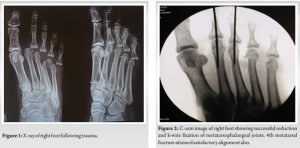
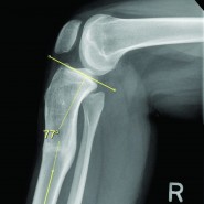
A 54-year-old male presented to the emergency department 5 days after a fall from height, complaining of pain and swelling in his right foot. Initial examination revealed dorsolateral dislocation of the second, third, and fifth MTP joints, coupled with a displaced fracture of the fourth metatarsal neck, while the fourth MTP joint remained intact (Fig. 1). Despite an unsuccessful closed reduction attempt, the patient was scheduled for open reduction and K-wire fixation to address the fourth metatarsal fracture and potentially facilitate the reduction of other MTP dislocations. A dorsal approach with two planned incisions was selected for the procedure. The initial incision was made in the third intermetatarsal space, and a careful dissection was performed to identify and reduce the challenging fourth metatarsal fracture. However, the reduction proved difficult, and satisfactory alignment with K-wires could not be achieved. Consequently, an extension of the incision proximally was required to manipulate the fracture effectively. Subsequently, a dorsal incision in the first intermetatarsal space was made, revealing soft tissue interpositions hindering the reduction of the second MTP joint. Through meticulous dissection and the release of soft tissue interpositions, the reduction of the second MTP joint was facilitated. This, in turn, led to the simultaneous reduction of the other MTP dislocations and the fourth metatarsal fracture. During the procedure, it was noted that the capsule and plantar plate were buttonholed into the joint space. The surgeon carefully pushed the plantar plate and other soft tissues posteriorly while simultaneously applying traction and a medial push to the proximal phalanx. Alternating dorsiflexion and plantar flexion maneuvers were employed at the second MTP joint to aid in the reduction. Importantly, the plantar plate was preserved throughout the procedure. The second MTP joint was stabilized with a K-wire, and under image guidance, confirmation was obtained that all other dislocations were reduced and the fracture was in good alignment. Additional K-wire fixation was extended to stabilize the third MTP joint as well (Fig. 2).
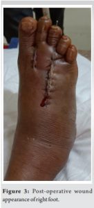
The wound was meticulously closed in layers after achieving hemostasis (Fig. 3), and the patient was immobilized with a below-knee plaster for duration of 1 month.
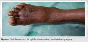
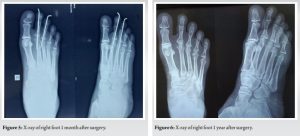
Post-operatively, the patient was monitored at regular intervals, and radiographic evaluations at 1 month (Fig. 4 and 5), 3 months, 6 months, and 1 year (Fig. 6) demonstrated satisfactory outcomes. The patient regained weight-bearing ability without pain, showcasing the success of the chosen intervention. He was able to return to his daily routine activities at the end of 3 months.
Reducing the second MTP joint during open reduction serves as a pivotal step, allowing for the restoration of the normal arc and creating adequate tension in the deep transverse metatarsal ligaments. This approach increases the likelihood of the other joints falling into place, contributing to a comprehensive reduction. It is important to note that multiple closed reduction attempts are not recommended, emphasizing the significance of prompt consideration for open reduction in cases of combined lesser MTP joint dislocations.
Understanding the intricate anatomy of the MTP joints is paramount when addressing MTP joint dislocations, as underscored by our experience in treating the presented case. The MTP joint, characterized as a synovial condyloid type of joint [1], plays a crucial role in the transverse arch of the foot and contributes significantly to the toe-off phase of the gait cycle [2]. Movements at these joints are limited to plantar flexion and dorsiflexion. The stability of the MTP joint is derived from a complex interplay of structures, including collateral ligaments, deep transverse metatarsal ligaments, the plantar plate, joint capsule, lumbricals, and the flexor and extensor tendons [5,6]. The plantar plate functions as the primary stabilizer of the MTP joint in the dorsal-plantar direction [6]. The plantar plate and deep transverse metatarsal ligaments, composed of fibrocartilage and forming a trapezoidal structure [7,8], act as static stabilizers [8], preventing dorsal translation and hyperextension of the proximal phalanx. Collateral ligaments contribute to sagittal plane stability. Notably, the second metatarsal, being the longest, assumes a pivotal role in the transverse arch. The plantar plate is attached to the distal fibers of the plantar fascia proximally, to the proximal phalanx distally, paired accessory collateral ligaments, the deep transverse metatarsal ligament, the fibrous sheath of flexor tendons, and the interosseous tendons [6]. Several factors contribute to the failure of closed reduction, and various mechanisms have been identified. The reduction process may encounter impediments due to the entrapment of the metatarsal head through the buttonhole of the capsule. In addition, the plantar plate can become entrapped between the dorsally displaced proximal phalanx and the metatarsal head, hindering the reduction process. Another potential obstacle arises from the incarceration of the metatarsal head under the flexor digitorum longus [9]. In our case, the patient presented with a combined dislocation, emphasizing the importance of restoring the normal arc connecting these joints for stability. Sudden dorsiflexion with considerable force can damage these supporting structures, leading to dorsal displacement [10], as observed in our patient. Notably, the order of reduction in combined MTP joint dislocations is not explicitly outlined in the literature. In the course of the procedure, a meticulous approach was taken to gently push the plantar plate posteriorly, with efforts made to avoid its release. This maneuver resulted in the successful reduction of the 2nd MTP joint. The significance lies in the attachment of the plantar plate to the deep transverse metatarsal ligaments. The reduction of the 2nd MTP joint played a crucial role in restoring tension between the deep transverse metatarsal ligaments. This adjustment not only facilitated the reduction of the specific joint but also contributed to the alleviation of other dislocations. The interconnected nature of the plantar plate and deep transverse metatarsal ligaments underscores the importance of their proper alignment in maintaining joint stability. Our experience revealed that attempting to reduce the fourth metatarsal fracture alone did not replicate the normal arc and failed to generate adequate tension in the deep transverse metatarsal ligaments. However, prioritizing the reduction of the second MTP joint proved effective. By reducing the second MTP joint first, the arc attained its normal length, providing sufficient pull in the supporting deep transverse metatarsal ligaments. Overall, the careful manipulation of the plantar plate proved instrumental in achieving the reduction of the 2nd MTP joint and addressing associated dislocations.
In the context of open reduction for combined dislocations of the lesser MTP joints, prioritizing the reduction of the second MTP joint is crucial for achieving simultaneous reduction of the other joints. Combined lesser MTP joint dislocation is a rare occurrence, often necessitating open reduction due to soft tissue interposition that impedes closed reduction. Nevertheless, attempts at closed reduction should be made, and a strategic focus on the second MTP joint can facilitate a more effective reduction process.
The prioritization of second MTP joint reduction during open reduction is highlighted as a key strategy, providing a more favorable biomechanical environment for achieving simultaneous reduction of the other affected joints. This approach aligns with the rarity and complexity of combined dislocations, emphasizing the importance of open reduction when closed reduction proves challenging due to soft tissue interposition.
References
- 1.Raj S, Subramani S, Babar SJ, Balaji MS, Anand V. Rare case report of closed traumatic dislocation of second to fifth metatarsophalangeal joints. Cureus 2020;12:e11745. [Google Scholar | PubMed]
- 2.Neogi DS, Bandekar SM, Sadekar V, Patnaik S, Bhat T, D’Mello Z. Irreducible dislocation of all the lesser metatarsophalangeal joints of the foot: A case report. Foot Ankle Spec 2012;5:324-6. [Google Scholar | PubMed]
- 3.Pai V. Irreducible dislocation of the metatarsophalangeal joints of the foot: A case report. Foot Ankle J 2008;1:5. [Google Scholar | PubMed]
- 4.Dhakshinamurthi Y, Meignanaguru M, Shetty GR. Management of acute traumatic irreducible metatarsophalangeal joint dislocation of the lesser toes: A case report. J Orthop Joint Surg 2023;5:37-9. [Google Scholar | PubMed]
- 5.Finney FT, Cata E, Holmes JR, Talusan PG. Anatomy and physiology of the lesser metatarsophalangeal joints. Foot Ankle Clin 2018;23:1-7. [Google Scholar | PubMed]
- 6.Nery C, Umans H, Baumfeld D. Etiology, clinical assessment, and surgical repair of plantar plate tears. Semin Musculoskelet Radiol 2013;20:205-13. [Google Scholar | PubMed]
- 7.Reeve A, Linklater JM, Dimmick S. Lesser metatarsophalangeal joint plantar plate degeneration and tear and acute first metatarsophalangeal joint capsuloligamentous injury: What the surgeon wants to know. Semin Ultrasound CT MR 2023;44:332-46. [Google Scholar | PubMed]
- 8.Maas NM, van der Grinten M, Bramer WM, Kleinrensink GJ. Metatarsophalangeal joint stability: A systematic review on the plantar plate of the lesser toes. J Foot Ankle Res 2016;9:32. [Google Scholar | PubMed]
- 9.De Palma L, Santucci A, Marinelli M. Traumatic dislocation of metatarsophalangeal joints: Report of three different cases. Foot Ankle Surg 2001;7:229-34. [Google Scholar | PubMed]
- 10.Brunet JA, Tubin S. Traumatic dislocations of the lesser toes. Foot Ankle Int 1997;18:406-11. [Google Scholar | PubMed]


Related Articles in Journal of Orthopaedic Case Reports
 August 10, 2021 Management of Symptomatic Isolated Congenital Anterior Cruciate Ligament Deficiency with Gradual Correction of Biplanar Proximal Tibial Deformity: A Case Report
 December 6, 2020 Dripping Wax Bone Disease – Melorheostosis – A Rare Case Scenario
 May 6, 2019 Reviewers Acknowledgement & Photo-gallery May-June 2019
 July 10, 2016 Dupuytren’s disease and needle aponeurotomy: rupture of a deep common flexor tendon: A case report and literature review.