
Flexible intramedullary nailing shows mostly successful outcomes; following basic surgical principles is required to avoid complications.
Dr. S Samundeeswari, Department of Orthopaedics, Sri Lakshmi Narayana Institute of Medical Sciences, Puducherry, India. E-mail: shastasamsas@gmail.com
Introduction: Diaphyseal forearm fractures pose a common challenge in children and adolescents, impacting forearm function due to rotational deformities and angulation. The landscape of pediatric forearm fracture treatment has seen limited progression, with increased surgical intervention adoption driven by factors such as functional implications, technological advancements, societal expectations, and legal concerns.
Materials and Methods: This study enrolled consecutive children aged 5–16 years with forearm fractures presenting between August 2018 and January 2020, requiring surgical intervention. The study assessed functional outcomes and complications in children treated with titanium elastic nailing.
Results: Sixteen patients underwent surgery for both-bone forearm fractures. Elastic nailing was the primary intervention, with 75% undergoing closed nailing. Patients’ ages ranged from 5 to 15 years, with 87.5% being male. The study evaluated fracture characteristics, surgical procedures, post-operative care, and complications.
Conclusion: The study demonstrates promising outcomes for flexible intramedullary nailing in pediatric forearm fractures. Despite the observed complications, the majority of cases achieved excellent results in fracture union and patient recovery, supporting the efficacy of this technique. Larger cohorts are needed for a comprehensive understanding of its applicability and outcomes in pediatric forearm fracture management.
Keywords: Pediatric forearm fractures, flexible intramedullary nailing, titanium elastic nailing, functional outcomes, surgical intervention.
Diaphyseal forearm fractures are prevalent among children [1-3] and adolescents [4-6], causing rotational deformities and angulation that impede forearm function [4,5]. Anatomical reduction is crucial, particularly for older children with limited remodeling potential [4,6]. Despite these concerns, the treatment landscape for pediatric forearm fractures has seen minimal progression. Factors such as functional implications of malunion, technological advancements, societal expectations, and legal concerns have driven the increased adoption of surgical intervention [7-9]. Open reduction and internal fixation (ORIF) methods involving stainless steel Kirschner wires, Steinmann’s pins, plates, screws, and intramedullary nails are common [4,5,8,10]. Among these, intramedullary nailing has gained traction due to the benefits it offers, including elasticity and stability from three-point fixation [11,12]. Intramedullary flexible nails, popularized by Metaizeau, provide good stability in long bone fractures in children [13,14], and in the forearm, they can adequately recreate the interosseous space [11,12]. Though they are not without complications [15], Titanium (Ti 6A114V) nails, favored over stainless steel, afford easier insertion, fixation, and fracture stability [5]. Although elastic nailing is established in the Western context, evidence remains scarce in the Indian population. Therefore, our study aims to address this gap by assessing the functional outcomes and complications in children with diaphyseal fractures of both forearm bones treated using titanium elastic nailing (TEN).
The study enrolled consecutive children aged 5–16 years with forearm fractures presenting between August 2018 and January 2020 and having unstable or unacceptable reduction. Institute Ethics Committee approval was obtained before initiating the study. Inclusion criteria encompassed children aged 5–16 years with forearm fractures displaying displacement, rotation, or reduction loss during conservative management. Type 3 compound fractures, pathological fractures, fractures associated with neurovascular injuries, and isolated fractures of either the radius or ulna were excluded from the study. The study proceeded with systematic data collection, encompassing pre-operative preparation, surgical procedures, and subsequent follow-up assessments.
Brief procedure
First, the fractures were assessed using digital radiographs and classified according to the AO classification system. Eligible patients underwent clinical and radiological evaluation before surgery, including routine blood tests and an assessment of their fitness for anesthesia. Next, the size of the nail needed was calculated based on the width of the forearm isthmus from the radiographs. Elastic nailing principles were used to ensure that the nail did not penetrate the physis. One nail per forearm bone was inserted, either retrograde for the radius or antegrade or retrograde for the ulna. The patient was lying on the operating table with the fractured limb on a radiolucent arm table, and a C-arm was used for imaging. The recommended nail diameter was 2/3 to 80% of the isthmus diameter, and similar diameters were used for balanced bending forces. For the radius, a dorsal approach was used on Lister’s tubercle with a perpendicular awl insertion angulated to 45° and a slightly larger opening than the nail diameter. The nail was introduced at 90°, rotated 180° to align with the medullary canal, and advanced with rocking motions to the fracture site. For the ulna, either an anterograde approach 2 cm distal to the olecranon or a retrograde approach 2 cm proximal to the joint line was used. The radius nail tip was aligned with the upper fragment medulla and advanced with reduction or open reduction, and the ulna nail advanced and docked the metaphysis with tips rotated toward the interosseous membrane. Finally, the nails were ensured to be properly positioned and cut 5 mm to 1 cm from the bone ends. Over-inserted nails were retracted with extraction pliers or capped, and the radial nail end was extended to avoid tendon friction. Image intensification was used to ensure that the reduction was correct and the nail length was appropriate.
Post-operative care
Immobilization was generally unnecessary post-operatively, allowing for the early initiation of active motion. In some cases, above-elbow plaster immobilization was preferred. Nail removal was done after complete fracture union and remodeling, typically occurring 6-month postoperatively, with the exact timing dependent on the patient’s age.
Post-operative assessment
All patients underwent a minimum of clinical and radiological assessments at 4, 8, 12, and 24 weeks post-surgery, followed by as needed thereafter. Clinical and radiological data were recorded using a pro forma and checklist. The results were evaluated using Daruwalla grading and et al. criteria [47,50]. A Student’s t-test was used to compare the means of the nail-to-canal ratio in relation to the presence or absence of major and/or minor complications.
Sixteen patients underwent surgery for two-bone fractures of the forearm. Twelve patients received elastic nailing for both bones, while four patients had the ulna nailed, while the radius was fixed with K-wires or plating due to their distal location. Patients’ ages ranged from 5 to 15 years, with a mean of 10 years. Seven were <10 years old and nine were >10 years old. Of the 16 patients, 14 (87.5%) were male and 2 (12.5%) were female (male: female ratio 7:1). Falling on an outstretched hand when playing was the primary causes (13 patients, 81.25%), while road-traffic accidents caused injuries in 3 patients (18.75%). The right forearm was affected in 9 patients (56.3%), and the left in 7 (43.8%). Most patients presented immediately after trauma, except one who presented 10 days later.
Fracture characteristics
All fractures were closed fractures (16 patients, 100%). Fracture distribution: middle one-third (8 patients, 50%), proximal one-third (5 patients, 31.3%), and distal one-third (3 patients, 18.8%). The most common pattern was transverse (9, 56.3%), followed by short-oblique (6, 37.5%), and partially comminuted (1, 6.3%). Overriding ranged from 6 mm to 18 mm (median = 13 mm). No major associated injuries or polytrauma were reported. Two patients (12.5%) had been treated by traditional bone setters before seeking orthopedic care.
Reduction and surgery
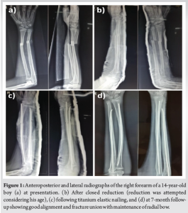
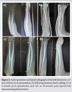
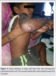
Most patients (11, 68.8%) had one reduction attempt, while 5 patients (31.3%) had two reduction attempts. Closed nailing of both bones was performed in 12 patients (75%), and open reduction in 4 (25%). Surgery was performed within 1–3 days (median = 1 day) from presentation, with surgical duration ranging from 60 to 180 min (median = 1.5 h). Hospital stays varied from 3 to 10 days (median = 5 days). The last follow-up ranged from 12 to 17-month postoperatively (mean = 15 months). C-arm images intraoperatively ranged from 8 to 40 (median = 22). In evaluating forearm fractures, the radius canal ranged from 3 mm to 5 mm (mean = 3.81), while the ulna canal measured 2 mm to 3 mm (mean = 2.4). The chosen diameter for titanium elastic nails was 2 mm to 3 mm, with 2.5 mm being the common choice for both the radius and ulna. The nail-to-canal ratio was determined as the bone isthmus diameter divided by the nail diameter. For the radius, the ratio ranged from 60.00 to 66.67 (mean = 64.81), and for the ulna, it ranged from 50.00 to 66.67 (mean = 60.39), revealing no significant relationship with complications (P > 0.05). During surgery, inserting nails into the distal fragment presented a common challenge, resulting in 25% of cases requiring open reduction. In post-surgery, below-elbow plaster support was provided for 2–4 weeks to ensure additional stability. Soft-tissue irritation manifested as nail extrusion in 18.75% of cases and skin irritation/bursitis in 12.5%; yet, these complications did not necessitate revision procedures. Most patients achieved full forearm rotations, elbows, and wrist movements by 12 weeks, except for one patient (6.25%) who experienced delayed union and 45° forearm pronation. Regarding angular and rotational malalignment, angular malalignment (>15° sagittal, >10° coronal) was observed in two patients (12.5%), with no significant movement restriction noted. Limb-length discrepancy was absent in all patients. The mean extraosseous nail lengths were 10.38 mm for the radius and 11.13 mm for the ulna, with nail end protrusion linked to lengths exceeding 10 mm. Fracture union, defined by bridging callus in ≥3 cortices, was achieved with varying times ranging from 8 weeks to 8 months (median = 12 weeks) (Fig. 1). Three patients had major complications (18.75%): a 15-year-old boy who needed open reduction for the radius had delayed union (Fig. 2); two patients (12.5%) developed malunion with loss of radial bow, one of them being the patient with delayed union; and one patient developed a deep infection in the ulnar nail entry site (Fig. 3), necessitating early removal of the nail at 2 months. Minor complications, including nail extrusion, prominences, and ulnar bursitis, were present in 37.5% of patients without necessitating unplanned revisions.
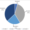
The final outcome was graded based on the price outcome score described by Price et al. as excellent, good, or poor. The outcome was “excellent” in 15 (93.8%), “good” in 1 (6.3%), and “poor” in 0 (0.0%). Similarly, the Daruwallah outcome score was “excellent” in 15 (93.8%) and “good” in 1 (6.3%). Nail removal was done post-fracture consolidation, occurring 6-month to 1-year post-surgery. None of the patients needed pre-union nail removal.
The epidemiology of forearm fractures in childhood reveals a prevalence of 18% by age 9, with a peak incidence occurring between 5 and 14 years, pre-dominantly affecting boys [16,17]. Forearm fractures constitute 40% of childhood fractures, with incidence peaks noted at 5–9 and 10–14 years with shaft fractures, accounting for 3–5% of pediatric fractures [17,18]. Forearm fracture treatment has shifted from common closed reduction, which had poor outcomes, to ORIF, associated with high infection and non-union rates [19]. While Sarmiento’s functional bracing approach exhibited superior results [20], Blount stressed the uniqueness of fractures in children due to their growth potential [21]. However, little is still known regarding the correction of rotational malalignment through remodeling [3]. Gandhi et al. pointed out that angular deformities persist in older children, prompting the emergence of alternative methods such as titanium elastic nails [22]. Surgical interventions are thus usually reserved for candidates who are generally non-responders to conservative management [2,22]. The remodeling potential of pediatric bones facilitates malunion correction, with effectiveness peaking below 10 years and declining thereafter [23-26]. Severe malunion correction is feasible in children below 8 years but diminishes in efficacy after 10 years [27]. The impact of age and fracture site on the remodeling of angular malalignment is evident, with sagittal malalignments showing better outcomes. Thus, Flynn’s acceptance of 15–20° angulation varies with age [28]. In contrast to angular malalignment, rotational malalignment lacks self-correction and is often associated with angulation [29,30]. Muscles play a crucial role in causing rotational deformity, impairing functionality, and leading to compensatory shoulder use, which can result in complications [31]. The primary goal of treatment should be restoring normal alignment, especially in complete shaft fractures [32,33]. While closed reduction with an above-elbow cast is common, malunion can compromise function [31,34,35]. Studies by Asadollahi identified a re-displacement rate of 11% due to poor reduction and increased angulation [36]. Price recommends angulations of ≤15° for children below 8 years and ≤10° for older children [37]. Non-operative approaches such as closed manipulation and casting have shown more favorable outcomes in age <10 years [38,39]. There is an increasing trend in surgical intervention, primarily for open fractures, instability, irreducibility, deformity, and post-manipulation re-displacement [12]. While elastic stable intramedullary nailing (ESIN) and plate-and-screw fixation offer similar outcomes [40], ESIN, being less invasive and tailored to pediatric anatomy, presents a more easily removable option [34,41]. Although plate fixation supports anatomic alignment and radial bow, allowing early motion [42], it may entail longer incisions and complications such as malunion and synostosis [42]. Flexible intramedullary nailing, introduced by Metaizeau, is a viable alternative using titanium alloy (Ti6Al14V) nails, known for their osseointegration, biocompatibility, and magnetic resonance imaging compatibility [43]. Curved nails enhance stability and radial bow [11,41]. Biomechanically, these curved nails boost interosseous membrane tension, providing rotational stability and radial bow [11,41]. Although complications such as extensor pollicis longus and extensor pollicis brevis ruptures, superficial radial nerve injury, and pin tract infections have been reported with elastic nailing, various studies report good functional outcomes [44-48]. Complication rates range from 14% to 21%, covering hardware issues, non-union, neurodeficits, infections, and compartment syndrome [25,34,41,49]. The management of pediatric forearm fractures has evolved from accepting deformity post-closed reduction to stringent alignment criteria [8,22]. Precision in angulation and rotation, aligned with the child’s age, is now demanded [3]. Internal fixation suits unstable fractures, malreduction, and older children [31,51,52]. In our study, patients specifically met these criteria, with 50–90% of internal fixations driven by instability and malreduction and no open fractures observed. Transverse fractures were pre-dominant, followed by short oblique fractures [27,37]. Although all patients underwent closed reduction, satisfactory alignment was not universally achieved [3]. The median age of patients was 11 years, with fracture union occurring within 8–12 weeks, except for one delayed union case. Healing rates after open reduction and internal fixation with plating have been shown to be slower than with elastic nailing [53]. Titanium nails’ elasticity allows up to 2° of movement, while their thicker periosteum offers stability and aids healing [31]. Nail selection is critical, with a nail-to-canal ratio ≥60–80% of the canal diameter required for stability [54]. The most common nail size used in our study was 2.5 mm for both the radius and the ulna. Immobilization post-TEN remains debatable, with patients commonly opting for plaster immobilization due to titanium nails’ relative flexibility and the risk of post-operative alignment loss [55]. No significant correlation was found between the nail-canal ratio and complications [56,57]. Surgery, ideally within 24–72 h, occurred on the nearest elective list [51], with a median hospital stay of 5 days for post-operative monitoring. Our series did not encounter any cases of non-union. Non-union has been reported primarily in open fractures [58]. Patient outcomes, evaluated using Price and Daruwallah scores, were pre-dominantly excellent or good [31,52,59]. However, the absence of open fractures may impact these outcomes. While TEN is not without complications, including nail extrusion, soft tissue irritation, and delayed union, our study indicates that flexible intramedullary nailing is a promising method for stabilizing unstable pediatric forearm diaphyseal fractures. Adhering to TEN principles and avoiding basic errors could prevent complications [41,49,60]. Neurovascular damage was absent in our study.
Our study has certain limitations. The sample size, although reflective of our institute’s patient load over 2 years, is small due to COVID-19 constraints. No open fractures were encountered, possibly due to regional trauma center distribution. This may account for our series’ favorable outcomes compared to the literature. A comparison with other treatments like conservative or plate osteosynthesis could yield stronger evidence, yet this was not our research aim [3].
Our study on flexible intramedullary nailing for pediatric forearm both-bone fractures revealed promising outcomes. The majority of cases underwent successful closed nailing, demonstrating overall excellent results in fracture union and patient recovery. Despite the observed complications, the study supports the efficacy of flexible intramedullary nailing in the management of pediatric forearm fractures. Further research with larger cohorts is warranted for a comprehensive understanding of this technique’s applicability and outcomes.
Flexible intramedullary nailing shows promise in pediatric forearm fractures, with mostly successful outcomes despite complications, emphasizing the need for further research with larger cohorts for a comprehensive understanding of its efficacy and applicability.
References
- 1.Waters PM, Skaggs DL, Flynn JM. Rockwood and Wilkins Fractures in Children. Philadelphia, PA: Lippincott Williams & Wilkins; 2019. [Google Scholar | PubMed]
- 2.Noonan KJ, Price CT. Forearm and distal radius fractures in children. J Am Acad Orthop Surg 1998;6:146-56. [Google Scholar | PubMed]
- 3.Madhuri V, Dutt V, Gahukamble AD, Tharyan P. Conservative interventions for treating diaphyseal fractures of the forearm bones in children. Cochrane Database Syst Rev 2013;4:CD008775. [Google Scholar | PubMed]
- 4.Westacott DJ, Jordan RW, Cooke SJ. Functional outcome following intramedullary nailing or plate and screw fixation of paediatric diaphyseal forearm fractures: A systematic review. J Child Orthop 2012;6:75-80. [Google Scholar | PubMed]
- 5.Vopat ML, Kane PM, Christino MA, Truntzer J, Mcclure P, Katarincic J, et al. Treatment of diaphyseal forearm fractures in children. Orthop Rev (Pavia) 2014;6:5325. [Google Scholar | PubMed]
- 6.Truntzer J, Vopat ML, Kane PM, Christino MA, Katarincic J, Vopat BG. Forearm diaphyseal fractures in adolescent population treatment and management. Eur J Orthop Surg Traumatol 2015;25:201-9. [Google Scholar | PubMed]
- 7.Shoemaker SD, Comstock CP, Mubarak SJ, Wenger DR, Chambers HG. Intramedullary Kirschner wire fixation of open or unstable forearm fractures in children. J Pediatr Orthop 1999;19:329-37. [Google Scholar | PubMed]
- 8.Sage FP. Medullary fixation of fractures of the forearm: A study of the medullary canal of the radius and a report of fifty fractures of the radius treated with a prebent triangular nail. J Bone Joint Surg Am 1959;41:1489-525. [Google Scholar | PubMed]
- 9.Thompson GH, Wilber JH, Marcus RE. Internal fixation of fractures in children and adolescents. A comparative analysis. Clin Orthop Relat Res 1984;188:10-20. [Google Scholar | PubMed]
- 10.Cruz AI Jr., Kleiner JE, DeFroda SF, Gil JA, Daniels AH, Eberson CP. Increasing rates of surgical treatment for paediatric diaphyseal forearm fractures: A national database study from 2000 to 2012. J Child Orthop 2017;11:201-9. [Google Scholar | PubMed]
- 11.Lascombes P, Prevot J, Ligier JN, Metaizeau JP, Poncelet T. Elastic stable intramedullary nailing in forearm shaft fractures in children: 85 cases. J Pediatr Orthop 1990;10:167-71. [Google Scholar | PubMed]
- 12.Furlan D, Pogorelic Z, Biocic M, Juric I, Budimir D, Todoric J, et al. Elastic stable intramedullary nailing for paediatric long bone fractures: Experience with 175 fractures. Scand J Surg 2011;100:208-15. [Google Scholar | PubMed]
- 13.Saseendar S, Panda S, Samundeeswari S, Kumar D. Titanium elastic nails in the management of fractures. In: Banerjee A, Biberthaler P, Shanmugasundaram S, editors. Handbook of Orthopaedic Trauma Implantology. Berlin: Springer, Singapore; 2022. [Google Scholar | PubMed]
- 14.Saseendar S, Menon J, Patro DK. Treatment of femoral fractures in children: Is titanium elastic nailing an improvement over hip spica casting? J Child Orthop 2010;4:245-51. [Google Scholar | PubMed]
- 15.Saseendar S, Menon J, Patro DK. Complications and failures of titanium elastic nailing in pediatric femur fractures. Eur J Orthop Surg Traumatol 2010;20:635-44. [Google Scholar | PubMed]
- 16.Sinikumpu JJ, Pokka T, Serlo W. The changing pattern of pediatric both-bone forearm shaft fractures among 86,000 children from 1997 to 2009. Eur J Pediatr Surg 2013;23:289-96. [Google Scholar | PubMed]
- 17.Lyons RA, Delahunty AM, Kraus D, Heaven M, McCabe M, Allen H, et al. Children’s fractures: A population based study. Inj Prev 1999;5:129-32. [Google Scholar | PubMed]
- 18.Ryan LM, Teach SJ, Searcy K, Singer SA, Wood R, Wright JL, et al. Epidemiology of pediatric forearm fractures in Washington, DC. J Trauma 2010;69:S200-5. [Google Scholar | PubMed]
- 19.Knight RA, Purvis GD. Fractures of both bones of the forearm in adults. J Bone Joint Surg Am 1949;31:755-64. [Google Scholar | PubMed]
- 20.Sarmiento A, Cooper JS, Sinclair WF. Forearm fractures. Early functional bracing-A preliminary report. J Bone Joint Surg Am 1975;57:297-304. [Google Scholar | PubMed]
- 21.Blount WP, Schaefer AA, Johnson JH. Fractures of the forearm in children. J Am Med Assoc 1942;120:111-6. [Google Scholar | PubMed]
- 22.Gandhi RK, Wilson P, Mason Brown JJ, Macleod W. Spontaneous correction of deformity following fractures of the forearm in children. Br J Surg 1962;50:5-10. [Google Scholar | PubMed]
- 23.Dormans JP, editor. Pediatric Orthopaedics: Core Knowledge in Orthopaedics. St. Louis: Mosby Incorporated; 2005. [Google Scholar | PubMed]
- 24.Tachdjian MO. Tachdjian’s Pediatric Orthopaedics. Philadelphia, PA: WB Saunders Company; 2002. [Google Scholar | PubMed]
- 25.Flynn JM, Jones KJ, Garner MR, Goebel J. Eleven years experience in the operative management of pediatric forearm fractures. J Pediatr Orthop 2010;30:313-9. [Google Scholar | PubMed]
- 26.Vittas DI, Larsen EI, Torp-Pedersen SO. Angular remodeling of midshaft forearm fractures in children. Clin Orthop Relat Res 1991;265:261-4. [Google Scholar | PubMed]
- 27.Fuller DJ, McCullough CJ. Malunited fractures of the forearm in children. J Bone Joint Surg Br 1982;64:364-7. [Google Scholar | PubMed]
- 28.Flynn JM, Sarwark JF, Waters PM, Bae DS, Lemke LP. The operative management of pediatric fractures of the upper extremity. J Bone Joint Surg 2002;84:2078-89. [Google Scholar | PubMed]
- 29.Rang M. Children’s Fractures. Philadelphia, PA: JB Lippincott; 1974. p. 126. [Google Scholar | PubMed]
- 30.Evans EM. Rotational deformity in the treatment of fractures of both bones of the forearm. J Bone Joint Surg 1945;27:373-9. [Google Scholar | PubMed]
- 31.Luhmann SJ, Gordon JE, Schoenecker PL. Intramedullary fixation of unstable both-bone forearm fractures in children. J Pediatr Orthop 1998;18:451-6. [Google Scholar | PubMed]
- 32.Tarr RR, Garfinkel AI, Sarmiento A. The effects of angular and rotational deformities of both bones of the forearm. An in vitro study. J Bone Joint Surg Am 1984;66:65-70. [Google Scholar | PubMed]
- 33.Schmittenbecher PP. State of the art treatment of forearm shaft fractures. Injury 2005;36:A25-34. [Google Scholar | PubMed]
- 34.Martus JE, Preston RK, Schoenecker JG, Lovejoy SA, Green NE, Mencio GA. Complications and outcomes of diaphyseal forearm fracture intramedullary nailing: A comparison of pediatric and adolescent age groups. J Pediatr Orthop 2013;33:598-607. [Google Scholar | PubMed]
- 35.Colaris JW, Allema JH, Reijman M, de Vries MR, Ulas Biter L, Bloem RM, et al. Which factors affect limitation of pronation/supination after forearm fractures in children? A prospective multicentre study. Injury 2014;45:696-700. [Google Scholar | PubMed]
- 36.Asadollahi S, Pourali M, Heidari K. Predictive factors for re-displacement in diaphyseal forearm fractures in children-role of radiographic indices. Acta Orthop 2017;88:101-8. [Google Scholar | PubMed]
- 37.Price CT. Acceptable Alingment of forearm fractures in children open reduction indications. J Pediatr Orthop 2010;30:S82-4. [Google Scholar | PubMed]
- 38.Tarmuzi NA, Abdullah S, Osman Z, Das S. Paediatric forearm fractures: Functional outcome of conservative treatment. Bratisl Lek Listy 2009;110:563-8. [Google Scholar | PubMed]
- 39.Zionts LE, Zalavras CG, Gerhardt MB. Closed treatment of displaced diaphyseal both-bone forearm fractures in older children and adolescents. J Pediatr Orthop 2005;25:507-12. [Google Scholar | PubMed]
- 40.Patel A, Li L, Anand A. Systematic review: Functional outcomes and complications of intramedullary nailing versus plate fixation for both-bone diaphyseal forearm fractures in children. Injury 2014;45:1135-43. [Google Scholar | PubMed]
- 41.Smith VA, Goodman HJ, Strongwater A, Smith B. Treatment of pediatric both-bone forearm fractures: A comparison of operative techniques. J Pediatr Orthop 2005;25:309-13. [Google Scholar | PubMed]
- 42.Holmes JH 4th, Wiebe DJ, Tataria M, Mattix KD, Mooney DP, Scaife ER, et al. The failure of nonoperative management in pediatric solid organ injury: A multi-institutional experience. J Trauma 2005;59:1309-13. [Google Scholar | PubMed]
- 43.Wall EJ, Jain V, Vora V, Mehlman CT, Crawford AH. Complications of titanium and stainless steel elastic nail fixation of pediatric femoral fractures. J Bone Joint Surg Am 2009;91:2041. [Google Scholar | PubMed]
- 44.Cumming D, Mfula N, Jones JW. Paediatric forearm fractures: The increasing use of elastic stable intramedullary nails. Int Orthop 2008;32:421-3. [Google Scholar | PubMed]
- 45.Weinberg AM, Castellani C, Amerstorfer F. Elastic stable intramedullary nailing (ESIN) of forearm fractures. Oper Orthop Traumatol 2008;20:285-96. [Google Scholar | PubMed]
- 46.Garg NK, Ballal MS, Malek IA, Webster RA, Bruce CE. Use of elastic stable intramedullary nailing for treating unstable forearm fractures in children. J Trauma 2008;65:109-15. [Google Scholar | PubMed]
- 47.Kapila R, Sharma R, Chugh A, Goyal M. Evaluation of clinical outcomes of management of paediatric bone forearm fractures using titanium elastic nailing system a prospective study of 50 cases. J Clin Diagn Res 2016;10:RC12-5. [Google Scholar | PubMed]
- 48.Kruppa C, Bunge P, Schildhauer TA, Dudda M. Low complication rate of elastic stable intramedullary nailing (ESIN) of paediatric forearm fractures. Medicine (Baltimore) 2017;96:e6669. [Google Scholar | PubMed]
- 49.Luhmann SJ, Schootman M, Schoenecker PL, Dobbs MB, Gordon JE. Complications of titanium elastic nails for pediatric femoral shaft fractures. J Pediatr Orthop 2003;23:443-7. [Google Scholar | PubMed]
- 50.Shivanna S, Maruthi CV. Pediatric forearm fractures with TENS: Freedom of movements. Int J Res Orthop 2016;2:142. [Google Scholar | PubMed]
- 51.Kapoor V, Theruvil B, Edwards SE, Taylor GR, Clarke NM, Uglow MG. Flexible intramedullary nailing of displaced diaphyseal forearm fractures in children. Injury 2005;36:1221-5. [Google Scholar | PubMed]
- 52.Richter D, Ostermann PA, Ekkernkamp A, Muhr G, Hahn MP. Elastic intramedullary nailing: A minimally invasive concept in the treatment of unstable forearm fractures in children. J Pediatr Orthop 1998;18:457-61. [Google Scholar | PubMed]
- 53.Shah AS, Lesniak BP, Wolter TD, Caird MS, Farley FA, Vander Have KL. Stabilization of adolescent both-bone forearm fractures: a comparison of intramedullary nailing versus open reduction and internal fixation. Journal of Orthopaedic Trauma. 2010 Jul 1;24(7):440-61. [Google Scholar | PubMed]
- 54.Dietz HG, Slongo T. AO Manual of Fracture Management-Elastic Stable Intramedullary Nailing in Children. New York: Thieme; 2006. [Google Scholar | PubMed]
- 55.Huber RI, Keller HW, Huber PM, Rehm KE. Flexible intramedullary nailing as fracture treatment in children. J Pediatr Orthop 1996;16:602-5. [Google Scholar | PubMed]
- 56.Pai VS, David GJ, Claude TJ. Femoral elastic nailing in the older child: Proceed with caution. Injury Extra 2005;36:185-9. [Google Scholar | PubMed]
- 57.Ozdemir HM, Yensel U, Senaran H, Mutlu M, Kutlu A. Immediate percutaneous intramedullary fixation and functional bracing for the treatment of pediatric femoral shaft fracture. J Pediatr Orthop 2003;23:453-7. [Google Scholar | PubMed]
- 58.Greenbaum B, Zionts LE, Ebramzadeh E. Open fractures of the forearm in children. J Orthop Trauma 2001;15:111-8. [Google Scholar | PubMed]
- 59.Jubel A, Andermahr J, Isenberg J, Issavand A, Prokop A, Rehm KE. Outcomes and complications of elastic stable intramedullary nailing for forearm fractures in children. J Pediatr Orthop B 2005;14:375-80. [Google Scholar | PubMed]
- 60.Slongo TF. Complications and failures of the ESIN technique. Injury 2005;36:A78-85. [Google Scholar | PubMed]


Related Articles in Journal of Orthopaedic Case Reports
 June 1, 2026 Survivorship and Functional Outcomes after Surgery for Metastatic Spinal Disease Including Cord Compression: Single-Surgeon Cohort Series from UK Tertiary Center
 June 1, 2026 Comparative Analysis of Functional Outcomes in Robotic-assisted versus Conventional Total Knee Arthroplasty
 May 1, 2026 Coronal Plane Alignment of the Knee Classification and Its Correlation with Post-operative Functional Outcomes in Total Knee Arthroplasty: A Prospective Cohort Study
 April 1, 2026 Outcomes of Total Hip Arthroplasty for Traumatic versus Non-Traumatic Hip Pathology: A Prospective Cohort Study