Conservative Management of Lateral Epicondylitis
Dr. M Sowbhikh Krishnan, Department of Orthopaedic Surgery, Chettinad Hospital and Research Institute, Rajiv Gandhi Salai, Kanchipuram, Tamil Nadu, India. E-mail: sowbhikh@gmail.com
Introduction: Lateral epicondylitis, or tennis elbow, affects 1%–3% of adults aged 35–50, causing pain and weakness in the dominant elbow due to chronic inflammation of the extensor tendon. While corticosteroid injections (CSI) are commonly used for treatment, they offer only short-term relief. Platelet-rich plasma (PRP) is a promising alternative with potential for long-term benefits. This study compares the efficacy of PRP and CSI in treating lateral epicondylitis.
Materials & Methods: A randomized controlled trial was conducted at Chettinad Hospital and Research Institute from February 2020 to March 2021, involving patients with lateral epicondylitis unresponsive to non-invasive treatments. Patients were randomly assigned to receive either PRP or CSI, with pre- and post-treatment pain and function assessed using VAS, PSFS, and PRTEE scores.
Results: PRP showed better long-term pain reduction and functional improvement than CSI. At 6 months, PRP-treated patients had significantly lower VAS and PRTEE scores, indicating superior outcomes.
Discussion: Although CSI provided quicker initial relief, PRP demonstrated sustained benefits at 3 and 6 months. PRP's effectiveness in promoting tissue healing may explain its long-term success.
Conclusion: PRP is more effective than CSI for long-term management of lateral epicondylitis, offering superior pain relief and functional improvement.
Keywords: Lateral epicondylitis, platelet-rich plasma injection, corticosteroid injection, Cozen’s test.
Lateral epicondylitis, also known as tennis elbow, affects 1%–3% of the general population between the ages of 35 and 50 [1]. It is caused by chronic inflammation of the extensor tendon affecting the ECRB [2]. The dominant elbow is usually affected due to repeated or forceful activities involving supination and extension of the wrist [1-3]. Symptoms include pain on the lateral side of the elbow, pain with wrist extension, and weakened grip strength. Treatment options include rest, NSAIDs, physiotherapy, splinting, shock wave diathermy therapy, injection therapies, and invasive surgery. However, their effectiveness is yet to be demonstrated [4,5]. Corticosteroid injection (CSI) is a commonly used therapy for tendinopathies, but its effects are temporary [6]. Ortho biological treatments, such as platelet-rich plasma (PRP), contain growth factors that help heal soft-tissue injuries [7,8]. While corticosteroids may provide short-term benefits, studies suggest that PRP may be more effective in the long term [9-11]. A recent study assessed the outcomes of people with lateral epicondylitis treated with PRP and CSI
This was a randomized control trial study which was conducted in Chettinad Hospital and Research Institute between February 2020 and March 2021 which involved all patients attending outpatient department at department of orthopedics, CHRI clinically diagnosed to have lateral epicondylitis. The Institutional Medical Ethics Committee approved this study. All the patients were selected for the research based on criteria for inclusion and exclusion and allotted to the respective groups using block randomization.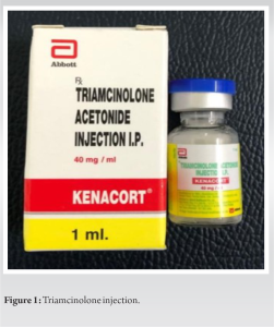
The Inclusion criteria for the study were as follows:
- Lateral epicondylitis for more than 3 months not responding to non-invasive treatment such as ice pack, medications, physiotherapy, and local ointments and gels.
- A pain score of at least 5 on a visual analog scale (VAS).
- Ruling out any bony injury after taking an X-Ray for confirmation.
- Positive Cozen’s test, Mill’s test, and Maudsley’s test
Exclusion criteria were as follows:
- Age below 18 years and above 50 years
- Carpal tunnel syndrome or cervical radiculopathy in the past
- History of old trauma or fracture over forearm
- Patient suffering from rheumatoid arthritis.
To ensure an identical number of instances in each category, block randomization was utilized. Each prospective participant with an odd serial number was assigned to the steroid category at random. Following the steroid case, the patient has been automatically assigned to the PRP category.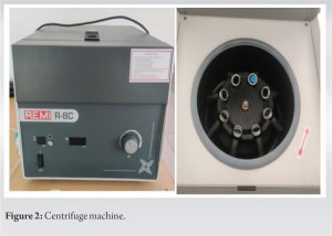
Proper history with occupation, age, sex, and side of the arm affected was noted. Patients were then clinically examined and the point of maximum tenderness was noted and diagnosis was confirmed by Cozen’s test, Mill’s test, and Maudsley’s test. VAS, PSFS, and patient-rated tennis elbow evaluation (PRTEE) scores were then assessed and recorded; patients gave written informed consent after explaining the study, benefits, and complications of the procedure to them. The procedure was done on an outpatient basis.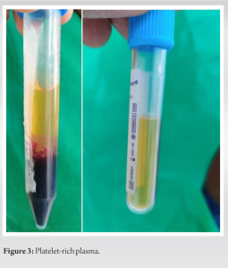
CSI
In the corticosteroid group of patients, 1 mL of 40 mg triamcinolone with 1 mL of lignocaine (2%, 10 mg/mL) was injected most tender point over the of humerus’s lateral epicondyle (ECRB tendon) using 22G needle, the peppering technique was used. After 15 min of observation, the patients were discharged.
PRP injection
To prepare PRP, 10 mL of the patient’s blood is drawn into a citrate tube and spun twice. The concentration of platelets is checked, and it is 5.5 times higher than in whole blood.
1–2 mL PRP collected in tube. Keeping the elbow parallel to the ground and hand mid pronated and the maximal tender point was identified by surgeon’s thumb. Under strict aseptic precautions, the PRP of 1 mL was injected into the tendon sheath of ECRB by clinical assessment of point of maximal tenderness using peppering technique.
The study compared PRP and steroid injections in treating tennis elbow among participants aged 18–40, with a majority of the female population. PRP showed better long-term pain reduction and functional improvement than steroids. Pre-injection scores for PRTEE score were 60.23 ± 10.464 and 72.50 ± 8.641, respectively, in PRP and steroid group and VAS were 6.77 ± 0.971 and 6.97 ± 0.964, respectively, in PRP and steroid group. However, at 4 weeks, the steroid group exhibited significantly lower PRTEE was 52.27 ± 10.201 and 42.70 ± 9.724, respectively, in PRP and steroid group with VAS scores 5.03 ± 0.928 and 3.97 ± 0.928, respectively, in PRP and steroid group, mean PRTEE score at 3 months was 28.57 ± 10.582 and 27.93 ± 10.99, respectively, in PRP and steroid group and mean PRTEE score at 6 months was 9.07 ± 4.201 and 23.30 ± 14.891, respectively, in PRP and steroid group and the mean VAS score at 6 months was 0.90 ± 1.561 and 2.43 ± 1.501, respectively, in PRP and steroid group, and the PRP group demonstrated substantially lower scores, suggesting superior long-term effectiveness. PSFS scores showed early functional benefits in the steroid group at 4 weeks. However, the PRP group had significantly higher PSFS scores at 6 months, suggesting better long-term functional outcomes. Complications, including recurrence, were minimal and similar between groups, with low rates of only 3.3% Both PRP and steroid injections provide relief for tennis elbow, but PRP offers better long-term benefits. Steroid injections may provide early relief but are not as effective as PRP over time. It is important to consider long-term outcomes when choosing treatment options for tennis elbow. (Graph 1-2).

Lateral epicondylitis is an inflammatory condition of the extensor tendon of the muscles of the forearm over the lateral epicondyle and is the most common chronic painful disabling condition. Our study compared PRP and CSIs in treating chronic lateral epicondylitis. After 4 weeks, corticosteroid showed more improvement. However, after 3 months, there was no significant difference, and after 6 months, PRP was significantly better in pain reduction and functional outcome. Omar AS et al. [10] showed that the mean age of the study participants was 40.5 and 37.5 in PRP and steroid group, respectively. The mean age of the participants in our study was 35 ± 7.3 and 35 ± 8.06 among the PRP and steroid group, respectively. In our study, the majority of the study participants in both groups were females comprising 63% and 63%. This is not in accordance with the study done by Sharma et al. [12], they showed that 38% and 40% of the study participants were female in PRP and steroid group, respectively. In our study, the right side (85%) most commonly involved similar to the results observed in the study conducted by Yadav et al [13]. In our study, the mean pre-injection VAS score among the PRP and steroid group was 6.77 ± 0.971 and 6.97 ± 0.964, respectively. Almost similar baseline VAS score was seen in a study done by Gawtham et al. [14]. The result in their study showed that the baseline VAS score was 7.1 and 7.0 in PRP and steroid group, respectively. In a study done by Sharma et al. [12], the results showed a slightly lower pre-injection VAS score was 5.78 and 5.46 among the PRP and steroid group, respectively. A higher baseline values were seen in a study done by Omar et al. [10], with the mean baseline VAS score as 8.0 and 8.6 in PRP and steroid group. Our study showed that although the mean VAS score got reduced post-injection in both groups, VAS score was higher among the PRP group, when compared to the steroid group post-injection at 4 weeks. The mean difference was higher among the steroid group at 4 weeks. The mean VAS score of PRP and steroid group at 4 weeks was 5.03 and 3.97, respectively. Similar report was shown in a study done by Gawtham et al. [14], they showed that the mean VAS score at 6 weeks was 2.7 and 1.4 in PRP and corticosteroid group, respectively. The mean VAS score at 3 months and 6 months post-injection was lower in the PRP group compared to steroid group. The mean difference than the pre-injection was also higher among the PRP group at 3 and 6 months. The mean VAS score at 3 months was 2.73 and 2.83 among the PRP and steroid group, respectively. This is almost similar to the study done by Yadav et al. [13], they showed that the mean VAS score was lower among the PRP group in comparison with those of the steroid group. The mean VAS score was 1.6 and 2.8 in the PRP and steroid group, respectively, in the later study. This is in contrast with the study done by Gawtham et al. [14], they showed that the mean VAS score was 1.8 and 1.7 in PRP and steroid group, respectively. The mean VAS score in our study at 6 months was 0.90 and 1.501 in the PRP and steroid group, respectively. Similar report has been shown in Varsheney et al. [15], they depicted that the mean VAS score was 0.69 and 4.61 at 6 months post-injection in the PRP and steroid group, respectively. A similar effect was shown by a study done by Khaliq et al. [16], they concluded that the PRP has a sustained effect on pain reduction. From pre-injection until follow-up for 6 months, a similar decreasing trend has been followed in Thanasas et al. [11] and Yadav et al. [13] for a steroid injection. The results thus showed us that though the mean VAS score was better at the earlier stage post-injection, PRP has a long-term effect reducing the pain more than the steroid at 3 and 6 months. Our study showed that the mean pre-injection PRTEE score was 69.23 and 72.50, respectively, in the PRP and steroid group. Likewise, a study done by Singh et al. [18] showed a pre-injection mean PRTEE score of 72.8 and 73.2, respectively, in PRP and steroid group, respectively. At 4 weeks post-injection, the mean PRTEE score was 52.27 and 42.70, respectively, in PRP and steroid group in the present study. Almost similar improvement has been shown in both groups. Nevertheless, the reduction was higher among the steroid group in the earlier week. A study done by Arik et al. [19] showed that the mean PRTEE score was 34.3 and 25 for autologous blood and steroid group. A study done by Arirachakaran et al. [7] showed that at around 2 months post-injection, the PRP and steroid group mean PRTEE score were 36.37 and 30.82, respectively. Our study showed that the mean PRTEE score was significantly lower among the PRP than the steroid group at 6 months after injection. Even though at the initial weeks, the mean PRTEE score was higher among the PRP group, at 6 months, the mean PRTEE score was significantly lower in the PRP group. A study done by Arirachakaran et al. [7] showed a similar trend at the final follow-up, they stated that the mean PRTEE score was 17.03 and 20.24, respectively, in PRP and steroid group. Likewise, a study done by Arik et al. [19] showed that at the final follow-up, the mean PRTEE score was 19.4 and 34.5 among the AB and steroid injection group, respectively. In our study, we have used PSFS score for the 1st time for an interventional study. This score has been used in various studies where different modalities of physiotherapy have been used to treat lateral epicondylitis. Our study showed that the mean pre-injection PSFS score was 2.83 and 2.90, respectively, in PRP and steroid group. Our study also showed that the mean post-injection PSFS score at 4 weeks was 4.53 and 5.30, respectively, in PRP and steroid group which shows a marginal difference in the functional outcome after PRP injection in comparison to steroid injection. In our study, the mean PSFS score at 3 months post-injection was 7.13 and 7.0 in PRP group and steroid group, respectively, which was not showing a significant difference between the groups. At 6 months post-injection the mean PSFS score was 9.17 and 7.43 in PRP group and steroid group, respectively, which showed a significant difference in the functional outcome between the two groups. It’s also shows that PRP injection has a better functional outcome at 6 months based on PSFS score used in our study. Corticosteroids have a high rate of relapsing and recurring, which is likely due to the fact that intra-tendinous injection can create permanent changes in the structure of the tendon, as well as the fact that patients tend to abuse the arm following injection due to immediate pain relief [10]. Autologous PRP, on the other hand, has been shown to improve early neotendon characteristics and tissue healing. The cells’ ability to recognize and react to mechanical stimulation at an early time point might be one explanation for PRP’s long-lasting impact [17]. Hence, at 3rd and 6th month post-injection, scores were better in PRP – group than the steroid – group. We encountered a complication of recurrence in our study. Although the mean recurrence was the same and not significant in both the study groups, common side effects found in other studies for steroid injection include steroid flare, tendon rupture, hypopigmentation, subcutaneous fat necrosis, and muscle atrophy [14].
We had a shorter follow-up and the subjects were not followed up after 6 months. Our sample size was small due to the COVID-19 pandemic situation. The injections in our patients were not given under ultrasound guidance. Ultrasound-guided injections may have an advantage as described in other studies. Some studies have given more than one dose of PRP/steroid injection for better outcome as well as to prevent recurrence, but the advantage multiple dose of injections still has not been established [17].
When comparing to CSI in the management of lateral epicondylitis, a single administration of autologous PRP offered greater long-term pain relief and improved functional ratings of the elbow. The improvement in the scores was maintained at 6 months in the PRP group when compared to the steroid group. In the steroid group, the pain and the functional scores started decreasing after 3 months. Hence, a single dose of PRP may be recommended in the treatment of lateral epicondylitis as it gives a better and more sustained outcome both in terms of pain and function.
PRP offers superior long-term pain relief and functional improvement over corticosteroid injections for treating lateral epicondylitis.
References
- 1.Nirschl RP, Pettrone FA. Tennis elbow. The surgical treatment of lateral epicondylitis. J Bone Joint Surg Am 1979;61:832-9. [Google Scholar | PubMed]
- 2.Jobe FW, Ciccotti MG. Lateral and medial epicondylitis of the elbow. J Am Acad Orthop Surg 1994;2:1-8. [Google Scholar | PubMed]
- 3.Levin D, Nazarian LN, Miller TT, O’Kane PL, Feld RI, Parker L, et al. Lateral epicondylitis of the elbow: US findings. Radiology 2005;237:230-4. [Google Scholar | PubMed]
- 4.Hart L. Corticosteroid and other injections in the management of tendinopathies: a review. Clinical journal of sport medicine. 2011 Nov 1;21(6):540-1. [Google Scholar | PubMed]
- 5.Edwards SG, Calandruccio JH. Autologous blood injections for refractory lateral epicondylitis. J Hand Surg 2003;28:272-8. [Google Scholar | PubMed]
- 6.Say F, Gürler D, Inkaya E, Bülbül M. Comparison of platelet-rich plasma and steroid injection in the treatment of plantar fasciitis. Acta Orthop Traumatol Turc 2014;48:667-72. [Google Scholar | PubMed]
- 7.Arirachakaran A, Sukthuayat A, Sisayanarane T, Laoratanavoraphong S, Kanchanatawan W, Kongtharvonskul J. Platelet-rich plasma versus autologous blood versus steroid injection in lateral epicondylitis: systematic review and network meta-analysis. Journal of Orthopaedics and Traumatology. 2016 Jun 1;17(2):101-12. [Google Scholar | PubMed]
- 8.Gosens T, Den Oudsten BL, Fievez E, Van’t Spijker P, Fievez A. Pain and activity levels before and after platelet-rich plasma injection treatment of patellar tendinopathy: A prospective cohort study and the influence of previous treatments. Int Orthop 2012;36:1941-6. [Google Scholar | PubMed]
- 9.Major HP. Lawn-tennis elbow [letter]. Br Med J 1883;2:557. [Google Scholar | PubMed]
- 10.Omar AS, Ibrahim ME, Ahmed AS, Said M. Local injection of autologous platelet rich plasma and corticosteroid in treatment of lateral epicondylitis and plantar fasciitis: randomized clinical trial. The Egyptian Rheumatologist. 2012 Apr 1;34(2):43-9. [Google Scholar | PubMed]
- 11.Thanasas C, Papadimitriou G, Charalambidis C, Paraskevopoulos I, Papanikolaou A. Platelet-rich plasma versus autologous whole blood for the treatment of chronic lateral elbow epicondylitis: a randomized controlled clinical trial. The American journal of sports medicine. 2011 Oct;39(10):2130-4. [Google Scholar | PubMed]
- 12.Sharma MK, Kumar R. Tennis Elbow Managed by Corticosteroid and Platelet Rich Plasma (PRP): A Prospective Comparative Study. [Google Scholar | PubMed]
- 13.Yadav R, Kothari SY, Borah D. Comparison of local injection of platelet rich plasma and corticosteroids in the treatment of lateral epicondylitis of humerus. J Clin Diagn Res 2015;9:RC05-7. [Google Scholar | PubMed]
- 14.Gautham VK, Verma S, Batra S, Bhatnagar N, Arora S. Platelet-rich plasma versus corticosteroid injection for recalcitrant lateral epicondylitis: clinical and ultrasonographic evaluation. Journal of Orthopaedic Surgery. 2015 Apr;23(1):1-5. [Google Scholar | PubMed]
- 15.Varshney A, Maheshwari R, Juyal A, Agrawal A, Hayer P. Autologous platelet-rich plasma versus corticosteroid in the management of elbow epicondylitis: A randomized study. Int J Appl Basic Med Res 2017;7:125-8. [Google Scholar | PubMed]
- 16.Khaliq A, Khan I, Inam M, Saeed M, Khan H, Iqbal MJ. Effectiveness of platelets rich plasma versus corticosteroids in lateral epicondylitis. J Pak Med Assoc 2015;65:S100-4. [Google Scholar | PubMed]
- 17.Krogh TP, Fredberg U, Stengaard-Pedersen K, Christensen R, Jensen P, Ellingsen T. Treatment of lateral epicondylitis with platelet-rich plasma, glucocorticoid, or saline: A randomized, double-blind, placebo-controlled trial. Am J Sports Med 2013;41:625-35. [Google Scholar | PubMed]
- 18.Singh A, Gangwar DS. Autologous blood versus corticosteroid local injection for treatment of lateral epicondylosis: A randomized clinical trial. Online J Health Allied Sci 2013;12:1-11. [Google Scholar | PubMed]
- 19.Arik HO, Kose O, Guler F, Deniz G, Egerci OF, Ucar M. Injection of autologous blood versus corticosteroid for lateral epicondylitis: A randomised controlled study. J Orthop Surg (Hong Kong) 2014;22:333-7. [Google Scholar | PubMed]


