

Early diagnosis of low-grade central osteosarcoma is crucial to prevent delayed treatment and high-grade transformation.
Dr. Avani M. Dangar, Department of Pathology, All India Institute of Medical Sciences, Rajkot, Gujarat, India. E-mail: avanidangaro68@gmail.com
Abstract
Introduction: Low-grade central osteosarcoma (LGCO) is a rare subtype of osteosarcoma, accounting for approximately 1–2% of cases. It typically exhibits indolent behavior but poses a diagnostic challenge due to its close radiologic and histologic resemblance to benign fibro-osseous lesions. Although LGCO generally has a favorable prognosis, it may undergo high-grade transformation, leading to more aggressive clinical behavior.
Case Report: We report a case of a 36-year-old male with type 2 diabetes mellitus who presented with recurrent right ankle pain following a remote history of trauma. Imaging revealed an expansile lytic lesion in the distal tibia. Initial biopsy suggested a benign fibroblastic lesion, favoring non-ossifying fibroma; however, repeat biopsy raised suspicion for LGCO based on spindle cell proliferation, irregular woven bone, mild nuclear pleomorphism, and focal osteoid formation. Positron emission tomography-computed tomography demonstrated a metabolically active lesion confined to the distal tibia with no evidence of metastasis. The patient underwent wide local excision followed by reconstruction with fibular tibialization and ankle arthrodesis. Final histopathology confirmed LGCO with high-grade transformation, demonstrating increased mitotic activity, marked nuclear atypia, vascular invasion, and osteoid production. Surgical margins were negative.
Discussion: This case is notable for its uncommon presentation in an adult patient, distal tibial involvement, and early high-grade transformation. The initial misdiagnosis highlights the critical importance of clinico-radiologic-pathologic correlation and the role of adjunct immunohistochemistry (MDM2/CDK4) in differentiating LGCO from benign mimickers. Limb salvage with fibular centralization proved to be an effective reconstructive strategy, achieving oncologic clearance while preserving function.
Conclusion: LGCO can present diagnostic difficulties and may exhibit unexpected aggressive behavior due to high-grade transformation. Early recognition, accurate diagnosis, complete surgical excision, and vigilant long-term follow-up are essential to optimize outcomes. Distal tibial reconstruction using fibular tibialization and ankle arthrodesis remains a viable limb-salvage option.
Keywords: Benign fibroblastic lesion, low-grade central osteosarcoma, high-grade transformation.

The most prevalent primary malignant bone tumor is osteosarcoma, which usually develops in the metaphysis of long bones in teenagers and young adults. It is distinguished histologically by the creation of malignant osteoid by tumor cells [1]. Among its subtypes, low-grade central osteosarcoma (LGCO) is extremely rare, representing only 1–2% of all osteosarcomas [2]. LGCO is often indolent, with slow growth, and has a much better prognosis compared to conventional high-grade osteosarcoma, provided complete surgical excision with negative margins is achieved [3]. A major diagnostic challenge of LGCO is its frequent radiologic and histologic resemblance to benign fibro-osseous lesions such as fibrous dysplasia, desmoplastic fibroma, or non-ossifying fibroma [4]. Misdiagnosis can delay definitive treatment, underscoring the importance of careful clinic-radio-pathological correlation and the use of adjunct immunohistochemistry. Amplification of MDM2 and CDK4 genes is a useful biomarker in distinguishing LGCO from benign mimickers [5]. Although LGCO is generally associated with favorable outcomes, it may undergo high-grade transformation (dedifferentiation) either at initial presentation or more commonly at recurrence [6]. This transformation confers aggressive biological behavior, higher rates of metastasis, and poorer prognosis and therefore alters management strategies [7]. Reported transformation rates vary from 10% to 36% in published series [6,7]. The distal tibia is an uncommon site for osteosarcoma overall, and its involvement presents unique reconstructive challenges due to limited soft-tissue coverage and the functional importance of the ankle joint [8]. Limb salvage procedures such as wide resection followed by fibular centralization (tibialization) and ankle arthrodesis have been reported with satisfactory oncological and functional outcomes [9]. The present case highlights the diagnostic challenges of LGCO arising in an unusual location with early high-grade transformation, emphasizing the importance of early recognition, accurate histopathological evaluation, and appropriate surgical management to improve patient outcomes [10].
A 36-year-old male patient with a known case of type 2 diabetes mellitus (on medications) presented to the orthopedic outpatient department with a history of a slip and fall about 5 years ago, after which he developed right ankle pain for 15 days; he had no history of swelling, redness, or deformity. The pain subsequently subsided; however, he continued to experience intermittent episodes of pain over the following years. About 1 month ago, symptoms recurred. Imaging (X-ray, magnetic resonance imaging) showed an expansile lytic lesion of the distal tibia (Fig. 1).
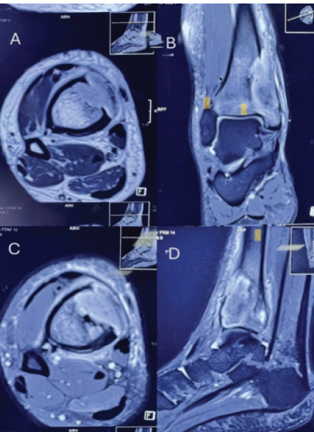
Figure 1: Axial T2-weighted image (a) and coronal proton density fat-suppressed (PDFS) (b) of the distal tibia demonstrate an eccentric, appearing hypointense on sagittal T1-weighted image (c) involving the medial epimetaphyseal region. The lesion causes cortical destruction and interrupted periosteal reaction and extends to the subarticular location without intra-articular extension. Post-contrast axial T1-weighted fat-saturated image (d) reveals heterogeneous enhancement. Perilesional marrow and regional periosseous soft tissues show mild edema, appearing hyperintense on PDFS images with corresponding mild post-contrast enhancement. Appearances are in keeping with an aggressive neoplastic lesion.
On examination, findings were mild swelling/tenderness over the right ankle, distal pulses palpable, and no obvious deformity. Initial needle biopsy showed spindle-shaped fibroblasts arranged in intersecting fascicles. Foci of reactive woven bone formation are also seen in the periphery. Fibroblastic proliferation with abundant collagen fibers is noted, extending up to and surrounding the bony trabeculae. Aggregates of foamy histiocytes are also seen focally. No significant giant cell proliferation. No nuclear pleomorphism or mitosis is seen. Features favor non-ossifying fibroma. After a multidisciplinary conference, a repeat biopsy was done; it shows mild to moderately cellular spindle cells in the fibrous sclerotic stroma. Mild-to-moderate nuclear pleomorphism is seen. Foci of irregular woven bone are present (Figs. 3d, 4a, and 4b). Mitotic activity is low. Focal osteoid matrix formation is also seen. The focal area shows evident nuclear pleomorphism with a few mitoses. The focal area resembles the appearance of a fibroma. Small fragments of skin and hemorrhage are also present. The impression is that the possibility of LGCO is favored over a benign fibroblastic tumor. Advice is clinico-radiological correlation and immunohistochemistry using CDK4 and MDM2 for further confirmation. The metastatic work-up of a positron emission tomography-computed tomography scan showed a lesion in the right distal tibia (cortex, cortex erosion, marrow infiltration, and joint extension), size 34 × 28 mm in axial, craniocaudal extent 51 mm, and maximum standardized uptake value 6.1. No evidence of metastasis or lymphadenopathy. Patient underwent wide local excision with reconstruction by tibialization of the fibula on the right side, using an 11-hole medial locking plate, plus two 20 mm K-wires (SS) (Fig. 2).
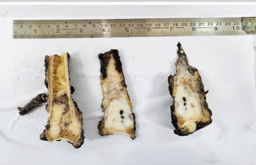
Figure 2: Gross image trisected into three shows the lower end of the tibia showing growth involving the epiphysis and metaphysis, involving both cortex and medulla, reaching up to the periosteum.
The histopathology report shows LGCO with high-grade transformation. Predominantly low-grade areas show mild to moderately cellular spindle cells in the fibrous sclerotic stroma. Mild-to-moderate nuclear pleomorphism is seen (Fig. 3c). Foci of irregular woven bone are present. Focal osteoid matrix formation is also seen (Fig. 3a).
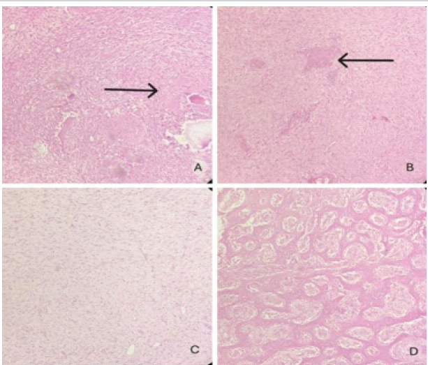
Figure 3: (a) High-grade osteosarcoma transformation with osteoid formation (black arrow marked) (Hematoxylin and Eosin [H&E]-stained slides, ×10). (b) Low-grade osteosarcoma component with osteoid formation and mild to moderate nuclear pleomorphism (black arrow marked), mild to moderately cellular spindle cells in the fibrous sclerotic stroma also seen (H&E-stained slides, ×10). (c) Predominantly low-grade areas show mild to moderately cellular spindle cells in the fibrous sclerotic stroma (H&E-stained slides, ×10). (d) Woven bone formation with osteoblasts showing mild to moderate pleomorphism (H&E-stained slides, ×10).
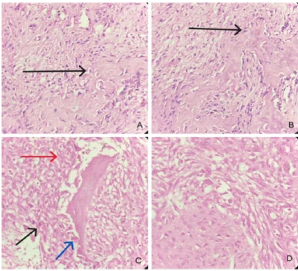
Figure 4: (a) Low-grade osteosarcoma component with mild pleomorphism and low-grade nuclei with focal new bone formation (black arrow marked) (Hematoxylin and Eosin [H&E]-stained slides, ×40). (b) Low-grade osteosarcoma component with mild pleomorphism and low-grade nuclei with focal new bone formation (black arrow marked) (H&E-stained slides, ×40). (c) High-grade transformation of osteosarcoma showing atypical mitosis (black arrow marked), marked nuclear pleomorphism, prominent nucleoli, and irregular nuclear border (red arrow marked) and occasional osteoclastic giant cells (blue arrow marked) with focal irregular woven bone formation (H&E-stained slide, ×40). (d) High-grade transformation with marked nuclear pleomorphism, prominent nucleoli, and irregular nuclear border (H&E-stained slides, ×40).
Young people most frequently develop LGCO, an uncommon subtype of osteosarcoma that makes up about 1% to 2% of cases, in the metaphysis of their long bones [2]. Our case is unusual as it occurred in a 36-year-old male with involvement of the distal tibia, an infrequent site for osteosarcoma [3,8]. Due to its overlap with benign fibro-osseous lesions such as fibrous dysplasia or desmoplastic fibroma, the LGCO diagnosis is still difficult [4]. This was evident in our instance, where a definitive diagnosis was delayed because the initial biopsy suggested a benign pathology. The literature has extensively described these problems, highlighting the significance of thorough clinico-radiologic and histological correlation [4]. The existence of high-grade transformation at the time of resection was another significant feature of this instance. The prognosis is considerably worsened when LGCO dedifferentiates into high-grade sarcoma, a well-known event. While Yadav et al. [7] emphasized its clinical implications, such as increased metastatic risk and worse outcomes, Choong et al. [6] documented this transition in a subgroup of patients throughout long-term follow-up. Features such as vascular invasion and significant mitotic activity in our patient demonstrated this aggressive change, highlighting the necessity of careful monitoring even for tumors that were initially deemed low-grade. Due to the ankle joint’s functional significance and the limited soft-tissue coverage, treating osteosarcoma of the distal tibia presents particular difficulties. Amputation was prevalent in the past, but limb-salvaging techniques are now more popular. Successful results with distal tibia excision followed by fibular centralization and ankle arthrodesis were reported by Wu et al. [8] and Kundu ZS et al. [9], offering both oncological safety and functional preservation. According to these accounts, our patient had ankle fusion, fibular tibialization, and broad local excision, resulting in negative margins despite close clearing. Compared to the review by Bertoni et al. [2], where most LGCOs behaved indolently with good outcomes after complete excision, our case demonstrates an atypical course with early high-grade transformation. This underlines that while LGCO generally carries a favorable prognosis, clinicians must remain alert to its potential for aggressive behavior.
LGCO is a rare tumor that can mimic benign fibro-osseous lesions, often leading to diagnostic delays. Although generally considered indolent with a favorable prognosis, our case illustrates that high-grade transformation can occur even at the time of initial diagnosis, conferring a more aggressive biological course. Distal tibial involvement poses additional surgical challenges, but limb salvage using fibular tibialization with ankle arthrodesis remains a viable reconstructive option. Careful long-term surveillance is essential, as recurrence and transformation may significantly influence outcomes.
Low-grade central osteosarcoma should always be considered in persistent bone lesions that appear benign but show clinical or radiological discordance. Misdiagnosis may result in inadequate treatment and tumor progression. Early multidisciplinary evaluation, appropriate biopsy sampling, and definitive wide excision remain the cornerstone of management. Awareness of the possibility of high-grade transformation is crucial, as it significantly alters prognosis and therapeutic planning. Limb-salvage procedures can successfully preserve function while achieving oncological clearance.
References
- 1. Mirabello L, Troisi RJ, Savage SA. Osteosarcoma incidence and survival rates from 1973 to 2004: Data from the surveillance, epidemiology, and end results program. Cancer 2009;115:1531-43. [Google Scholar] [PubMed]
- 2. Bertoni F, Bacchini P, Staals EL. Low-grade central osteosarcoma: A review of 45 cases. Am J Clin Pathol 2005;124:711-6. [Google Scholar] [PubMed]
- 3. Campanacci M, Picci P, Gherlinzoni F. Osteosarcoma of the distal tibia: Results of 29 cases. Cancer 1981;48:1569-82. [Google Scholar] [PubMed]
- 4. De Andrea CE, San-Julian M, Bovée JV. Low-grade central osteosarcoma: Diagnostic pitfalls and differential diagnosis. Sarcoma 2012;2012:756710. [Google Scholar] [PubMed]
- 5. Dujardin F, Binh MB, Bouvier C, Gomez-Brouchet A, Larousserie F, Muret AD, et al. MDM2 and CDK4 immunohistochemistry is a valuable tool in the differential diagnosis of low-grade osteosarcomas and other primary fibro-osseous lesions of the bone. Mod Pathol 2011;24:624-37. [Google Scholar] [PubMed]
- 6. Choong PF, Pritchard DJ, Rock MG, Sim FH, Frassica FJ. Low-grade central osteosarcoma: A long-term followup of 20 patients. J Bone Joint Surg Am 1996;78:1363-72. [Google Scholar] [PubMed]
- 7. Yadav AK, Tewari S, Singh A, Misra V. Low grade central osteosarcoma transforming to high grade sarcoma-clinical implications and brief review of literature. Indian J Orthop Surg 2021;7:344-7. [Google Scholar] [PubMed]
- 8. Zhao Z, Yan T, Tang X, Guo W, Yang R, Tang S. Novel “double-strut” fibula ankle arthrodesis for large tumor-related bone defect of distal tibia. BMC Musculoskelet Disord. 2019 Aug 9;20(1):367. doi: 10.1186/s12891-019-2742-6. PMID: 31399083; PMCID: PMC6689168. [Google Scholar] [PubMed] [CrossRef]
- 9. Kundu ZS, Gogna P, Gupta V, Singla R, Sangwan SS, Mohindra M, Singh A. Ankle fusion with centralisation of the fibula after distal tibia bone tumour resection. J Orthop Traumatol. 2014 Jun;15(2):95-101. doi: 10.1007/s10195-013-0279-2. Epub 2013 Dec 15. PMID: 24337812; PMCID: PMC4033796. [Google Scholar] [PubMed] [CrossRef]
- 10. Kurt AM, Unni KK, McLeod RA, Pritchard DJ. Low-grade intraosseous osteosarcoma. Cancer 1990;65:1418-28. [Google Scholar] [PubMed]
Related Articles in Journal of Orthopaedic Case Reports
 June 1, 2025 Rare Case of Subscapularis Myositis Ossificans with Intra-articular Extension
 April 1, 2025 Recurrent Intra-articular Lipoma of Ankle Joint; A Rare Case Report
 July 10, 2021 Success Story of Ilizarov in Chronic Non-union of Tibia with 18 Years of Morbidity: A Case Report
 June 10, 2022 Ancient Schwannoma with Hemorrhagic Degeneration Causing Chronic Intramuscular Hematoma: A Case Report