

Plastic deformation of the tibia in pediatric fractures is exceedingly rare, though taught commonly in orthopedic and medical textbooks. This case highlights the unambiguous documentation of a tibial plastic deformation, emphasizing the importance of clinical vigilance and comprehensive radiographic assessment when evaluating pediatric lower extremity trauma.
Maria Luiso, Division of Orthopedic Surgery and Sports Medicine, Ann and Robert H. Lurie Children’s Hospital of Chicago, Chicago, Illinois, USA. E-mail: maluiso@luriechildrens.org
Abstract
Introduction: Although taught everywhere and in textbooks as a common fracture condition in orthopedics, plastic deformation of the lower extremities in pediatrics has been exceedingly rarely reported.
Case Report: We present unambiguous documentation of a case of a 9-year-old female who sustained an isolated plastic deformation of the tibia and fibula. The diagnosis and treatment course are described, with emphasis on this documentation with serendipitous full-length leg radiographs obtained before and images obtained after injury that demonstrate the plastic deformation pattern.
Conclusion: This case provides novel and unambiguous documentation of plastic deformation of the tibia for the medical literature given its rarity in the pediatric orthopedic population. This case presentation provides valuable radiographic documentation.
Keywords: Tibia, fracture, plastic deformation, lower extremity, tibial fracture, lower extremity fracture, trauma.

Plastic deformation of long bones is a known unique pediatric orthopedic condition, likely secondary to increased circumferential lamellar bone, fewer osteons and osteon fragments, and less mineralization compared to adults [1,2,3]. This culmination of factors results in decreased modulus of elasticity, increasing the possibility of persistent plastic deformation before cortex failure [4]. We present a case of an isolated plastic deformation of the tibial shaft in a 9-year-old female, with emphasis on the rare documentation with unique full-length radiographic images, which include pre-injury and post-healing. Of the cases published, lower extremity cases are exceedingly rare, with only three previous reports of pediatric tibial involvement and none with unambiguous documentation [5,6,7,8]. A case with complete pre- and post-event documentation is presented here.
We report the case of a 9-year-old female who presented to the orthopedic clinic for assessment of a recent right lower extremity injury sustained during a family soccer game in November 2023. The patient reported that her adult cousin had slid and fallen on her ankle during play. Despite the injury, the patient was able to bear weight and walk, though her mother noted that she had developed a limp. The patient’s past medical history was significant for adolescent idiopathic scoliosis and genu valgum, for which she had undergone an unrelated contralateral left medial proximal tibia hemiepiphysiodesis on July 14, 2023. Her most recent follow-up before this injury was in October 2023, where standing leg radiographs showed neutral alignment (Fig. 1).
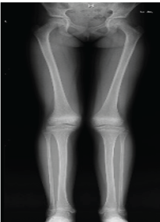
Figure 1: Full-length legs standing anteroposterior X-ray, pre-injury, demonstrating native lower extremity alignment.
On recent physical examination for the current injury, her right lower extremity showed mild bruising anterior to the medial malleolus without significant swelling or deformity. There was tenderness to palpation over the mid-anterior tibia and with a lateral squeeze. Neurovascular status was intact, with brisk capillary refill and normal sensation to light touch in the tibial, deep peroneal, and superficial peroneal nerve distributions. She demonstrated a full range of motion at the ankle. Gait examination revealed an antalgic gait. Radiographs of the right lower extremity taken in November 2023 revealed a plastic bowing deformity of the tibia and fibula (Fig. 2), which were compared to known pre-injury films.
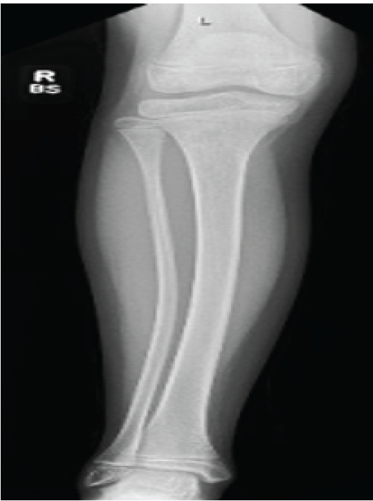
Figure 2: R tibia/fibula anteroposterior X-ray, days after injury, demonstrating right tibia and fibula plastic deformation.
Given the stability of the plastic deformity and the patient’s ability to bear weight, conservative management was initiated. The patient was fitted with a controlled ankle motion boot for 10 days, with instructions to weight-bear as tolerated. Follow-up to monitor healing and ensure proper alignment included repeat standing lower extremity radiographs showing near full restoration of the tibia and fibula morphology at 2-year follow-up (Fig. 3).
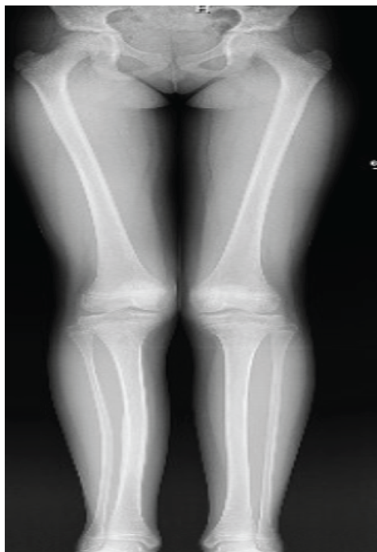
Figure 3: Full-length legs standing anteroposterior X-ray, at follow-up, demonstrating near native right tibia and fibula curvature with possible lateral tibial shaft cortical reaction.
Plastic deformation of long bones in pediatric patients is a biomechanical phenomenon arising from the unique histological characteristics of immature bone, which confer greater elasticity and the capacity to absorb energy without immediate cortical failure [1,2,3,4]. While this property accounts for the relative frequency of plastic deformation injuries in children, the anatomical distribution of such injuries is not uniform across the extremities, with the radius and ulna being almost exclusively reported [5]. The forearm with two parallel bones of similar diameter, each with intrinsic curvature, has a broad zone of plastic deformation before fracture occurs [9,10]. Moreover, the forearm is frequently subjected to longitudinal and rotational forces during falls or direct trauma, mechanisms that readily induce plastic bowing [9]. In contrast, the tibia is a single, relatively straight and robust bone and is less commonly exposed to the specific loading patterns that facilitate plastic deformation. The absence of intrinsic curvature in the tibia likely reduces the likelihood of plastic deformation, as the energy required to induce such a change in a straight bone is substantially higher [10]. Further, the mechanisms of injury in those rare reported cases of tibial plastic deformation often involve atypical force vectors, such as a fulcrum-like mechanism or direct longitudinal compression, which may be less common in typical pediatric trauma [5,6,7,8]. Radiographically, plastic deformation of the tibia can be subtle, as there may be no overt fracture line or periosteal reaction. The diagnosis relies on the identification of smooth, persistent bowing on full-length radiographs and careful clinical correlation. Failure to recognize and appropriately manage these injuries may result in angular deformity, altered biomechanics, and an increased risk of the requirement of later surgical intervention [11,12,13]. Management strategies are informed by the degree of deformity, patient age, and remodeling potential. Younger children may exhibit spontaneous correction, but remodeling is limited in older patients, and residual deformities >10° may necessitate surgical intervention, particularly if symptomatic, underscoring the importance of early diagnosis and individualized treatment planning to optimize functional outcomes and minimize long-term sequelae [8,13].
In summary, documented plastic deformation of the tibia is rare in the orthopedic literature. Plastic deformation is attributable to differences in anatomy, loading patterns, and biomechanical properties. This case presentation highlights the unambiguous documentation for the literature of tibial plastic deformation and demonstrates the ability of younger children for fracture remodeling of plastic deformation of the long bones.
Documented plastic deformation of the tibia is rare in the orthopedic literature. This case presentation highlights the unambiguous documentation for the literature of tibial plastic deformation and presents a case example for future reference, emphasizing the importance of clinical vigilance and comprehensive radiographic assessment when evaluating pediatric lower extremity trauma.
References
- 1. Mabrey JD, Fitch RD. Plastic deformation in pediatric fractures: Mechanism and treatment. J Pediatr Orthop 1989;9:310-4. [Google Scholar] [PubMed]
- 2. Currey JD. The mechanical consequences of variation in the mineral content of bone. J Biomech 1969;2:1-11. [Google Scholar] [PubMed]
- 3. Kerley ER. The microscopic determination of age in human bone. Am J Phys Anthropol 1965;23:149-63. [Google Scholar] [PubMed]
- 4. Currey JD, Butler G. The mechanical properties of bone tissue in children. J Bone Joint Surg Am 1975;57:810-4. [Google Scholar] [PubMed]
- 5. Stenström R, Gripenberg L, Bergius A. Traumatic bowing of forearm and lower leg in childen. Acta Radiol Diagn (Stockh) 1978;19:243-9. [Google Scholar] [PubMed]
- 6. Dhillon PS, Currall V, Shannon MN. Traumatic plastic deformation of the tibia: Case report and literature review. Ann R Coll Surg Engl 2012;94:e30-2. [Google Scholar] [PubMed]
- 7. Neumann L. Acute plastic bowing fractures of both the tibia and the fibula in a child. Injury 1990;21:122-3. [Google Scholar] [PubMed]
- 8. Orenstein E, Dvonch V, Demos T. Acute traumatic bowing of the tibia without fracture. Case report. J Bone Joint Surg Am 1985;67:965-7. [Google Scholar] [PubMed]
- 9. Borden S. Traumatic bowing of the forearm in children. J Bone Joint Surg Am 1974;56:611-6. [Google Scholar] [PubMed]
- 10. Borden S 4th. Roentgen recognition of acute plastic bowing of the forearm in children. Am J Roentgenol Radium Ther Nucl Med 1975;125:524-30. [Google Scholar] [PubMed]
- 11. Van Der Schoot DK, Den Outer AJ, Bode PJ, Obermann WR, Van Vugt AB. Degenerative changes at the knee and ankle related to malunion of tibial fractures: 15-Year follow-up of 88 patients. J Bone Joint Surg Br 1996;78:722-5. [Google Scholar] [PubMed]
- 12. Kettelkamp DB, Hillberry BM, Murrish DE, Heck DA. Degenerative arthritis of the knee secondary to fracture malunion. Clin Orthopaed Relat Res 1988;234:159-69. [Google Scholar] [PubMed]
- 13. Heinrich SD, Mooney JF. Rockwood CA, Wilkins KE, Beaty JH. Fractures of the shaft of the tibia and fibula. In: Rockwood and Wilkins’ Fractures in Children. Vol. 4. Philadelphia, USA: Lippincott-Raven Publishers; 2006. p. 1033-76. [Google Scholar] [PubMed]



Related Articles in Journal of Orthopaedic Case Reports
 September 1, 2025 Transverse Periprosthetic Fracture With a Bent Stem Following Reverse Total Shoulder Arthroplasty – A Case Report
 July 10, 2023 A Type IV Tibial Tuberosity Avulsion Fracture in an Adolescent – A Case Report
 August 10, 2022 Acute Reconstruction of Traumatic Zone IV Re-tear of Extensor Hallucis Longus Tendon using Extensor Hallucis Brevis loop Technique – A Mid-term Follow-up Case Report
 September 1, 2026 Limb Salvage for Entire Tibial Ewing Sarcoma in a 4-year-Old Child Using Extracorporeal Irradiation and Free Flap Reconstruction: A Case Report and Surgical Technique